



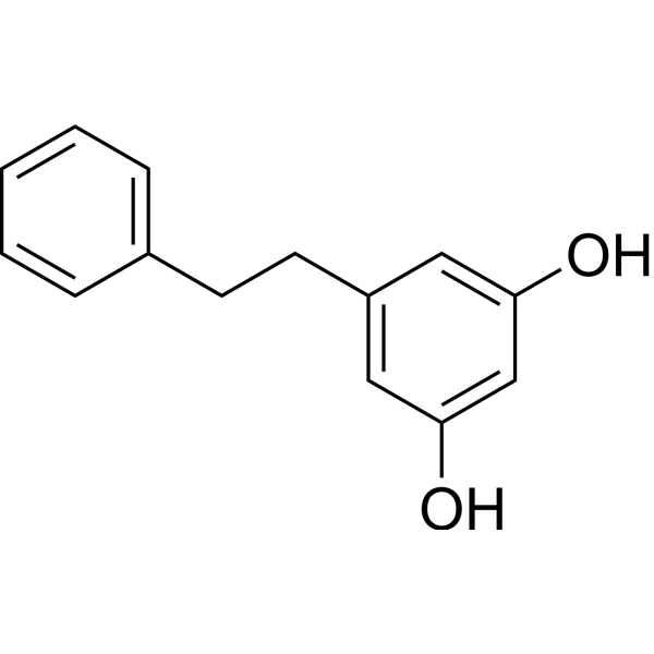
Lunularin

Based on 1 Customer Validation
Lunularin is an inhibitor of 11β-hydroxysteroid dehydrogenase 1, with an IC50 of 45.44 μM and a Ki of 35.8 μM against human 11β-HSD1, and an IC50 of 17.39 μM and a Ki of 10.31 μM against rat 11β-HSD1. Lunularin upregulates the transcription levels of Sirt1 and Hmox1 genes in the liver. Lunularin reduces food intake and body weight gain, and decreases blood glucose levels in mice fed a high-fat diet. Lunularin inhibits LPS-induced TLR4-mediated NF-κB pathway activation and nitric oxide production. Lunularin inhibits the proliferation and colony formation of renal cancer and colon cancer cells, and exhibits cancer cell-specific cytotoxicity. Lunularin binds to the steroid-binding site of human 11β-HSD1 and the steroid/NADPH-binding region of rat 11β-HSD1, but does not inhibit 11β-HSD2 or mouse 11β-HSD1. Lunularin can be used in research related to diet-induced obesity, renal cancer, colorectal cancer, inflammatory diseases and metabolic syndrome.
For research use only. We do not sell to patients.
- Purity : 99.87%
- CAS No.: 37116-80-6
- Formula: C14H14O2
- Molecular Weight:214.26
-
Storage:
4°C, stored under nitrogen
* In solvent : -80°C, 6 months; -20°C, 1 month (stored under nitrogen)


To place orders, for customer services and technical support, please contact: MedChemExpress USA
Tel: 609-228-6898 E-mail: [email protected] [email protected]


-
 Biological Activity
-
Chemical Information
-
Solvent & Solubility
- Protocols
- Purity & Documentation
- References
-
 Help & FAQs


All Endogenous Metabolite Isoforms
More

Biological Activity
Description
Cellular Effect
|
Cell Line

|
Type | Value | Description | References |
|---|---|---|---|---|
| A549 | IC50 |
150 μM
Compound: Lunularin
|
Cytotoxic activity against human A549 cells assessed as reduction in cell viability incubated for 24 hrs by MTT assay
Cytotoxic activity against human A549 cells assessed as reduction in cell viability incubated for 24 hrs by MTT assay
|
[PMID: 30848895] |
| J774 | IC50 |
>512 μM
Compound: HO21
|
Cytotoxicity against mouse J774 cells assessed as cell viability after 48 hrs by resazurin assay
Cytotoxicity against mouse J774 cells assessed as cell viability after 48 hrs by resazurin assay
|
[PMID: 25884114] |
| MRC5 | IC50 |
200 μM
Compound: Lunularin
|
Cytotoxic activity against human MRC5 cells assessed as reduction in cell viability incubated for 24 hrs by MTT assay
Cytotoxic activity against human MRC5 cells assessed as reduction in cell viability incubated for 24 hrs by MTT assay
|
[PMID: 30848895] |
In Vitro
Lunularin (0.5×-1.5×; 26.8-80.4 nmol/g) dose-dependently inhibits the proliferation of 786-O human renal adenocarcinoma cells, with greater activity than dihydroresveratrol alone at concentrations relevant to kidney tissue levels[2].
Lunularin (0.5×-1.5×; 26.8-80.4 nmol/g) dose-dependently inhibits the proliferation of A498 human renal carcinoma cells, with greater activity than dihydroresveratrol alone at concentrations relevant to kidney tissue levels[2].
Lunularin (1×; 53.6 nmol/g) significantly inhibits the clonogenic growth of 786-O human renal adenocarcinoma cells at concentrations relevant to kidney tissue levels, with enhanced activity when combined with dihydroresveratrol[2].
Lunularin (1×100; 53.6 nmol/g) significantly inhibits the clonogenic growth of A498 human renal carcinoma cells at concentrations relevant to kidney tissue levels, with enhanced activity when combined with dihydroresveratrol[2].
Lunularin (0.5×-1.5×; 30.25-90.75 nmol/g) inhibits the proliferation of HCT-116 human colorectal carcinoma cells, with enhanced activity when combined with dihydroresveratrol at concentrations relevant to colonic tissue levels[2].
Lunularin (1×; 60.5 nmol/g; 12 days) significantly inhibits the clonogenic growth of HT-29 human colon adenocarcinoma cells at concentrations relevant to colonic tissue levels, with enhanced activity when combined with dihydroresveratrol[2].
Lunularin (0.5×-1.5×; 30.25-90.75 nmol/g) dose-dependently inhibits LPS-induced NO production in RAW264.7 mouse macrophages, with enhanced activity when combined with dihydroresveratrol at concentrations relevant to colonic tissue levels[2].
Lunularin (0.5×-1.5×; 30.25-90.75 nmol/g) dose-dependently inhibits LPS-induced SEAP production in HEK-Blue mTLR-4 cells (targeting the TLR-4-mediated NF-κB pathway), with greater activity than resveratrol alone at concentrations relevant to colonic tissue levels[2].
Lunularin (20-100 μM; 30 min to standard assay conditions) competitively inhibits human liver microsomal 11β-HSD1 with an IC50 of 45.44 μM and a Ki of 35.8 μM by binding to the enzyme's steroid-binding site[4].
Lunularin (12.5-100 μM; 30 min to standard assay conditions) acts as a mixed inhibitor of rat liver microsomal 11β-HSD1 with an IC50 of 17.39 μM and a Ki of 10.31 μM by binding to the enzyme's combined NADPH and steroid-binding region[4].
Lunularin does not inhibit mouse liver microsomal 11β-HSD1 activity[4].
MedChemExpress (MCE) has not independently confirmed the accuracy of these methods. They are for reference only.
In Vivo
Lunularin is a gut microbiota-derived metabolite of resveratrol, is highly abundant in mouse tissues, biological fluids, and gastrointestinal tract compared to resveratrol itself, and is eliminated when gut microbiota is depleted via antibiotics[2].
MedChemExpress (MCE) has not independently confirmed the accuracy of these methods. They are for reference only.
-
Animal Model:C57BL/6JRj (13-month-old, diet-induced obesity model)[1]
-
Dosage:24 mg/kg
-
Administration:intraperitoneal injection; 3 times per week; 8 weeks
-
Result:Slightly reduced feed intake to 2.81 ± 0.1 g per day.
Showed a highly significant reduction in body weight gain over the study period.
Had slightly lower blood glucose levels after 8 weeks of treatment.
Had significantly elevated hepatic Sirt1 mRNA levels
Had significantly elevated hepatic Hmox1 mRNA level.
Reached mean liver tissue concentrations of 28.8 pmol/g liver.
Did not alter plasma levels of cholesterol, insulin, leptin, or adiponectin; did not alter hepatic phosphorylated AMPK levels, major urinary protein levels, or hepatic mRNA levels of Pck1, Pgc1α, Foxo3, or Tnfα.


Chemical Information
-
CAS No. 37116-80-6
-
Appearance Solid
-
Molecular Weight 214.26
-
Formula C14H14O2
-
Color White to off-white
-
SMILES
OC1=CC=C(CCC2=CC=CC(O)=C2)C=C1
-
Structure Classification
-
Shipping
Room temperature in continental US; may vary elsewhere.
-
Storage
4°C, stored under nitrogen
* In solvent : -80°C, 6 months; -20°C, 1 month (stored under nitrogen)


Solvent & Solubility
In Vitro:
DMSO : 100 mg/mL (466.72 mM; Need ultrasonic; Hygroscopic DMSO has a significant impact on the solubility of product, please use newly opened DMSO)
Please refer to the solubility information to select the appropriate solvent. Once prepared, please aliquot and store the solution to prevent product inactivation from repeated freeze-thaw cycles.
Storage method and period of stock solution: -80°C, 6 months; -20°C, 1 month (stored under nitrogen). When stored at -80°C, please use it within 6 months. When stored at -20°C, please use it within 1 month.
Please refer to the solubility information to select the appropriate solvent. Once prepared, please aliquot and store the solution to prevent product inactivation from repeated freeze-thaw cycles.
Storage method and period of stock solution: -80°C, 6 months; -20°C, 1 month (stored under nitrogen). When stored at -80°C, please use it within 6 months. When stored at -20°C, please use it within 1 month.
Concentration (start) × Volume (start) = Concentration (final) × Volume (final)
In Vivo:
Select the appropriate dissolution method based on your experimental animal and administration route.
- For the following dissolution methods, please ensure to first prepare a clear stock solution using an In Vitro approach and then sequentially add co-solvents:
- To ensure reliable experimental results, the clarified stock solution can be appropriately stored based on storage conditions. As for the working solution for In Vivo experiments, it is recommended to prepare freshly and use it on the same day.
- The percentages shown for the solvents indicate their volumetric ratio in the final prepared solution. If precipitation or phase separation occurs during preparation, heat and/or sonication can be used to aid dissolution.
Add each solvent one by one: 10% DMSO 40% PEG300 5% Tween-80 45% Saline
Solubility: ≥ 2.5 mg/mL (11.67 mM); Clear solution
This protocol yields a clear solution of ≥ 2.5 mg/mL (saturation unknown).
Taking 1 mL working solution as an example, add 100 μL DMSO stock solution (25.0 mg/mL) to 400 μL PEG300, and mix evenly; then add 50 μL Tween-80 and mix evenly; then add 450 μL Saline to adjust the volume to 1 mL.
Preparation of Saline: Dissolve 0.9 g sodium chloride in ddH₂O and dilute to 100 mL to obtain a clear Saline solution.
Add each solvent one by one: 10% DMSO 90% (20% SBE-β-CD in Saline)
Solubility: ≥ 2.5 mg/mL (11.67 mM); Clear solution
This protocol yields a clear solution of ≥ 2.5 mg/mL (saturation unknown).
Taking 1 mL working solution as an example, add 100 μL DMSO stock solution (25.0 mg/mL) to 900 μL 20% SBE-β-CD in Saline, and mix evenly.
Preparation of 20% SBE-β-CD in Saline (4°C, storage for one week): 2 g SBE-β-CD powder is dissolved in 10 mL Saline, completely dissolve until clear.
In Vivo Dissolution Calculator
Please enter the basic information of animal experiments:
-
-
-
-
Recommended: Prepare an additional quantity of animals to account for potential losses during experiments.
Please enter your animal formula composition:
-
%DMSO +
Recommended: Keep the proportion of DMSO in working solution below 2% if your animal is weak.
-
%+
-
+%Tween-80 + +
-
%Saline +
The co-solvents required include: DMSO, . All of co-solvents are available by MedChemExpress (MCE). , Tween 80. All of co-solvents are available by MedChemExpress (MCE).
Working solution concentration: 0.22 mg/mL
Method for preparing stock solution: mg drug dissolved in μL DMSO. Stock solution concentration: mg/mL. * In solvent : -80°C, 6 months; -20°C, 1 month (stored under nitrogen)
1. Take μL DMSO stock solution;
2. Add μL .
μL , mix evenly;
3. Then add μL Tween 80, mix evenly;
4. Then add μL
Please ensure that the stock solution in the first step is dissolved to a clear state, and add co-solvents in sequence. You can use ultrasonic heating (ultrasonic cleaner, recommended frequency 20-40 kHz), vortexing, etc. to assist dissolution.


Purity & Documentation
-
Data Sheet (277 KB)
-
SDS (394 KB)
- English - EN (394 KB)
- Français - FR (394 KB)
- Deutsch - DE (394 KB)
- Norwegian - NO (394 KB)
- Español - ES (394 KB)
- Swedish - SV (394 KB)
- Italian - IT (394 KB)
- Korean - KR (394 KB)
- Portuguese - PT (394 KB)
-
Handling Instructions (2659 KB)
References
[1]. Pallauf K, et al. Resveratrol, lunularin and dihydroresveratrol do not act as caloric restriction mimetics when administered intraperitoneally in mice. Sci Rep. 2019;9(1):4445. Published 2019 Mar 14. [Content Brief]
[2]. Li F, et al. Gut Microbiota-Derived Resveratrol Metabolites, Dihydroresveratrol and Lunularin, Significantly Contribute to the Biological Activities of Resveratrol. Front Nutr. 2022;9:912591. Published 2022 May 11. [Content Brief]
[4]. Hu C, et al. Resveratrol analogues and metabolites selectively inhibit human and rat 11β-hydroxysteroid dehydrogenase 1 as the therapeutic drugs: structure-activity relationship and molecular dynamics analysis. SAR QSAR Environ Res. 2024;35(7):641-663. [Content Brief]


Complete Stock Solution Preparation Table
Please refer to the solubility information to select the appropriate solvent. Once prepared, please aliquot and store the solution to prevent product inactivation from repeated freeze-thaw cycles.
Storage method and period of stock solution: -80°C, 6 months; -20°C, 1 month (stored under nitrogen). When stored at -80°C, please use it within 6 months. When stored at -20°C, please use it within 1 month.
| Optional Solvent | Concentration Solvent Mass | 1 mg | 5 mg | 10 mg | 25 mg |
|---|---|---|---|---|---|
| DMSO | 1 mM | 4.6672 mL | 23.3361 mL | 46.6723 mL | 116.6807 mL |
| 5 mM | 0.9334 mL | 4.6672 mL | 9.3345 mL | 23.3361 mL | |
| 10 mM | 0.4667 mL | 2.3336 mL | 4.6672 mL | 11.6681 mL | |
| 15 mM | 0.3111 mL | 1.5557 mL | 3.1115 mL | 7.7787 mL | |
| 20 mM | 0.2334 mL | 1.1668 mL | 2.3336 mL | 5.8340 mL | |
| 25 mM | 0.1867 mL | 0.9334 mL | 1.8669 mL | 4.6672 mL | |
| 30 mM | 0.1556 mL | 0.7779 mL | 1.5557 mL | 3.8894 mL | |
| 40 mM | 0.1167 mL | 0.5834 mL | 1.1668 mL | 2.9170 mL | |
| 50 mM | 0.0933 mL | 0.4667 mL | 0.9334 mL | 2.3336 mL | |
| 60 mM | 0.0778 mL | 0.3889 mL | 0.7779 mL | 1.9447 mL | |
| 80 mM | 0.0583 mL | 0.2917 mL | 0.5834 mL | 1.4585 mL | |
| 100 mM | 0.0467 mL | 0.2334 mL | 0.4667 mL | 1.1668 mL |


Lunularin Related Classifications
HY-124529 Related Classifications
 Powered by Bioz