



HRX-0233

Based on 1 Customer Validation
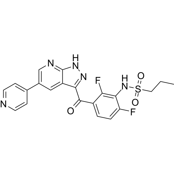
HRX-0233 is an orally potent MAP2K4 inhibitor. HRX-0233 inhibits the MAP2K4-JNK-JUN signaling pathway, blocks the feedback activation of receptor tyrosine kinases, and sustains the inhibitory effect on KRAS signaling. HRX-0233 exerts synergistic inhibition on the mTORC1 and MAPK pathways. When combined with Ipatasertib (HY-15186) or Sotorasib (HY-114277), it produces synergistic antiproliferative, pro-apoptotic (apoptosis) and antitumor effects, and shows good tolerability in mouse models. HRX-0233 can be used in CRISPR knockout screens to identify vulnerability-related genes. HRX-0233 is applicable to research related to prostate cancer, non-small cell lung cancer and colorectal cancer.
For research use only. We do not sell to patients.
- Purity : 98.92%
- CAS No.: 2409140-12-9
- Formula: C24H21F2N5O3S
- Molecular Weight:497.52
-
Storage:Powder -20°C, 3 years ; In solvent -80°C, 6 months , -20°C, 1 month


To place orders, for customer services and technical support, please contact: MedChemExpress USA
Tel: 609-228-6898 E-mail: [email protected] [email protected]


-
 Biological Activity
-
Chemical Information
-
Solvent & Solubility
- Protocols
- Purity & Documentation
- References
-
 Help & FAQs


All MEK Isoforms
More

Biological Activity
Description
IC50 & Target
|
MAP2K4 |
mTOR1 |
In Vitro
Combination treatment with HRX-0233 and Ipatasertib (HY-15186) synergistically reduces cell fitness and inhibits the activities of the mTORC1 and MAPK pathways in AR-negative, PTEN-deficient prostate cancer cells[1].
HRX-0233 (0-10 μM; 10 to 14 d) exhibits highly synergistic effects with Sotorasib (HY-114277), RMC-6236 (HY-148439), Trametinib (HY-10999) and SCH772984 (HY-50846) in KRASG12C-mutant H358, H2122, A549, SW837, HCT116 and DLD1 cancer cells, with the average Bliss synergy score reaching up to 69.89[2].
HRX-0233 (6 μM; 6-72 h) inhibits the MAP2K4-JNK-JUN signaling pathway, prevents the feedback activation of ERBB2/ERBB3, and sustains the inhibition of downstream RAS signaling pathways (phosphorylated ERK, phosphorylated RSK, phosphorylated S6) in H358, SW837, H2122 and DLD1 cells treated with Sotorasib, RMC-6236 or Trametinib[2].
HRX-0233 (6 μM) exhibits weak antiproliferative activity, but exerts synergistic effects with Sotorasib or Trametinib in wild-type H358 and SW837 cells; however, its activity is reduced in MAP2K4 knockout cells, indicating that this effect is mainly a target-specific one[2].
HRX-0233 (6 μM; 3-14 d) acts synergistically with Sotorasib to inhibit proliferation and induce apoptosis in H358 and SW837 cells; it also exerts a synergistic effect with RMC-6236 to suppress proliferation of DLD1 cells[2].
MedChemExpress (MCE) has not independently confirmed the accuracy of these methods. They are for reference only. Further protocols information, click here.
-
Cell Line:KRAS-mutant non-small cell lung cancer (NSCLC) cell lines H358 (KRASG12C), H2122 (KRASG12C), A549 (KRASG12S) and colorectal cancer (CRC) cell lines SW837 (KRASG12C), HCT116 (KRASG13D), DLD1 (KRASG13D)
-
Concentration:0, 2, 4, 6, 8 and 10 μM
-
Incubation Time:10 to 14 d, media refreshed every 2 to 3 d
-
Result:Exhibited minimal single-agent antiproliferative activity in all tested cell lines.
Produced average Bliss synergy scores of 69.89 (H358), above the 10 threshold for synergy, when combined with sotorasib, with scores also above 10 in H2122 and SW837.
Yielded an average Bliss synergy score of 46.41 (DLD1), with scores above 10 across all tested cell lines, when combined with RMC-6236.
Produced average Bliss synergy scores above 10 in all six cell lines tested when combined with trametinib or SCH772984.
-
Cell Line:KRAS-mutant NSCLC cell lines H358 (KRASG12C), SW837 (KRASG12C) and CRC cell line DLD1 (KRASG13D)
-
Concentration:6 μM; 6 μM combined with 50 nM sotorasib (H358, SW837) or 100 nM RMC-6236 (DLD1) (apoptosis readout)
-
Incubation Time:10-14 d, media refreshed twice weekly; 72 h (apoptosis readout)
-
Result:Had a modest impact on cell proliferation and minimal caspase 3/7 activity when administered alone in all tested cell lines.
Caused sustained suppression of cell proliferation and strong synergistic induction of caspase 3/7 activity in H358 and SW837 cells when combined with sotorasib.
Caused sustained proliferation suppression but only a modest increase in caspase 3/7 activity in DLD1 cells when combined with RMC-6236.
-
Cell Line:KRAS-mutant NSCLC cell lines H358 (KRASG12C), SW837 (KRASG12C), H2122 (KRASG12C) and CRC cell line DLD1 (KRASG13D)
-
Concentration:6 μM; 6 μM combined with 25 nM sotorasib, 100 nM RMC-6236, or trametinib
-
Incubation Time:6, 48 and 72 h; 6 and 48 h (with sotorasib); 6 h, 72 h (with RMC-6236); 6 and 72 h (with trametinib)
-
Result:Decreased phospho-JUN and total JUN levels in all tested cell lines when administered alone.
Prevented the upregulation of phospho-JUN, phospho-ERBB2, phospho-ERBB3, and total ERBB2/ERBB3 levels induced by single-agent KRAS/MAPK inhibitor treatment at 48 to 72 h when combined with sotorasib, RMC-6236, or trametinib.
Sustained suppression of phospho-ERK, phospho-RSK, and phospho-S6 levels at 48 to 72 h when combined with sotorasib, RMC-6236, or trametinib, whereas single-agent KRAS/MAPK inhibitor treatment showed rebound activation of these signaling markers.
Had no effect on phospho-ERK or phospho-RSK levels when administered alone.
-
Cell Line:MAP2K4 wild-type and knockout KRASG12C-mutant NSCLC cell line H358 and CRC cell line SW837
-
Concentration:6 μM
-
Incubation Time:5 d, media refreshed after 2 d
-
Result:Induced mild sensitivity in wild-type H358 and SW837 cells, while MAP2K4 knockout cells showed significantly less sensitivity when administered alone.
Enhanced the antiproliferative effect of sotorasib or trametinib in wild-type cells but not in MAP2K4 knockout cells.
In Vivo
HRX-0233 (250 mg/kg; p.o.; daily; up to 60 days) in combination with trametinib 0.25 mg/kg provides more sustained tumor suppression and reduces tumor cell proliferation in H2122 KRASG12C-mutant non-small cell lung cancer xenografts without causing toxicity[2].
MedChemExpress (MCE) has not independently confirmed the accuracy of these methods. They are for reference only.
-
Animal Model:6- to 8-week-old NOD-SCID IL-2Rg(null) (NSG) mice were subcutaneously injected in the right flank with 5 × 10⁶ H358 cells (mixed 1:1 with Matrigel), and treatment was initiated when tumor volumes reached 200-250 mm3[2]
-
Dosage:250 mg/kg (combined with Sotorasib (HY-114277) 10 mg/kg)
-
Administration:p.o.; daily; up to 60 days
-
Result:Showed no effect on tumor growth compared to vehicle.
Induced durable tumor shrinkage (with individual mice showing >30% tumor volume reduction from baseline, meeting RECIST criteria for partial response) over 60 days when combined with sotorasib.
Reduced the percentage of Ki67-positive cells to a significantly lower level than vehicle, HRX-0233 monotherapy, and sotorasib monotherapy when combined with Sotorasib (HY-114277).
Reduced ERBB2 H-scores compared to vehicle as monotherapy.
Trended toward reversing sotorasib-induced ERBB2 upregulation when co-administered with sotorasib.
Caused no weight loss when combined with Sotorasib.
-
Animal Model:6- to 8-week-old Balb/c nude mice were subcutaneously injected in the right flank with 5 × 10⁶ H2122 cells (mixed 1:1 with Matrigel), and treatment was initiated when tumor volumes reached approximately 200–250 mm3.[2]
-
Dosage:250 mg/kg (combined with Trametinib (HY-10999) 0.25 mg/kg)
-
Administration:p.o.; daily; up to 60 days
-
Result:Monotherapy data was not reported.
Provided more sustained tumor suppression over 60 days compared to Trametinib (HY-10999) monotherapy when combined with Trametinib.
Significantly reduced the percentage of Ki67-positive cells compared to vehicle and Trametinib monotherapy when combined with Trametinib.
Reduced ERBB2 H-scores compared to vehicle as monotherapy.
Significantly reversed Trametinib-induced ERBB2 upregulation when co-administered with trametinib.
Caused no weight loss when combined with Trametinib.


Chemical Information
-
CAS No. 2409140-12-9
-
Appearance Solid
-
Molecular Weight 497.52
-
Formula C24H21F2N5O3S
-
Color White to off-white
-
SMILES
O=C(C1=CNC2=NC=C(C3=CN=C(C4CC4)N=C3)C=C21)C5=CC=C(C(NS(=O)(CCC)=O)=C5F)F
-
Shipping
Room temperature in continental US; may vary elsewhere.
-
Storage
Powder -20°C 3 years In solvent -80°C 6 months -20°C 1 month


Solvent & Solubility
In Vitro:
DMSO : 17.5 mg/mL (35.17 mM; Need ultrasonic; Hygroscopic DMSO has a significant impact on the solubility of product, please use newly opened DMSO)
Please refer to the solubility information to select the appropriate solvent. Once prepared, please aliquot and store the solution to prevent product inactivation from repeated freeze-thaw cycles.
Storage method and period of stock solution: -80°C, 6 months; -20°C, 1 month. When stored at -80°C, please use it within 6 months. When stored at -20°C, please use it within 1 month.
Please refer to the solubility information to select the appropriate solvent. Once prepared, please aliquot and store the solution to prevent product inactivation from repeated freeze-thaw cycles.
Storage method and period of stock solution: -80°C, 6 months; -20°C, 1 month. When stored at -80°C, please use it within 6 months. When stored at -20°C, please use it within 1 month.
Concentration (start) × Volume (start) = Concentration (final) × Volume (final)

Protocols
-
Gene Editing
Gene editing modify specific sites within the genome through gene deletions, insertions or conversions to study functionally unknown genes or conduct gene therapy. It is also used to change the biological traits of organisms to establish new varieties. Gene editing techniques include zinc finger nuclease (ZFN), transcription activator-like effector nuclease (TALEN), and clustered regularly interspaced short palindromic repeats (CRISPR)/CRISPR-associated protein 9 (Cas 9) (CRISPR/Cas9).
-
CRISPR-Cas9 knockout in cultured mammalian cells
CRISPR-Cas9 knockout in cultured mammalian cells uses an sgRNA to direct Cas9 to a complementary genomic sequence adjacent to a compatible PAM; Cas9 creates a targeted DNA double-strand break, and repair by non-homologous end joining can introduce insertions or deletions that disrupt the coding sequence or functional genomic element. The readout of knockout is detection of edited alleles and loss of gene product or phenotype, commonly by PCR/Sanger-sequence trace decomposition, targeted sequencing, immunoblotting, immunostaining, or flow cytometry when the target protein is detectable at the cell surface.
-
Apoptosis
Apoptosis, also called programmed cell death, is generally characterized by distinct morphological characteristics.
-
TUNEL staining for apoptotic DNA fragmentation
TUNEL staining detects DNA strand breaks by using terminal deoxynucleotidyl transferase to add labeled nucleotides to exposed 3′-OH DNA termini, generating either microscopic staining in fixed cells or tissue sections, or fluorescence/cytometric signal in cell suspensions. TUNEL positivity reflects DNA fragmentation but should not be interpreted alone as definitive apoptosis, because TUNEL can also label necrotic, autolytic, mechanically damaged, or DNA-repair-associated DNA breaks.
-
Annexin V plus membrane-impermeant dye apoptosis staining
Annexin V-based apoptosis assays rely on the detection of phosphatidylserine (PS) externalization from the inner leaflet of the plasma membrane to the outer leaflet, an early biochemical hallmark of apoptosis. Fluorescently labeled Annexin V binds PS in a calcium-dependent manner, enabling identification of early apoptotic cells by flow cytometry or fluorescence microscopy. When combined with a membrane-impermeant DNA-binding dye (e. g. , propidium iodide), this approach allows discrimination between viable (Annexin V−/dye−), early apoptotic (Annexin V+/dye−), and late apoptotic or necrotic (Annexin V+/dye+) cell populations by assessing membrane integrity and PS exposure.
-
Apoptosis Solutions
Apoptosis is a regulated, generally non-lytic cell-death pathway that removes unwanted, damaged, infected, or abnormal cells through coordinated morphological changes, caspase activation, DNA fragmentation, and membrane remodeling. The intrinsic apoptosis pathway is controlled mainly by mitochondrial outer membrane permeabilization, BCL-2 family proteins, cytochrome c release, apoptosome formation, caspase-9 activation, and downstream executioner caspase-3/7 activation. The extrinsic apoptosis pathway is initiated by death receptors such as Fas, TNFR, and TRAIL receptors, which recruit adaptor proteins and activate caspase-8 before engaging executioner caspases or mitochondrial amplification through BID cleavage. Apoptosis is linked to many phenotypes, including cancer cell killing, tissue homeostasis, immune regulation, neurodegeneration, infection response, and treatment-induced cytotoxicity; unresolved questions include how apoptosis interacts with necroptosis, pyroptosis, ferroptos
-
Cell Viability Determination by MTT Colorimetric Assay
The following protocol uses the MTT colorimetric assay as a classic literature-established method for assessing cell viability/metabolic activity in cultured mammalian cells. MTT[3-(4,5-dimethylthiazol-2-yl)-2,5-diphenyltetrazolium bromide] is reduced by metabolically active cells to a colored formazan product; the amount of formazan is quantified spectrophotometrically and provides an indirect measure of metabolically active viable cells. Importantly, MTT reduction reflects cellular oxidoreductase/metabolic activity rather than an absolute direct count of living cells, so changes in cellular metabolism can alter the signal independently of cell number.


Purity & Documentation
-
Data Sheet (293 KB)
-
SDS (252 KB)
- English - EN (252 KB)
- Français - FR (252 KB)
- Deutsch - DE (252 KB)
- Norwegian - NO (252 KB)
- Español - ES (252 KB)
- Swedish - SV (252 KB)
- Italian - IT (252 KB)
- Korean - KR (252 KB)
- Portuguese - PT (252 KB)
-
Handling Instructions (2659 KB)
References


Complete Stock Solution Preparation Table
Please refer to the solubility information to select the appropriate solvent. Once prepared, please aliquot and store the solution to prevent product inactivation from repeated freeze-thaw cycles.
Storage method and period of stock solution: -80°C, 6 months; -20°C, 1 month. When stored at -80°C, please use it within 6 months. When stored at -20°C, please use it within 1 month.
| Optional Solvent | Concentration Solvent Mass | 1 mg | 5 mg | 10 mg | 25 mg |
|---|---|---|---|---|---|
| DMSO | 1 mM | 2.0100 mL | 10.0498 mL | 20.0997 mL | 50.2492 mL |
| 5 mM | 0.4020 mL | 2.0100 mL | 4.0199 mL | 10.0498 mL | |
| 10 mM | 0.2010 mL | 1.0050 mL | 2.0100 mL | 5.0249 mL | |
| 15 mM | 0.1340 mL | 0.6700 mL | 1.3400 mL | 3.3499 mL | |
| 20 mM | 0.1005 mL | 0.5025 mL | 1.0050 mL | 2.5125 mL | |
| 25 mM | 0.0804 mL | 0.4020 mL | 0.8040 mL | 2.0100 mL | |
| 30 mM | 0.0670 mL | 0.3350 mL | 0.6700 mL | 1.6750 mL |


HRX-0233 Related Classifications
HY-159190 Related Classifications
 Powered by Bioz
Keywords
- HRX-0233
- 2409140-12-9
- HRX0233
- HRX 0233
- MEK
- Apoptosis
- MAPKAPK2 (MK2)
- mTOR
- MAP2K4
- KRAS
- mouse xenograft models
- KRAS-mutant colorectal cancer
- CRISPR knockout screen
- mTORC1
- MAPK pathways
- MAP2K4-JNK-JUN signaling pathway
- androgen receptor-negative metastatic castration-resistant prostate cancer
- KRAS-mutant non-small cell lung cancer
- Inhibitor
- inhibitor
- inhibit