



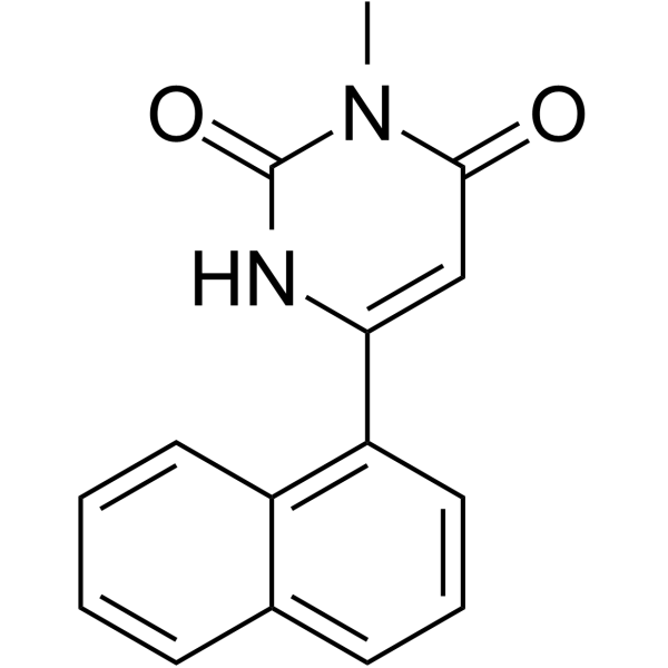
MNK8

Based on 1 Customer Validation
MNK8 is a potent STAT3 (signal transducer and activator of transcription 3) inhibitor. MNK8 inhibits STAT3 activation and reduced its DNA binding ability. MNK8 shows good growth inhibition against hepatocellular carcinoma (HCC) cells. MNK8 induces apoptosis in HCC cells. MNK8 reduces prosurvival proteins expression and migration/invasion of HCC cells.
商品は「研究用試薬」です。人や動物の医療用・臨床診断用・食品用の製品ではありません。
研究用途以外に使用した場合、当社は一切の責任を負いかねます。
- 純度 : 99.81%
- CAS 番号: 2055078-49-2
- 分子式: C15H12N2O2
- 分子量:252.27
-
保管条件:
4°C, protect from light
* In solvent : -80°C, 6 months; -20°C, 1 month (protect from light)

To place orders, for customer services and technical support, please contact: MedChemExpress USA
Tel: 609-228-6898 E-mail: [email protected] [email protected]


-
 生物活性
生物活性
-
化学情報
-
溶剤 & 溶解度
- プロトコル
- 純度とドキュメンテーション
- 参考文献
-
 Help & FAQs




生物活性
製品説明
IC50 & Target
|
STAT3 |
Bcl-2 |
Cellular Effect
|
Cell Line

|
Type | Value | Description | References |
|---|---|---|---|---|
| HCCLM3 | IC50 |
42.6 μM
Compound: MNK8
|
Cytotoxicity against human HCCLM3 cells assessed as reduction in cell viability measured upto 72 hrs by MTT assay
Cytotoxicity against human HCCLM3 cells assessed as reduction in cell viability measured upto 72 hrs by MTT assay
|
[PMID: 34418571] |
| HepG2 | IC50 |
20.9 μM
Compound: MNK8
|
Cytotoxicity against human HepG2 cells assessed as reduction in cell viability measured upto 72 hrs by MTT assay
Cytotoxicity against human HepG2 cells assessed as reduction in cell viability measured upto 72 hrs by MTT assay
|
[PMID: 34418571] |
体外実験
MNK8 (50 μM, 0-48 h) increases Sub-G1 cells in a time-dependent fashion, and decreases the expression of antiapoptotic proteins such as Bcl-2, survivin, and cyclin D1[1].
MedChemExpress (MCE) has not independently confirmed the accuracy of these methods. They are for reference only.


化学情報
-
CAS 番号 2055078-49-2
-
性状 Solid
-
分子量 252.27
-
分子式 C15H12N2O2
-
Color White to off-white
-
SMILES
O=C1N(C)C(C=C(C2=C3C=CC=CC3=CC=C2)N1)=O
-
輸送条件
Room temperature in continental US; may vary elsewhere.
-
保管条件
4°C, protect from light
* In solvent : -80°C, 6 months; -20°C, 1 month (protect from light)


溶剤 & 溶解度
体外:
DMSO : 41.67 mg/mL (165.18 mM; Need ultrasonic; Hygroscopic DMSO has a significant impact on the solubility of product, please use newly opened DMSO)
Please refer to the solubility information to select the appropriate solvent. Once prepared, please aliquot and store the solution to prevent product inactivation from repeated freeze-thaw cycles.
Storage method and period of stock solution: -80°C, 6 months; -20°C, 1 month (protect from light). When stored at -80°C, please use it within 6 months. When stored at -20°C, please use it within 1 month.
Please refer to the solubility information to select the appropriate solvent. Once prepared, please aliquot and store the solution to prevent product inactivation from repeated freeze-thaw cycles.
Storage method and period of stock solution: -80°C, 6 months; -20°C, 1 month (protect from light). When stored at -80°C, please use it within 6 months. When stored at -20°C, please use it within 1 month.
濃度 (開始) × 体積 (開始) = 濃度 (終了) × 体積 (終了)


純度とドキュメンテーション
-
データシート (276 KB)
-
SDS (252 KB)
- English - EN (252 KB)
- Français - FR (252 KB)
- Deutsch - DE (252 KB)
- Norwegian - NO (252 KB)
- Español - ES (252 KB)
- Swedish - SV (252 KB)
- Italian - IT (252 KB)
- Korean - KR (252 KB)
- Portuguese - PT (252 KB)
-
取扱説明書 (2659 KB)
参考文献


Complete Stock Solution Preparation Table
Please refer to the solubility information to select the appropriate solvent. Once prepared, please aliquot and store the solution to prevent product inactivation from repeated freeze-thaw cycles.
Storage method and period of stock solution: -80°C, 6 months; -20°C, 1 month (protect from light). When stored at -80°C, please use it within 6 months. When stored at -20°C, please use it within 1 month.
| Optional Solvent | Concentration Solvent Mass | 1 mg | 5 mg | 10 mg | 25 mg |
|---|---|---|---|---|---|
| DMSO | 1 mM | 3.9640 mL | 19.8200 mL | 39.6401 mL | 99.1002 mL |
| 5 mM | 0.7928 mL | 3.9640 mL | 7.9280 mL | 19.8200 mL | |
| 10 mM | 0.3964 mL | 1.9820 mL | 3.9640 mL | 9.9100 mL | |
| 15 mM | 0.2643 mL | 1.3213 mL | 2.6427 mL | 6.6067 mL | |
| 20 mM | 0.1982 mL | 0.9910 mL | 1.9820 mL | 4.9550 mL | |
| 25 mM | 0.1586 mL | 0.7928 mL | 1.5856 mL | 3.9640 mL | |
| 30 mM | 0.1321 mL | 0.6607 mL | 1.3213 mL | 3.3033 mL | |
| 40 mM | 0.0991 mL | 0.4955 mL | 0.9910 mL | 2.4775 mL | |
| 50 mM | 0.0793 mL | 0.3964 mL | 0.7928 mL | 1.9820 mL | |
| 60 mM | 0.0661 mL | 0.3303 mL | 0.6607 mL | 1.6517 mL | |
| 80 mM | 0.0496 mL | 0.2478 mL | 0.4955 mL | 1.2388 mL | |
| 100 mM | 0.0396 mL | 0.1982 mL | 0.3964 mL | 0.9910 mL |


MNK8 関連分類
HY-147187 関連分類
 Powered by Bioz