



SK1-I
Based on 1 publication(s) in Google Scholar
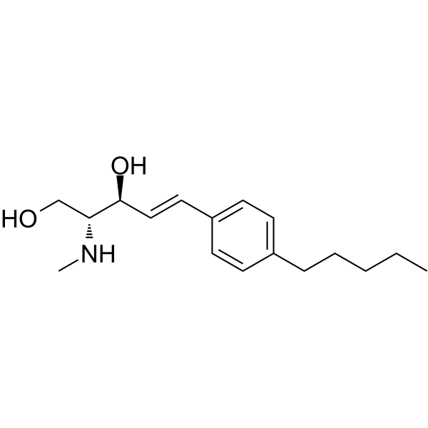
SK1-I (BML-258), an analog of sphingosine, is an isozyme-specific competitive SPHK1 inhibitor with a Ki value of 10 μM. SK1-I shows no activity at SPHK1 PKCα, PKCδ, PKA, AKT1, ERK1, EGFR, CDK2, IKKβ or CamK2β. SK1-I enhances autophagy and has antitumor activity.
商品は「研究用試薬」です。人や動物の医療用・臨床診断用・食品用の製品ではありません。
研究用途以外に使用した場合、当社は一切の責任を負いかねます。
- 純度 : 98.83%
- CAS 番号: 1072443-89-0
- 分子式: C17H27NO2
- 分子量:277.40
-
保管条件:Powder -20°C, 3 years , 4°C, 2 years ; In solvent -80°C, 6 months , -20°C, 1 month

To place orders, for customer services and technical support, please contact: MedChemExpress USA
Tel: 609-228-6898 E-mail: [email protected] [email protected]


-
 生物活性
生物活性
-
化学情報
-
溶剤 & 溶解度
- プロトコル
- 純度とドキュメンテーション
- 参考文献
-
 Help & FAQs


-
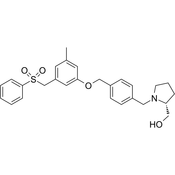
HY-15425
PF-543SPHK1 Inhibitor -
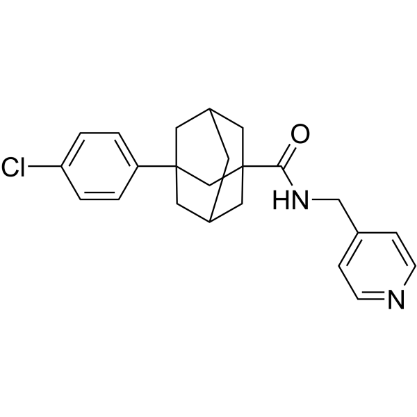
HY-16015
Opaganib -
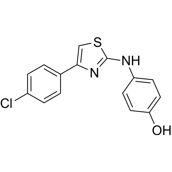
HY-13822
SKI IISphK/DES1 Inhibitor -
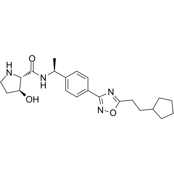
HY-101805
SK1-IN-1SPHK1 Inhibitor -
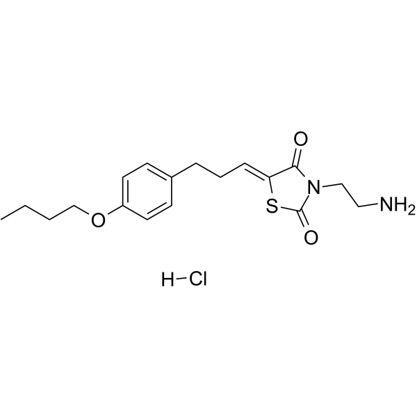
HY-15779A
K145 hydrochlorideSphK2 Inhibitor -

HY-18739
Phorbol 12-myristate 13-acetatePKC Activator -
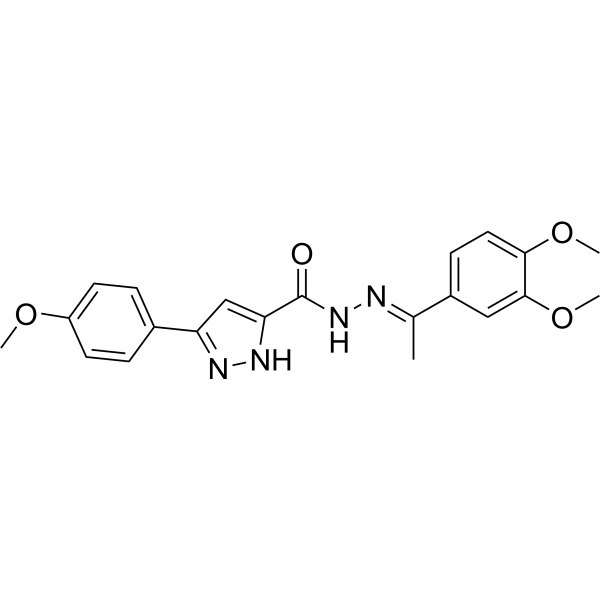
HY-12892
SKI-178SphK1/SphK2 Inhibitor -
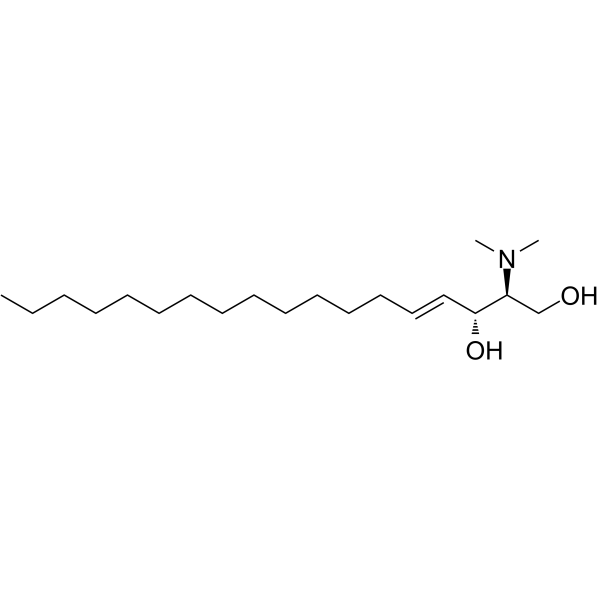
HY-108491
N,N-DimethylsphingosineSphingosine Kinase Inhibitor -

HY-16015R
Opaganib (Standard) -

HY-18739R
Phorbol 12-myristate 13-acetate (Standard)PKC Activator -
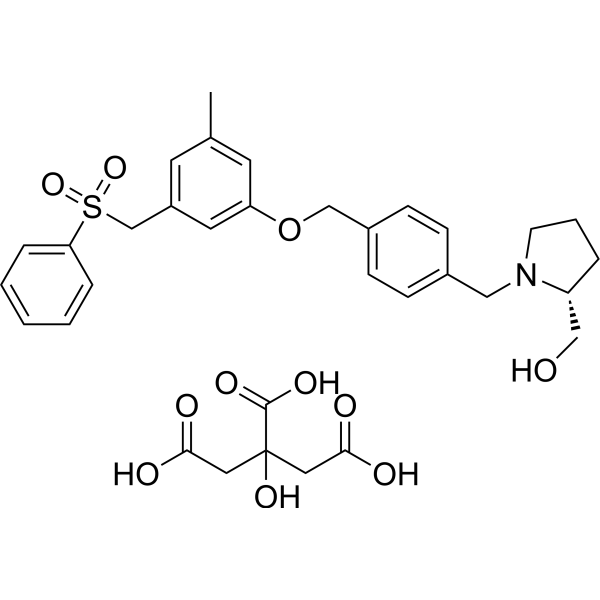
HY-15425A
PF-543 CitrateSPHK1 Inhibitor -
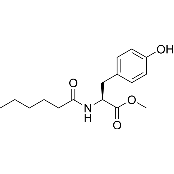
HY-101572
MHPSPHK1 Activator
MedChemExpress(MCE)の使用を引用している文献 SK1-I
More

生物活性
製品説明
IC50 & Target
|
SphK1 |
Cellular Effect
|
Cell Line

|
Type | Value | Description | References |
|---|---|---|---|---|
| HEK293 | IC50 |
10 μM
Compound: 7, SK1-I
|
Competitive inhibition of V5-His-tagged human SphK2 expressed in HEK293 cells assessed as S1P production using sphingosine and [gamma-32P]ATP as substrate by thin layer chromatography
Competitive inhibition of V5-His-tagged human SphK2 expressed in HEK293 cells assessed as S1P production using sphingosine and [gamma-32P]ATP as substrate by thin layer chromatography
|
[PMID: 24471412] |
体外実験
SK1-I (0-10 μM; 24 hours) attenuates cancer cell growth and survival in a TP53-dependent manner in HCT116 cells and HCT116 cells bearing TP53 null cancer[2].
SK1-I (0-20 μM; 12 hours) induces more CASP3 cleavage in HCT116 cells, compared to HCT116 cells lacking TP53, leading to a hallmark of apoptosis[2].
MedChemExpress (MCE) has not independently confirmed the accuracy of these methods. They are for reference only.
-
Cell Line:HCT116 cells and HCT116 cells bearing TP53 null cancer
-
Concentration:0 µM, 2.5 µM, 5 µM, 7.5 µM, 10 µM
-
Incubation Time:24 hours
-
Result:Decreased cancer cell growth and survival.
-
Cell Line:HCT116 cells and HCT116 cells bearing TP53 null cancer
-
Concentration:0 µM, 5 µM, 10 µM, 20 µM
-
Incubation Time:12 hours
-
Result:Induced more CASP3 cleavage in HCT116 cells, compared to HCT116 cells lacking TP53.
体内実験
MedChemExpress (MCE) has not independently confirmed the accuracy of these methods. They are for reference only.
-
Animal Model:Male C57BL/6 mice (24 ± 3.5 g) [3]
-
Dosage:75 mg/kg
-
Administration:Intraperitoneal (i.p.) injection; once; 24 hours prior to baseline MAP measurement
-
Result:Significantly lowered baseline mean arterial blood pressure (MAP).


化学情報
-
CAS 番号 1072443-89-0
-
性状 Solid
-
分子量 277.40
-
分子式 C17H27NO2
-
Color Off-white to light yellow
-
SMILES
OC[C@H]([C@H](/C=C/C1=CC=C(C=C1)CCCCC)O)NC
-
別名
BML-258
-
輸送条件
Room temperature in continental US; may vary elsewhere.
-
保管条件
Powder -20°C 3 years 4°C 2 years In solvent -80°C 6 months -20°C 1 month


Publications (1)
-
Journal Impact Factor
-
Most Recent
-
PLoS Pathog
Enzymatic independent role of sphingosine kinase 2 in regulating the expression of type I interferon during influenza A virus infection. [Abstract]2022 Sep 7;18(9):e1010794. PMID: 36070294


溶剤 & 溶解度
体外:
DMSO : 100 mg/mL (360.49 mM; Need ultrasonic; Hygroscopic DMSO has a significant impact on the solubility of product, please use newly opened DMSO)
Please refer to the solubility information to select the appropriate solvent. Once prepared, please aliquot and store the solution to prevent product inactivation from repeated freeze-thaw cycles.
Storage method and period of stock solution: -80°C, 6 months; -20°C, 1 month. When stored at -80°C, please use it within 6 months. When stored at -20°C, please use it within 1 month.
Please refer to the solubility information to select the appropriate solvent. Once prepared, please aliquot and store the solution to prevent product inactivation from repeated freeze-thaw cycles.
Storage method and period of stock solution: -80°C, 6 months; -20°C, 1 month. When stored at -80°C, please use it within 6 months. When stored at -20°C, please use it within 1 month.
濃度 (開始) × 体積 (開始) = 濃度 (終了) × 体積 (終了)


純度とドキュメンテーション
-
データシート (271 KB)
-
SDS (251 KB)
- English - EN (251 KB)
- Français - FR (251 KB)
- Deutsch - DE (251 KB)
- Norwegian - NO (251 KB)
- Español - ES (251 KB)
- Swedish - SV (251 KB)
- Italian - IT (251 KB)
- Korean - KR (251 KB)
- Portuguese - PT (251 KB)
-
取扱説明書 (2659 KB)
参考文献
[1]. Melissa R Pitman, et al. Inhibitors of the sphingosine kinase pathway as potential therapeutics. Curr Cancer Drug Targets. 2010 Jun;10(4):354-67. [Content Brief]
[2]. Santiago Lima, et al. TP53 is required for BECN1- and ATG5-dependent cell death induced by sphingosine kinase 1 inhibition. Autophagy. 2018;14(6):942-957. [Content Brief]
[3]. Fiona H Greig, et al. Requirement for sphingosine kinase 1 in mediating phase 1 of the hypotensive response to anandamide in the anaesthetised mouse. Eur J Pharmacol. 2019 Jan 5;842:1-9. [Content Brief]


Complete Stock Solution Preparation Table
Please refer to the solubility information to select the appropriate solvent. Once prepared, please aliquot and store the solution to prevent product inactivation from repeated freeze-thaw cycles.
Storage method and period of stock solution: -80°C, 6 months; -20°C, 1 month. When stored at -80°C, please use it within 6 months. When stored at -20°C, please use it within 1 month.
| Optional Solvent | Concentration Solvent Mass | 1 mg | 5 mg | 10 mg | 25 mg |
|---|---|---|---|---|---|
| DMSO | 1 mM | 3.6049 mL | 18.0245 mL | 36.0490 mL | 90.1226 mL |
| 5 mM | 0.7210 mL | 3.6049 mL | 7.2098 mL | 18.0245 mL | |
| 10 mM | 0.3605 mL | 1.8025 mL | 3.6049 mL | 9.0123 mL | |
| 15 mM | 0.2403 mL | 1.2016 mL | 2.4033 mL | 6.0082 mL | |
| 20 mM | 0.1802 mL | 0.9012 mL | 1.8025 mL | 4.5061 mL | |
| 25 mM | 0.1442 mL | 0.7210 mL | 1.4420 mL | 3.6049 mL | |
| 30 mM | 0.1202 mL | 0.6008 mL | 1.2016 mL | 3.0041 mL | |
| 40 mM | 0.0901 mL | 0.4506 mL | 0.9012 mL | 2.2531 mL | |
| 50 mM | 0.0721 mL | 0.3605 mL | 0.7210 mL | 1.8025 mL | |
| 60 mM | 0.0601 mL | 0.3004 mL | 0.6008 mL | 1.5020 mL | |
| 80 mM | 0.0451 mL | 0.2253 mL | 0.4506 mL | 1.1265 mL | |
| 100 mM | 0.0360 mL | 0.1802 mL | 0.3605 mL | 0.9012 mL |


SK1-I 関連分類
HY-119016 関連分類
 Powered by Bioz