



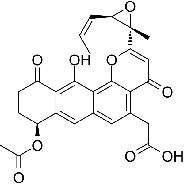
Kapurimycin A3
Kapurimycin A3 is an antitumor and antibacterial antibiotic. Kapurimycin A3 has DNA binding and breakage activity. Kapurimycin A3 can be isolated from Streptomyces.
For research use only. We do not sell to patients.
- CAS No.: 129966-45-6
- Formula: C27H24O9
- Molecular Weight:492.47
-
Storage:
Please store the product under the recommended conditions in the Certificate of Analysis.

To place orders, for customer services and technical support, please contact: MedChemExpress USA
Tel: 609-228-6898 E-mail: [email protected] [email protected]


-
 Biological Activity
-
Chemical Information
- Protocols
- Purity & Documentation
- References
-
 Help & FAQs


All Antibiotic Isoforms
More

Biological Activity
Description


Chemical Information
-
CAS No. 129966-45-6
-
Molecular Weight 492.47
-
Formula C27H24O9
-
SMILES
O=C(O)CC(C=C1C=C2[C@@H](OC(C)=O)CC3)=C(C(C=C([C@@]4(C)O[C@@H]4/C=C\C)O5)=O)C5=C1C(O)=C2C3=O
-
Structure Classification
-
Initial Source
Streptomyces
-
Shipping
Room temperature in continental US; may vary elsewhere.
-
Storage
Please store the product under the recommended conditions in the Certificate of Analysis.

Protocols
-
Protocol for Electrophoretic Mobility Shift Assay (EMSA)
Electrophoretic mobility shift assay detects protein-nucleic acid binding by incubating a labeled DNA or RNA probe with purified protein or cell extract, then separating free probe from slower-migrating protein-probe complexes on a native gel. For cancer cells, primary neurons, mouse tumor samples, intestinal organoids, inflammatory macrophages, or drug-treated samples, EMSA can measure transcription-factor DNA binding or RNA-binding protein activity in extracts, but it does not directly measure transcription, protein expression, or chromatin occupancy in intact cells. Specificity is judged by competition with unlabeled wild-type probe, failure of mutated or unrelated competitors to compete, and antibody supershift or disruption when the binding protein identity must be confirmed.
-
Research Protocol for Infectious Diseases
Infectious-disease experiments test how pathogens interact with host barriers, innate immune receptors, inflammatory signaling, pathogen replication, and tissue injury; pattern-recognition receptors such as TLRs, RIG-I-like receptors, NOD-like receptors, and inflammasomes detect microbial molecules and activate NF-κB, interferon, and cytokine responses. The central hypothesis is that infection severity reflects the balance between pathogen burden and host response: protective inflammation restricts pathogen growth, whereas excessive or mislocalized inflammation contributes to tissue damage and disease phenotype. Unresolved questions include which host pathways are protective versus pathogenic, why some infection models fail to translate to human disease, and which combined readouts best predict clinically relevant infection outcomes.


Purity & Documentation
References

Calculators
Concentration (start) × Volume (start) = Concentration (final) × Volume (final)


Kapurimycin A3 Related Classifications
HY-125633 Related Classifications
 Powered by Bioz