



QC6352
Based on 12 publication(s) in Google Scholar
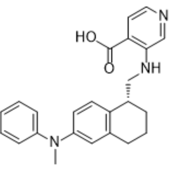
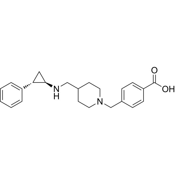
QC6352 is a DHODH and KDM4 inhibitor. The IC50 values of QC6352 for human DHODH, KDM4A, KDM4B, KDM4C, and KDM4D are 0.3 nM, 104 nM, 56 nM, 35 nM, and 104 nM, respectively. QC6352 inhibits JmjC domain Fe (II)/2-ketoglutarate-dependent histone demethylases and blocks DHODH-mediated de novo pyrimidine biosynthesis through ubiquinone binding, reducing H3K9me3/H3K36me3 demethylation and increasing histone methylation marks. QC6352 induces G0/G1 phase arrest, senescence, apoptosis (apoptosis), DNA damage, and interferon/chemokine responses, and reduces stemness markers and BMI1+ cancer stem cells, exhibiting selective antiproliferative/cytotostatic activity against glioblastoma cells with limited effects on normal astrocytes. QC6352 can be used for research on glioblastoma, breast cancer, squamous cell carcinoma, and TP53-mutant acute myeloid leukemia.
연구목적의 판매만을 진행합니다. 환자를 대상으로 한 판매는 하지 않습니다.
- Purity : 99.13%
- CAS No.: 1851373-36-8
- 화학식: C24H25N3O2
- 분자량:387.47
-
보관:
4°C, protect from light
* In solvent : -80°C, 2 years; -20°C, 1 year (protect from light)

To place orders, for customer services and technical support, please contact: MedChemExpress USA
Tel: 609-228-6898 E-mail: [email protected] [email protected]


-
 Biological Activity
-
Chemical Information
-
용액&용해도
- Protocol
- 순도&문서
- References
-
 Help & FAQs


-
HY-100421
CPI-455KDM5 Inhibitor -
HY-15648B
GSK-J4JMJD3/UTX Inhibitor -
HY-13953
JIB-04Histone Demethylase Inhibitor -
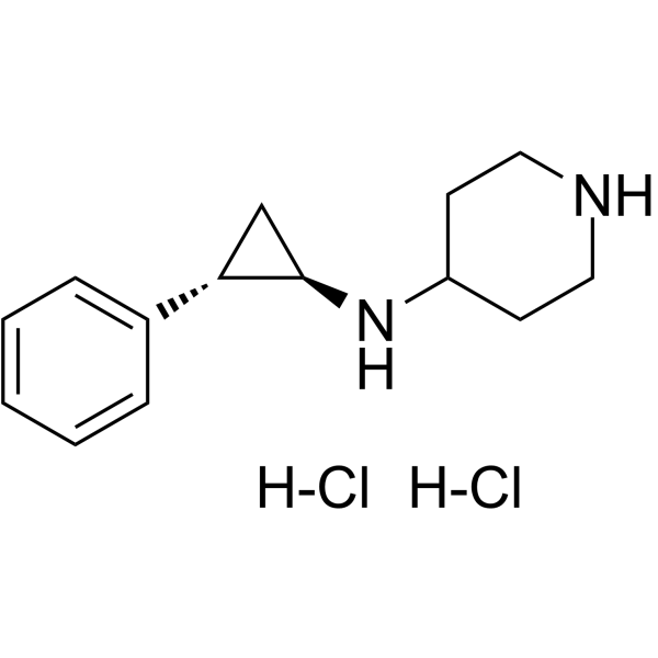
HY-100546A
GSK-LSD1 dihydrochlorideLSD1/KDM1A Inhibitor -
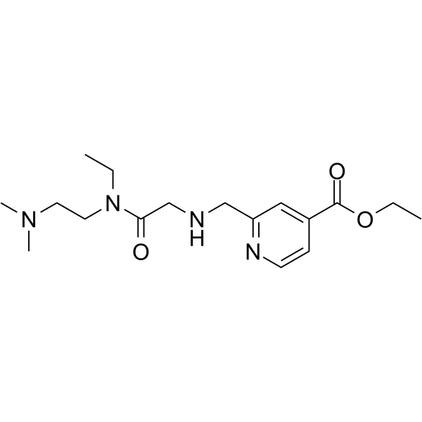
HY-120400
KDM5-C70KDM5 Inhibitor -
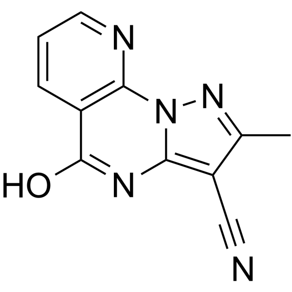
HY-101928
KDM4D-IN-1KDM4D Inhibitor -
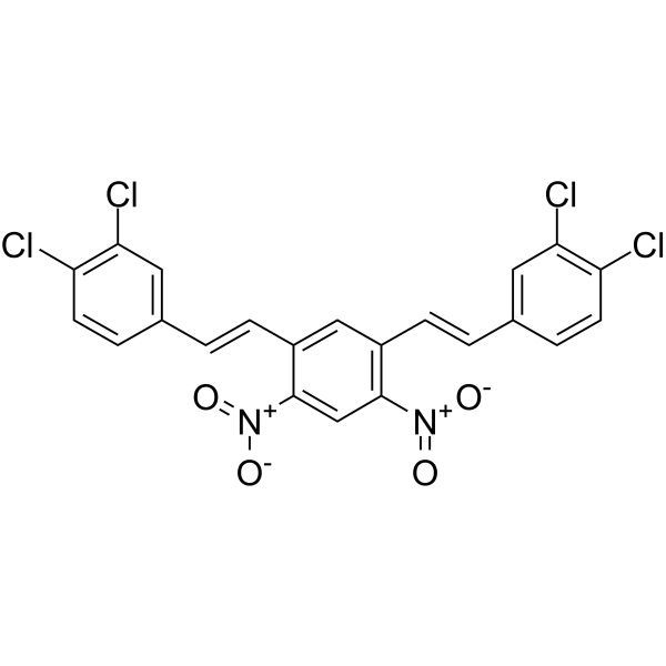
HY-110154
NSC636819KDM4A/KDM4B Inhibitor -
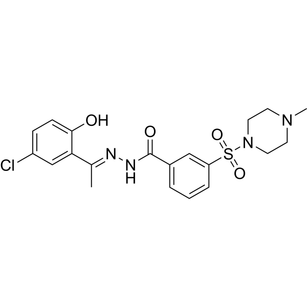
HY-103713
SeclidemstatLSD1 Inhibitor -
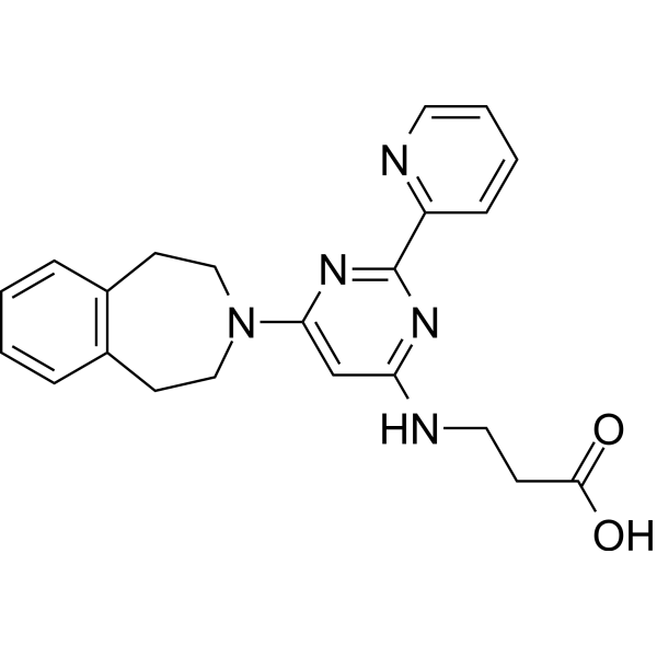
HY-15648
GSK-J1JMJD3/KDM6B Inhibitor -
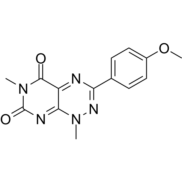
HY-146148
KDM4C-IN-1KDM4C Inhibitor -
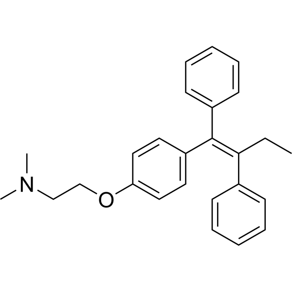
HY-13757A
TamoxifenEstrogen Receptor Modulator -
HY-18632
GSK2879552LSD1 Inhibitor
Publications Citing Use of MedChemExpress (MCE) QC6352
More- Nat Commun. 2026 Feb 12;17(1):1214. [Abstract]
- Mol Cell. 2021 May 20;81(10):2148-2165.e9. [Abstract]
- Acta Pharmacol Sin. 2022 Feb;43(2):457-469. [Abstract]
- Aging Dis. 2026 Aug 9.
- J Med Chem. 2024 Mar 28;67(6):4525-4540. [Abstract]
- RSC Med Chem. 2025 Jul 1. [Abstract]
- Clin Exp Med. 2025 Nov 25;26(1):33. [Abstract]
- BMC Bioinformatics. 2026 Feb 19.
- bioRxiv. 2025 Sep 16:2025.09.16.676586. [Abstract]
- bioRxiv. 2025 Feb 5:2025.01.31.635709. [Abstract]
- bioRxiv. 2024 Sep 16:2024.09.16.613328. [Abstract]
- bioRxiv. 2022 May 14.491739.
 Customer Validation & Images

-
In Vivo Efficacy Study
-
Apoptosis Analysis
-
IF

-
Histological Imaging/Staining

-
IHC


Biological Activity
제품 설명
|
DHODH 0.3 nM (IC50) |
KDM4A 104 nM (IC50) |
KDM4B 56 nM (IC50) |
KDM4C 35 nM (IC50) |
KDM4D 104 nM (IC50) |
Cellular Effect
|
Cell Line

|
Type | Value | Description | References |
|---|---|---|---|---|
| KYSE-150 cell line | EC50 |
3.5 nM
|
Antiproliferative activity against human KYSE-150 esophageal cancer cells assessed by BrdU incorporation.
Antiproliferative activity against human KYSE-150 esophageal cancer cells assessed by BrdU incorporation.
|
28835804 |
| KYSE-150 cell line | EC50 |
1.3 nM
|
Induction of H3K36me3 in KYSE-150^+KDM4C cells overexpressing KDM4C assessed by HTRF after 24 hrs incubation.
Induction of H3K36me3 in KYSE-150^+KDM4C cells overexpressing KDM4C assessed by HTRF after 24 hrs incubation.
|
28835804 |
| IMR-90 | EC50 |
30000 nM
|
Cytotoxicity against human IMR-90 normal fibroblasts assessed as viability/antiproliferation with EC50 greater than 30000 nM.
Cytotoxicity against human IMR-90 normal fibroblasts assessed as viability/antiproliferation with EC50 greater than 30000 nM.
|
28835804 |
In Vitro
QC6352 (0.1 nM-10 μM; 5 days) inhibits RKI1, MMK1, JK2, GBM6, FPW1 and HW1 glioblastoma stem cell growth with GR50 values of 3-133 nM while sparing NHAs up to 1,000 nM[1].
QC6352 (50-100 nM; 10 days) induces senescence-associated β-galactosidase in FPW1 and RKI1 glioblastoma cells[1].
QC6352 (50-100 nM; 10-day exposure plus 10-day washout) produces sustained inhibition of colony formation in FPW1 and RKI1 cells[1].
QC6352 (50-100 nM; 0-14 days) inhibits FPW1 and RKI1 glioblastoma spheroid growth[1].
QC6352 (5-100 nM; 10 days) abolishes colony formation in FPW1, RKI1, JK2, SB2 and HW1 glioblastoma cells at 50 nM[1].
QC6352 (10-100 nM; 72 h) increases H3K9me3 and H3K36me3 in RKI1 and JK2 glioblastoma cells[1].
QC6352 (C5 days) efficacy is not changed by KDM4B depletion in A172 glioblastoma cells[1].
QC6352 (5 days) sensitivity is not affected by individual KDM4A, KDM4B or KDM4C knockout in RKI1 glioblastoma cells[1].
QC6352 (25-70 nM; 5 days per exposure round; two exposure rounds) chemogenomic screening in RKI1 cells does not identify KDM4 enzymes as mediators of QC6352 efficacy[1].
QC6352 inhibits KYSE-150 cell proliferation with an EC50 of 3.5 nM[2].
QC6352 (5 days) inhibits breast (BR0869f) and colon (SU60) organoids with IC50 values of 5 nM and 13 nM, respectively[2].
QC6352 increases HP1γ puncta formation in SCC cells[3].
QC6352 (15-60 nM; days 1-4) suppresses THP-1 and U937 proliferation over 4 days[4].
QC6352 (30-60 nM; 48 h) promotes myeloid differentiation marker expression in primary TP53-mutated AML cells[4].
QC6352 (15-60 nM; 48 h) induces SA-β-gal-associated senescence in primary TP53-mutated AML cells, THP-1, and U937 cells[4].
QC6352 (30 nM; 48 h) increases DNA damage signaling and cytosolic dsDNA accumulation in THP-1 and U937 cells[4].
QC6352 (30 nM; 48 h pre-exposure; 4 h co-culture) increases NK-cell-mediated killing of primary TP53-mutated AML cells and THP-1 and U937 cells[4].
QC6352 (50-100 nM; 10 days) induces apoptosis in FPW1 and RKI1 glioblastoma cells[1].
QC6352 (100 nM; 7 days) inhibits growth and induces apoptosis in FPW1, RKI1, A172 and HEK293T cells[1].
QC6352 (15-240 nM; 48 h) induces apoptosis in THP-1 and U937 TP53-mutated AML cells after 48 h[4].
QC6352 (30-60 nM; 48 h) induces G0/G1 arrest in THP-1 and U937 cells[4].
QC6352 (30 nM; 48 h) upregulates cyclin-dependent kinase inhibitor genes P21 and P16 in THP-1 and U937 cells[4].
QC6352 (30 nM; 48 h) induces SASP-associated inflammatory gene expression, including IL8 and IL1B, in THP-1 and U937 cells, with IL6 induction in U937 but not THP-1 cells[4].
QC6352 (15-60 nM; 48 h) increases NKG2D ligand surface expression in THP-1, U937, and primary TP53-mutated AML cells[4].
QC6352 (24-48 h co-culture) pre-exposure of THP-1 and U937 cells enhances the activation phenotype of co-cultured NK cells[4].
QC6352 (30-60 nM; 48 h pre-exposure; 6 h co-culture) enhances NK-cell degranulation and polyfunctional effector responses after pre-exposure of THP-1 and U937 cells[4].
QC6352 (100 µM uridine; 5 days) growth inhibition is completely rescued by exogenous uridine in FPW1 and RKI1 glioblastoma cells[1].
QC6352 (50 nM; 24 h) inhibits DHODH in FPW1 glioblastoma cells, causing dihydroorotate accumulation and pyrimidine nucleotide depletion[1].
QC6352 (5 days) efficacy is strongly rescued by DHODH A58T expression in FPW1 glioblastoma cells, supporting DHODH as the functional target[1].
QC6352 (1 mM; 1 h; 6-10 days at 20°C) binds human DHODH in a hydrophobic channel and blocks the active site, explaining DHODH inhibition[1].
QC6352 (15 min preincubation; 45 min enzyme reaction) inhibits recombinant KDM4A in the AlphaScreen assay with an IC50 of 355 nM[1].
QC6352 (1 µM; 2 h) does not alter KDM4A chromatin mobility in HeLa cells[1].
QC6352 (0.02-0.2 mg/mL BSA) is a potent KDM4-family inhibitor with KDM4C IC50 35 nM and retained KDM5B activity (IC50 750 nM)[2].
QC6352 (24 h) induces H3K36me3 in KYSE-150+KDM4C cells with EC50 1.3 nM[2].
QC6352 shows no general cytotoxicity in IMR-90 normal fibroblasts (EC50 >30,000 nM)[2].
MedChemExpress (MCE) has not independently confirmed the accuracy of these methods. They are for reference only.
-
Cell Line:A172-FUCCI glioblastoma cells
-
Concentration:50 and 100 nM
-
Incubation Time:5 days
-
Result:Nearly 80% of cells arrested in G0/G1 phase.
-
Cell Line:FPW1 glioblastoma cells
-
Concentration:100 nM
-
Incubation Time:5 and 7 days
-
Result:Increased expression of senescence and apoptotic genes was observed, particularly after 5 days.
GFAP, TUBB2A and TUBB3 expression decreased; MBP and OLIG2 increased.
Stemness markers NES, SOX2 and SOX9 were reduced after 7 days.
-
Cell Line:FPW1 and RKI1 glioblastoma cells
-
Concentration:50 and 100 nM
-
Incubation Time:10 days
-
Result:Increased Annexin V fluorescence in FPW1 and RKI1 cells, with a more pronounced effect in FPW1.
-
Cell Line:FPW1 and RKI1 glioblastoma cells
-
Concentration:50 and 100 nM
-
Incubation Time:10-day exposure plus 10-day washout
-
Result:Colony area was reduced at 50 and 100 nM.
After washout, confluence did not exceed 25%.
-
Cell Line:FPW1 and RKI1 glioblastoma cells
-
Concentration:50 and 100 nM
-
Incubation Time:days 0, 7 and 14
-
Result:Substantially inhibited FPW1 and RKI1 spheroid growth over 14 days.
-
Cell Line:FPW1, RKI1, JK2, SB2 and HW1 glioblastoma cells
-
Concentration:5, 10, 50 and 100 nM
-
Incubation Time:10 days
-
Result:Completely abolished colony formation at 50 nM.
-
Cell Line:RKI1 and JK2 glioblastoma cells
-
Concentration:10, 50 and 100 nM
-
Incubation Time:72 h
-
Result:Increased H3K9me3 and H3K36me3 abundance in RKI1 and JK2 cells, with up to a three-fold increase.
-
Cell Line:A172 glioblastoma cells
-
Concentration:Concentration-response
-
Incubation Time:5 days
-
Result:KDM4B knockdown did not alter the antiproliferative efficacy.
-
Cell Line:RKI1 glioblastoma cells
-
Concentration:Concentration-response
-
Incubation Time:5 days
-
Result:Individual KDM4A, KDM4B or KDM4C knockout did not change sensitivity.
-
Cell Line:FPW1 and RKI1 glioblastoma cells
-
Concentration:Concentration-response; 100 µM uridine
-
Incubation Time:5 days
-
Result:Uridine supplementation completely rescued growth arrest in FPW1 and RKI1 cells.
-
Cell Line:FPW1 glioblastoma cells
-
Concentration:Concentration-response
-
Incubation Time:5 days
-
Result:Expression of DHODH A58T strongly rescued antiproliferative efficacy.
Weak efficacy at doses >1 µM was rescued by exogenous uridine.
-
Cell Line:THP-1 and U937 cells
-
Concentration:15, 30, 60 nM
-
Incubation Time:days 1, 2, 3, and 4
-
Result:Reduced relative cell viability in both THP-1 and U937 cells, inhibiting growth of both TP53-mutated AML cell lines.
-
Cell Line:THP-1 and U937 cells
-
Concentration:15, 30, 60, 120, 240 nM
-
Incubation Time:48 h
-
Result:Increased the proportion of Annexin V+ cells in THP-1 and U937 cells in a concentration-dependent manner.
-
Cell Line:THP-1 and U937 cells
-
Concentration:30, 60 nM
-
Incubation Time:48 h
-
Result:Increased the G0/G1 fraction: THP-1 G0/G1 was 76.5% at 30 nM and 80.8% at 60 nM; U937 G0/G1 was 71.1% at 30 nM and 72.4% at 60 nM.
-
Cell Line:THP-1 and U937 cells
-
Concentration:30 nM
-
Incubation Time:48 h
-
Result:Increased P21 and P16 mRNA expression in THP-1 and U937 cells; P27 did not show a comparable increase.\nIncreased IL8 and IL1B mRNA expression in THP-1 and U937 cells; IL6 mRNA increased in U937 cells but was unchanged in THP-1 cells.
Parmacokinetics
In Vivo
QC6352 (10-50 mg/kg; BID on a 5-on/2-off schedule; 22 days) reduces tumor growth dose-dependently in the BR0869f breast cancer PDX model[2].
QC6352 (50 mg/kg; BID on a 5-on/2-off schedule; 22 days) reduces tumor-initiating cell frequency by greater than 14-fold in BR0869f xenograft cells[2].
QC6352 (10 mg/kg; i.p.; every other day (Qod); starting immediately after Day 6 engraftment confirmation) reduces U937-Luc leukemia burden and prolongs survival, and its combination with NK cells produces the slowest leukemia progression in a partly STING-dependent manner[4].
QC6352 (10 mg/kg; i.p.; every other day (Qod); starting Day 7 after THP-1 injection) plus PBMC infusion improves antileukemic efficacy in THP-1 xenografts, and NK cells contribute to this effect[4].
MedChemExpress (MCE) has not independently confirmed the accuracy of these methods. They are for reference only.
-
Animal Model:BALB/c nude mice were injected with 3 × 10⁵ GBM6 cells in the cortex[1]
-
Dosage:100 mg/kg
-
Administration:intraperitoneal; 5 days on/2 days off; 2 weeks
-
Result:Attenuated growth of orthotopically engrafted GBM6 xenografts as measured by bioluminescence.
Survival was extended to 34.5 days versus 28.5 days with vehicle control.
-
Animal Model:Seven-week-old female NOD SCID gamma mice were inoculated
subcutaneously in the flank region with 5x10⁶
patient-derived BR0869f tumor cells suspended in 100uL
Matrigel.[2] -
Dosage:10, 25, and 50 mg/kg
-
Administration:BID on a 5-on/2-off schedule; 22 days
-
Result:Tumor growth was diminished dose-dependently versus vehicle.
Tumor growth inhibitions were 22% at 10 mg/kg, 36% at 25 mg/kg, and 61% at 50 mg/kg.
-
Animal Model:C57BL/6J (Bmi1Cre-ERT2;tdTomato reporter; male and female; 8 weeks old; CSC lineage-tracing cohort)[3]
-
Dosage:8 mg/kg (i.p.); 100 μL of 1 mg/mL (topical)
-
Administration:i.p. every other day; topical over the whole tongue once daily
-
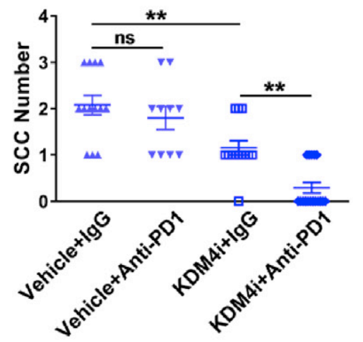
Result:Reduced SCC number, SCC size, and invasive degree.
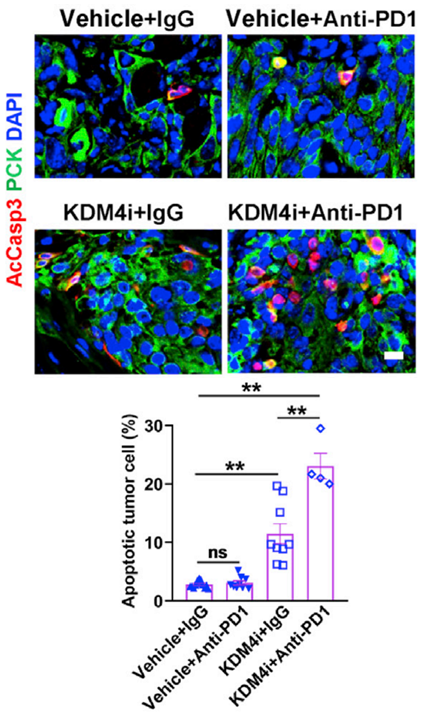
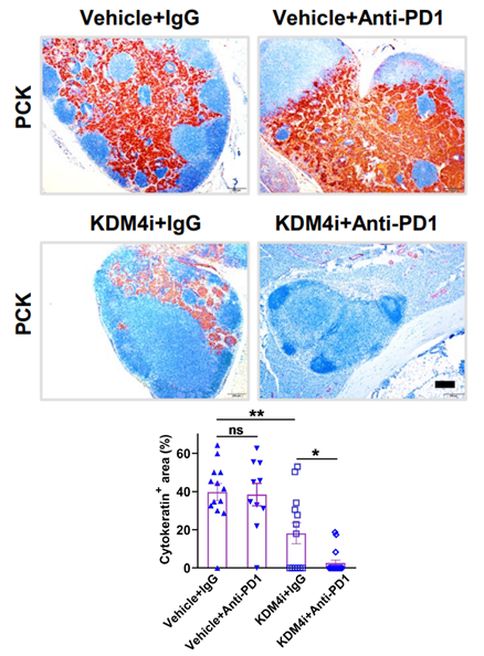
Induced more apoptosis, further increased CD8+ T cells and GZMB+CD8+ T cells, eliminated lymph node metastasis, and more drastically reduced BMI1+ CSCs.
-
Animal Model:NOD/ShiLtJGpt-Prkdc^em26Cd52 IL-2rg^em26Cd22/Gpt (NCG) (female, 6 weeks old)[4]
-
Dosage:10 mg/kg
-
Administration:i.p.; every other day (Qod); starting Day 7 after THP-1 injection
-
Result:On Day 30, bone marrow hCD45+ cells were 36.8% versus 61.5% in control, and 24.3% with combined administration versus 42.2% with NK cells alone.
The combination group had the lowest leukemia burden among all groups.
SA-β-gal and MICA/B on THP-1 cells increased in vivo.
NK cells recovered from combination-group mice showed increased CD69, NKG2D, CD107a, IFN-γ, and granzyme B compared with NK-cell-only mice.
-
Animal Model:NOD/ShiLtJGpt-Prkdc^em26Cd52 IL-2rg^em26Cd22/Gpt (NCG) (female, 6 weeks old)[4]
-
Dosage:10 mg/kg
-
Administration:i.p.; every other day (Qod); starting immediately after Day 6 engraftment confirmation
-
Result:Reduced U937-Luc leukemia burden by bioluminescence imaging and prolonged survival compared with vehicle.
The combination group showed the slowest leukemia progression among PBS, single-agent, and NK cells alone.
STING inhibition partially attenuated the enhanced antileukemic effect of the combination.
-
Animal Model:NOD/ShiLtJGpt-Prkdc^em26Cd52 IL-2rg^em26Cd22/Gpt (NCG) (female, 6 weeks old)[4]
-
Dosage:10 mg/kg
-
Administration:i.p.; every other day (Qod); starting immediately after Day 9 engraftment confirmation
-
Result:Combined PBMC transfer slowed U937-Luc leukemia progression compared with PBMC transfer alone.
-
Animal Model:NOD/ShiLtJGpt-Prkdc^em26Cd52 IL-2rg^em26Cd22/Gpt (NCG) (female, 6 weeks old)[4]
-
Dosage:10 mg/kg
-
Administration:i.p.; every other day (Qod); starting Day 7 after THP-1 injection
-
Result:Combined PBMC transfer enhanced antileukemic activity; this effect was attenuated when NK cells were depleted from PBMCs before transfer.
- Detection of Apoptosis
- TUNEL staining for apoptotic DNA fragmentation
- Flow cytometric DNA-content cell-cycle staining
- Annexin V plus membrane-impermeant dye apoptosis staining
- Apoptosis Solutions
- Protocol for Cell Cycle
- Breast Cancer Modeling
- Senescence-associated β-galactosidase staining
- Genotoxicity/Mutagenicity Study
- Cell Viability Determination by MTT Colorimetric Assay


Chemical Information
-
CAS No. 1851373-36-8
-
Appearance Solid
-
분자량 387.47
-
화학식 C24H25N3O2
-
Color Light yellow to yellow
-
SMILES
O=C(C1=C(NC[C@@H]2CCCC3=C2C=CC(N(C)C4=CC=CC=C4)=C3)C=NC=C1)O
-
선적
Room temperature in continental US; may vary elsewhere.
-
보관
4°C, protect from light
* In solvent : -80°C, 2 years; -20°C, 1 year (protect from light)


Publications (12)
-
Journal Impact Factor
-
Most Recent
-
Nat Commun
Human iPSC-based Modeling of Pulmonary Fibrosis Reveals p300/CBP Inhibition Suppresses Alveolar Transitional Cell State. [Abstract]2026 Feb 12;17(1):1214. PMID: 41680175 -
Mol Cell
Targeting KDM4A epigenetically activates tumor-cell-intrinsic immunity by inducing DNA replication stress. [Abstract]2021 May 20;81(10):2148-2165.e9. PMID: 33743195
QC6352 purchased from MedChemExpress. Usage Cited in: Mol Cell. 2021 May 20;81(10):2148-2165.e9. [Abstract]
Quantification of primary SCC number in mice treated with anti-PD1, KDM4i (QC6352; 8 mg/kg i.p. every other day; 100 μL of a 1 mg/mL solution applied over the whole tongue once daily) or their combination. The treatment with KDM4i alone significantly reduced SCC number.
QC6352 purchased from MedChemExpress. Usage Cited in: Mol Cell. 2021 May 20;81(10):2148-2165.e9. [Abstract]
Apoptotic tumor cells were analyzed by immunofluorescence staining and quantification in primary SCCs from mice treated with KDM4i (QC6352; 8 mg/kg i.p. every other day; 100 μL of a 1 mg/mL solution applied over the whole tongue once daily), anti-PD1, or their combination. KDM4i significantly induced apoptosis in SCCs, and the combination further increased apoptosis.
QC6352 purchased from MedChemExpress. Usage Cited in: Mol Cell. 2021 May 20;81(10):2148-2165.e9. [Abstract]
GZMB+CD8+ T cells within and around tumor islets were analyzed by immunofluorescence staining and quantification in primary SCCs from mice treated with KDM4i (QC6352; 8 mg/kg i.p. every other day; 100 μL of a 1 mg/mL solution applied over the whole tongue once daily), anti-PD1, or their combination. KDM4i increased CD8+ T-cell infiltration as well as activating GZMB+CD8+ T cells, and the combination further enhanced these responses.
QC6352 purchased from MedChemExpress. Usage Cited in: Mol Cell. 2021 May 20;81(10):2148-2165.e9. [Abstract]
H&E staining and area quantification of primary SCCs in mice treated with anti-PD1, KDM4i (QC6352; 8 mg/kg i.p. every other day; 100 μL of a 1 mg/mL solution applied over the whole tongue once daily), or their combination. The treatment with KDM4i alone significantly reduced SCC size compared with vehicle control and its combination with anti-PD1 produced greater inhibition.
QC6352 purchased from MedChemExpress. Usage Cited in: Mol Cell. 2021 May 20;81(10):2148-2165.e9. [Abstract]
PCK immunohistochemistry and quantification of metastatic areas were performed in lymph nodes from mice treated with KDM4i (QC6352; 8 mg/kg i.p. every other day; 100 μL of a 1 mg/mL solution applied over the whole tongue once daily), anti-PD1, or their combination. The combination treatment of KDM4i and PD1 blockade also collaboratively eliminated lymph node metastasis of SCCs.
-
Acta Pharmacol Sin
2022 Feb;43(2):457-469. PMID: 33850273 -
-
J Med Chem
Focused Screening Identifies Different Sensitivities of Human TET Oxygenases to the Oncometabolite 2-Hydroxyglutarate. [Abstract]2024 Mar 28;67(6):4525-4540. PMID: 38294854 -
RSC Med Chem
2025 Jul 1. PMID: 40656835 -
Clin Exp Med
Corin: a dual inhibitor for KDM1A/HDAC1, suppresses hepatocellular carcinoma by triggering cuproptosis. [Abstract]2025 Nov 25;26(1):33. PMID: 41286164 -
-
bioRxiv
2025 Sep 16:2025.09.16.676586. PMID: 41000952 -
bioRxiv
Heterochromatin fidelity is a therapeutic vulnerability in lymphoma and other human cancers. [Abstract]2025 Feb 5:2025.01.31.635709. PMID: 39975048 -
bioRxiv
Distinct H3K9me3 heterochromatin maintenance dynamics govern different gene programs and repeats in pluripotent cells. [Abstract]2024 Sep 16:2024.09.16.613328. PMID: 39345615 -


용액&용해도
In Vitro:
DMSO : 16.67 mg/mL (43.02 mM; Need ultrasonic; Hygroscopic DMSO has a significant impact on the solubility of product, please use newly opened DMSO)
Please refer to the solubility information to select the appropriate solvent. Once prepared, please aliquot and store the solution to prevent product inactivation from repeated freeze-thaw cycles.
Storage method and period of stock solution: -80°C, 2 years; -20°C, 1 year (protect from light). When stored at -80°C, please use it within 2 years. When stored at -20°C, please use it within 1 year.
Please refer to the solubility information to select the appropriate solvent. Once prepared, please aliquot and store the solution to prevent product inactivation from repeated freeze-thaw cycles.
Storage method and period of stock solution: -80°C, 2 years; -20°C, 1 year (protect from light). When stored at -80°C, please use it within 2 years. When stored at -20°C, please use it within 1 year.
Concentration (start) × Volume (start) = Concentration (final) × Volume (final)
In Vivo:
Select the appropriate dissolution method based on your experimental animal and administration route.
- For the following dissolution methods, please ensure to first prepare a clear stock solution using an In Vitro approach and then sequentially add co-solvents:
- To ensure reliable experimental results, the clarified stock solution can be appropriately stored based on storage conditions. As for the working solution for In Vivo experiments, it is recommended to prepare freshly and use it on the same day.
- The percentages shown for the solvents indicate their volumetric ratio in the final prepared solution. If precipitation or phase separation occurs during preparation, heat and/or sonication can be used to aid dissolution.
Add each solvent one by one: 10% DMSO 40% PEG300 5% Tween-80 45% Saline
Solubility: ≥ 1.67 mg/mL (4.31 mM); Clear solution
This protocol yields a clear solution of ≥ 1.67 mg/mL (saturation unknown).
Taking 1 mL working solution as an example, add 100 μL DMSO stock solution (16.7 mg/mL) to 400 μL PEG300, and mix evenly; then add 50 μL Tween-80 and mix evenly; then add 450 μL Saline to adjust the volume to 1 mL.
Preparation of Saline: Dissolve 0.9 g sodium chloride in ddH₂O and dilute to 100 mL to obtain a clear Saline solution.
Add each solvent one by one: 10% DMSO 90% (20% SBE-β-CD in Saline)
Solubility: 1.67 mg/mL (4.31 mM); Suspended solution; Need ultrasonic
This protocol yields a suspended solution of 1.67 mg/mL. Suspended solution can be used for oral and intraperitoneal injection.
Taking 1 mL working solution as an example, add 100 μL DMSO stock solution (16.7 mg/mL) to 900 μL 20% SBE-β-CD in Saline, and mix evenly.
Preparation of 20% SBE-β-CD in Saline (4°C, storage for one week): 2 g SBE-β-CD powder is dissolved in 10 mL Saline, completely dissolve until clear.
For the following dissolution methods, please prepare the working solution directly:
It is recommended to prepare fresh solutions and use them promptly within a short period of time.
The percentages shown for the solvents indicate their volumetric ratio in the final prepared solution. If precipitation or phase separation occurs during preparation, heat and/or sonication can be used to aid dissolution.
Add each solvent one by one: 50% PEG300 50% Saline
Solubility: 10 mg/mL (25.81 mM); Suspended solution; Need ultrasonic
In Vivo Dissolution Calculator
Please enter the basic information of animal experiments:
-
-
-
-
Recommended: Prepare an additional quantity of animals to account for potential losses during experiments.
Please enter your animal formula composition:
-
%DMSO +
Recommended: Keep the proportion of DMSO in working solution below 2% if your animal is weak.
-
%+
-
+%Tween-80 + +
-
%Saline +
The co-solvents required include: DMSO, . All of co-solvents are available by MedChemExpress (MCE). , Tween 80. All of co-solvents are available by MedChemExpress (MCE).
Working solution concentration: 0.22 mg/mL
Method for preparing stock solution: mg drug dissolved in μL DMSO. Stock solution concentration: mg/mL. * In solvent : -80°C, 2 years; -20°C, 1 year (protect from light)
1. Take μL DMSO stock solution;
2. Add μL .
μL , mix evenly;
3. Then add μL Tween 80, mix evenly;
4. Then add μL
Please ensure that the stock solution in the first step is dissolved to a clear state, and add co-solvents in sequence. You can use ultrasonic heating (ultrasonic cleaner, recommended frequency 20-40 kHz), vortexing, etc. to assist dissolution.


순도&문서
-
Data Sheet (310 KB)
-
SDS (393 KB)
- English - EN (393 KB)
- Français - FR (393 KB)
- Deutsch - DE (393 KB)
- Norwegian - NO (393 KB)
- Español - ES (393 KB)
- Swedish - SV (393 KB)
- Italian - IT (393 KB)
- Korean - KR (393 KB)
- Portuguese - PT (393 KB)
-
Handling Instructions (2659 KB)
References
[2]. Chen YK, et al. Design of KDM4 Inhibitors with Antiproliferative Effects in Cancer Models. ACS medicinal chemistry letters. 2017 Aug 10;8(8):869-874. [Content Brief]
[3]. Zhang W, et al. Targeting KDM4A epigenetically activates tumor-cell-intrinsic immunity by inducing DNA replication stress. Molecular cell. 2021 May 20;81(10):2148-2165.e9. [Content Brief]


Complete Stock Solution Preparation Table
Please refer to the solubility information to select the appropriate solvent. Once prepared, please aliquot and store the solution to prevent product inactivation from repeated freeze-thaw cycles.
Storage method and period of stock solution: -80°C, 2 years; -20°C, 1 year (protect from light). When stored at -80°C, please use it within 2 years. When stored at -20°C, please use it within 1 year.
| Optional Solvent | Concentration Solvent Mass | 1 mg | 5 mg | 10 mg | 25 mg |
|---|---|---|---|---|---|
| DMSO | 1 mM | 2.5808 mL | 12.9042 mL | 25.8084 mL | 64.5211 mL |
| 5 mM | 0.5162 mL | 2.5808 mL | 5.1617 mL | 12.9042 mL | |
| 10 mM | 0.2581 mL | 1.2904 mL | 2.5808 mL | 6.4521 mL | |
| 15 mM | 0.1721 mL | 0.8603 mL | 1.7206 mL | 4.3014 mL | |
| 20 mM | 0.1290 mL | 0.6452 mL | 1.2904 mL | 3.2261 mL | |
| 25 mM | 0.1032 mL | 0.5162 mL | 1.0323 mL | 2.5808 mL | |
| 30 mM | 0.0860 mL | 0.4301 mL | 0.8603 mL | 2.1507 mL | |
| 40 mM | 0.0645 mL | 0.3226 mL | 0.6452 mL | 1.6130 mL |


QC6352 Related Classifications
HY-104048 Related Classifications
 Powered by Bioz