



Kudinoside D
Based on 1 publication(s) in Google Scholar
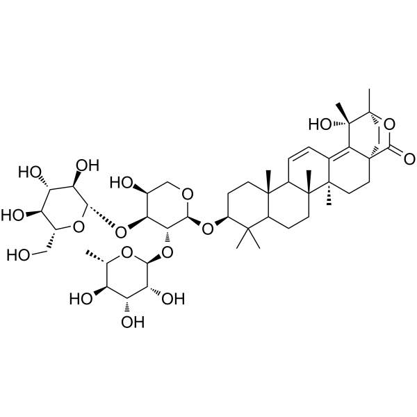
Kudinoside D is a triterpenoid saponin. Kudinoside D is isolated from the leaves of Ilex kudingcha. Kudinoside D enhances the phosphorylation levels of AMPK and its downstream molecule ACC, and inhibits the expression of PPARγ, C/EBPα, SREBP-1c as well as the target genes of adipogenic transcription factors. Kudinoside D inhibits the activity of ANO1. Kudinoside D possesses hypolipidemic activity. Kudinoside D can be used in obesity-related research.
Nur für Forschungszwecke. Wir verkaufen nicht an Patienten.
- Reinheit : 99.80%
- CAS. Nr.: 173792-61-5
- Formel: C47H72O17
- Molecular Weight:909.06
-
Speicherung:
4°C, protect from light
* In solvent : -80°C, 6 months; -20°C, 1 month (protect from light)

To place orders, for customer services and technical support, please contact: MedChemExpress USA
Tel: 609-228-6898 E-mail: [email protected] [email protected]


-
 Biologische Aktivität
-
Chemical Information
-
Lösungsmittel & Löslichkeit
- Protokoll
- Reinheit & Dokumentation
- Verweise
-
 Help & FAQs


Publications Citing Use of MedChemExpress (MCE) Kudinoside D
MoreAlle AMPK Isoform-spezifische Produkte anzeigen
More

Biologische Aktivität
Beschreibung
IC50 & Target
[1]|
PPARγ |
In Vitro
Kudinoside D (0-40 μM; 7 days) shows no cytotoxicity against 3T3-L1 preadipocytes, with a cell viability IC50 of 59.49 μM[1].
Kudinoside D (30 μM) inhibits ANO1 activity in FRT cells expressing ANO1 and YFP, with an inhibition rate of 26.2% at the concentration of 30 μM[2].
Kudinoside D (20-40 μM; 7 days) dose-dependently inhibits lipid droplet accumulation in differentiated 3T3-L1 adipocytes[1].
Treatment with Kudinoside D (20-40 μM; 7 days) throughout the 7-day differentiation period of 3T3-L1 adipocytes reduces intracellular triglyceride levels in differentiated cells: a 32.4% reduction at 20 μM and a 61.6% reduction at 40 μM[1].
Kudinoside D (20-40 μM; 7 days) alters the fatty acid composition of differentiated 3T3-L1 adipocytes and reduces SCD-1 activity (evidenced by a decreased C18:1 n-9/C18:0 ratio), with this treatment applied throughout the 7-day differentiation process[1].
Kudinoside D (20-40 μM) dose-dependently downregulates the protein expression levels of key adipogenic transcription factors (C/EBPβ, SREBP-1c, PPARγ, C/EBPα) and their target proteins (FABP4, FAS) during adipogenic differentiation of 3T3-L1 cells[1].
Kudinoside D (40 μM; 24 h) suppresses the protein expression of PPARγ and C/EBPα in differentiated 3T3-L1 adipocytes in vitro in an AMPK-dependent manner, as this effect is blocked by pretreatment with the AMPK inhibitor Compound C (10 μM; 30 min)[1].
MedChemExpress (MCE) has not independently confirmed the accuracy of these methods. They are for reference only.
-
Cell Line:3T3-L1 preadipocytes
-
Concentration:0-40 μM; 50 μM (cytotoxicity assessment)
-
Incubation Time:7 days
-
Result:Showed no cytotoxicity at concentrations up to 40 μM.
Exhibited a cell viability IC50 of 59.49 μM, with cytotoxicity starting around 50 μM.
-
Cell Line:differentiated 3T3-L1 adipocytes
-
Concentration:40 μM (kudinoside D); 10 μM (Compound C, pre-incubation)
-
Incubation Time:24 h (kudinoside D); 30 min (Compound C, pre-incubation)
-
Result:Inhibited the protein expression of PPARγ and C/EBPα without Compound C pretreatment.
Lost inhibitory effects on PPARγ and C/EBPα protein expression after pretreatment with Compound C.
In Vivo
MedChemExpress (MCE) has not independently confirmed the accuracy of these methods. They are for reference only.


Chemical Information
-
CAS. Nr. 173792-61-5
-
Appearance Solid
-
Molecular Weight 909.06
-
Formel C47H72O17
-
Color White to off-white
-
SMILES
CC1([C@H](CC[C@]2(C1CC[C@@]3(C2C=CC4=C5[C@@](C(O6)=O)(CC[C@@]34C)CC[C@]6([C@@]5(C)O)C)C)C)O[C@H]7[C@@H]([C@H]([C@H](CO7)O)O[C@H]8[C@@H]([C@H]([C@@H]([C@H](O8)CO)O)O)O)O[C@H]9[C@@H]([C@@H]([C@H]([C@@H](O9)C)O)O)O)C
-
Structure Classification
-
Initial Source
-
Versand
Room temperature in continental US; may vary elsewhere.
-
Speicherung
4°C, protect from light
* In solvent : -80°C, 6 months; -20°C, 1 month (protect from light)


Publications (1)
-
Journal Impact Factor
-
Most Recent


Lösungsmittel & Löslichkeit
In Vitro:
DMSO : 100 mg/mL (110.00 mM; Need ultrasonic; Hygroscopic DMSO has a significant impact on the solubility of product, please use newly opened DMSO)
Please refer to the solubility information to select the appropriate solvent. Once prepared, please aliquot and store the solution to prevent product inactivation from repeated freeze-thaw cycles.
Storage method and period of stock solution: -80°C, 6 months; -20°C, 1 month (protect from light). When stored at -80°C, please use it within 6 months. When stored at -20°C, please use it within 1 month.
Please refer to the solubility information to select the appropriate solvent. Once prepared, please aliquot and store the solution to prevent product inactivation from repeated freeze-thaw cycles.
Storage method and period of stock solution: -80°C, 6 months; -20°C, 1 month (protect from light). When stored at -80°C, please use it within 6 months. When stored at -20°C, please use it within 1 month.
Konzentration (Stammlösung) × Volumen (Stammlösung) = Konzentration (Ziellösung) × Volumen (Ziellösung)


Reinheit & Dokumentation
-
Data Sheet (292 KB)
-
SDS (252 KB)
- English - EN (252 KB)
- Français - FR (252 KB)
- Deutsch - DE (252 KB)
- Norwegian - NO (252 KB)
- Español - ES (252 KB)
- Swedish - SV (252 KB)
- Italian - IT (252 KB)
- Korean - KR (252 KB)
- Portuguese - PT (252 KB)
-
Handling Instructions (2659 KB)
Verweise
[1]. Che Y, et al. Kudinoside-D, a triterpenoid saponin derived from Ilex kudingcha suppresses adipogenesis through modulation of the AMPK pathway in 3T3-L1 adipocytes. Fitoterapia. 2018 Mar;125:208-216. [Content Brief]
[2]. Mai NT, et al. Triterpenoid glycosides from the rhizomes of Allium ascalonicum and their anoctamin-1 inhibitory activity. Nat Prod Res. 2021 Nov;35(22):4338-4346. [Content Brief]


Complete Stock Solution Preparation Table
Please refer to the solubility information to select the appropriate solvent. Once prepared, please aliquot and store the solution to prevent product inactivation from repeated freeze-thaw cycles.
Storage method and period of stock solution: -80°C, 6 months; -20°C, 1 month (protect from light). When stored at -80°C, please use it within 6 months. When stored at -20°C, please use it within 1 month.
| Optional Solvent | Concentration Solvent Mass | 1 mg | 5 mg | 10 mg | 25 mg |
|---|---|---|---|---|---|
| DMSO | 1 mM | 1.1000 mL | 5.5002 mL | 11.0004 mL | 27.5009 mL |
| 5 mM | 0.2200 mL | 1.1000 mL | 2.2001 mL | 5.5002 mL | |
| 10 mM | 0.1100 mL | 0.5500 mL | 1.1000 mL | 2.7501 mL | |
| 15 mM | 0.0733 mL | 0.3667 mL | 0.7334 mL | 1.8334 mL | |
| 20 mM | 0.0550 mL | 0.2750 mL | 0.5500 mL | 1.3750 mL | |
| 25 mM | 0.0440 mL | 0.2200 mL | 0.4400 mL | 1.1000 mL | |
| 30 mM | 0.0367 mL | 0.1833 mL | 0.3667 mL | 0.9167 mL | |
| 40 mM | 0.0275 mL | 0.1375 mL | 0.2750 mL | 0.6875 mL | |
| 50 mM | 0.0220 mL | 0.1100 mL | 0.2200 mL | 0.5500 mL | |
| 60 mM | 0.0183 mL | 0.0917 mL | 0.1833 mL | 0.4583 mL | |
| 80 mM | 0.0138 mL | 0.0688 mL | 0.1375 mL | 0.3438 mL | |
| 100 mM | 0.0110 mL | 0.0550 mL | 0.1100 mL | 0.2750 mL |


Kudinoside D Related Classifications
HY-N4253 Related Classifications
 Powered by Bioz