Mitochondria serve as central hubs of cellular energy metabolism and signal transduction and are essential for maintaining cellular homeostasis. Mitophagy, a form of selective autophagy, is critical for mitochondrial quality control and intracellular homeostasis through the clearence of damaged or dysfunctional mitochondria. Studies have shown that mitophagy not only participates in physiological processes such as development, differentiation, and metabolic regulation, but also exerts protective effects against oxidative stress and cellular damage[1].
Notably, dysregulated mitophagy is widely recognized as a key pathogenic mechanism in various diseases, including neurodegenerative diseases, cardiovascular diseases, metabolic disorders, and cancer, contributing to disease progression through alterations in mitochondrial function and metabolic reprogramming. With increasing insights into its molecular mechanisms (such as PINK1/Parkin-dependent and receptor-mediated pathways), mitophagy is emerging as a promising therapeutic target. Current research focuses on restoring mitochondrial homeostasis through precise modulation of mitophagy, thereby providing new intervention strategies for related diseases[2].
This article reviews the regulatory mechanisms of mitophagy, analyzes its roles in the pathogenesis of major diseases, and highlights recent advances in drug development and therapeutic strategies targeting mitophagy, aiming to provide clearer insights and strategic guidance for future research.
- Mechanisms of Mitophagy Regulation
- Mitophagy in Disease Research
- Therapeutic Strategies Targeting Mitophagy

Figure 1. The main processes of mitophagy[3].
Mitophagy is crucial for maintaining mitochondrial and cellular homeostasis. Under stress conditions such as reactive oxygen species (ROS) accumulation, nutrient deprivation, and cellular senescence, mitochondria can suffer depolarization and damage. To preserve the stability of the mitochondrial network and the overall cellular environment, cells selectively engulf and degrade dysfunctional mitochondria through autophagy. This process involves the following four key steps: 1) Depolarization of damaged mitochondria, resulting in loss of membrane potential. 2) Encapsulation of mitochondria by autophagosomes to form mitophagosomes. 3) Fusion of mitophagosomes with lysosomes. 4) Degradation of mitochondrial contents by lysosomes. These lysosomal or vacuolar acidic hydrolases facilitate the breakdown of damaged mitochondria[3].
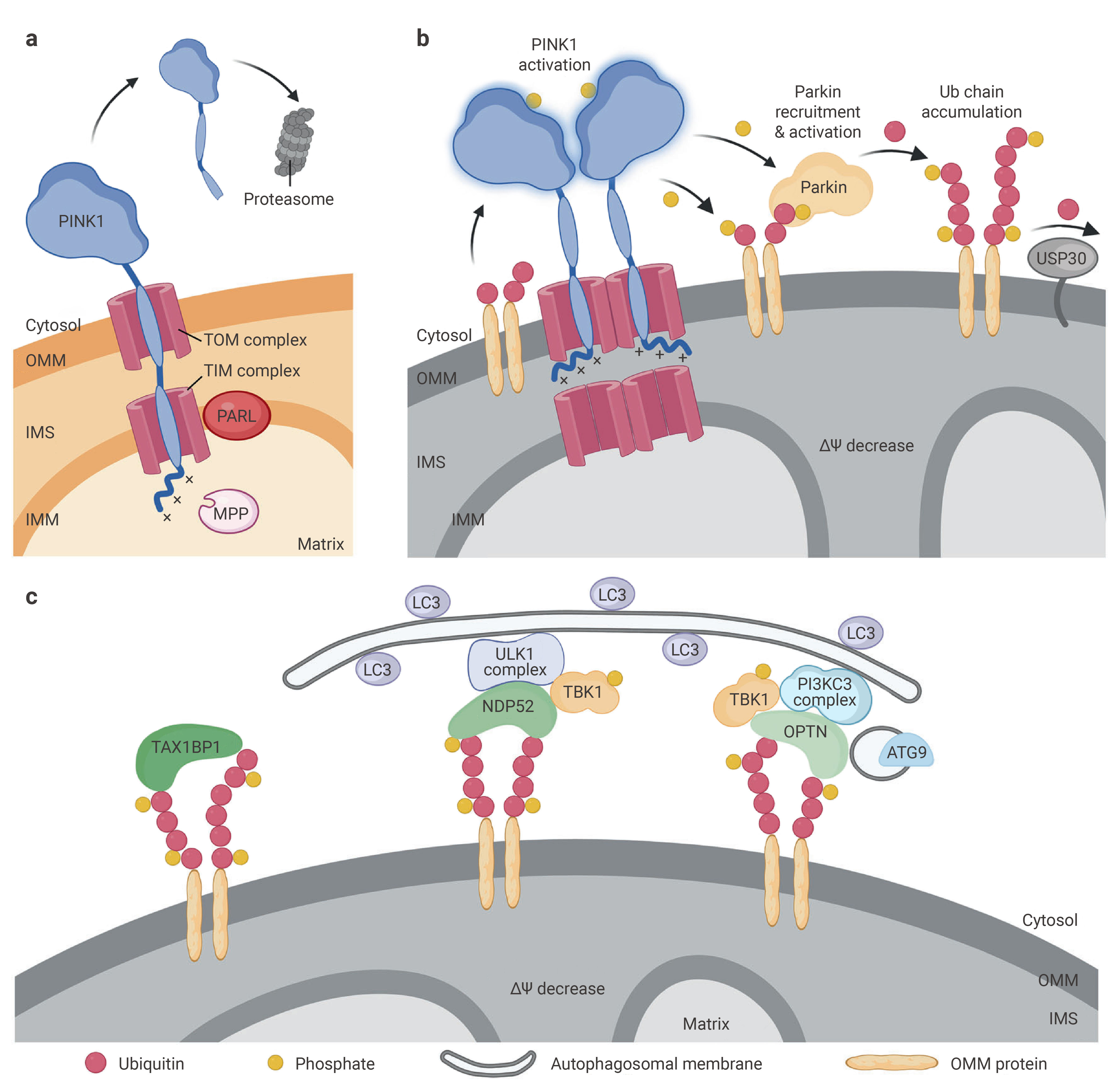
Figure 2. The main processes of mitophagy[2].
The PINK1/Parkin pathway is the most extensively studied mechanism of mitophagy. In healthy mitochondria, PINK1 is continuously imported into the inner mitochondrial membrane and degraded. However, upon loss of mitochondrial membrane potential, PINK1 accumulates on the outer mitochondrial membrane (OMM), where it becomes activated. Activated PINK1 phosphorylates ubiquitin and the E3 ligase Parkin, leading to Parkin recruitment to the mitochondrial surface.
Once activated, Parkin catalyzes the formation of various types of ubiquitin chains (K6, K11, K48, K63) on outer mitochondrial membrane proteins (such as MFN1/2, VDAC, etc.), creating an amplification loop that enhances ubiquitin signaling. These ubiquitin chains are recognized by autophagy receptors (OPTN, NDP52, TAX1BP1, etc.), which simultaneously recruit autophagosomes through the LC3-interacting region (LIR), facilitating the selective clearance of damaged mitochondria[2].
Although the PINK1/Parkin pathway is widely regarded as the core mechanism of mitophagy, increasing evidence suggests that other E3 ubiquitin ligases can also mediate mitophagy, particularly in Parkin-deficient cell. These alternative E3 ligases can ubiquitinate specific mitochondrial proteins, thereby recruiting autophagy receptors and initiating mitophagy. Moreover, ubiquitin modifications regulate the stability and expression of receptor pathways like BNIP3/NIX, highlighting the integrative role of the ubiquitin system across various mitophagy pathways[4].
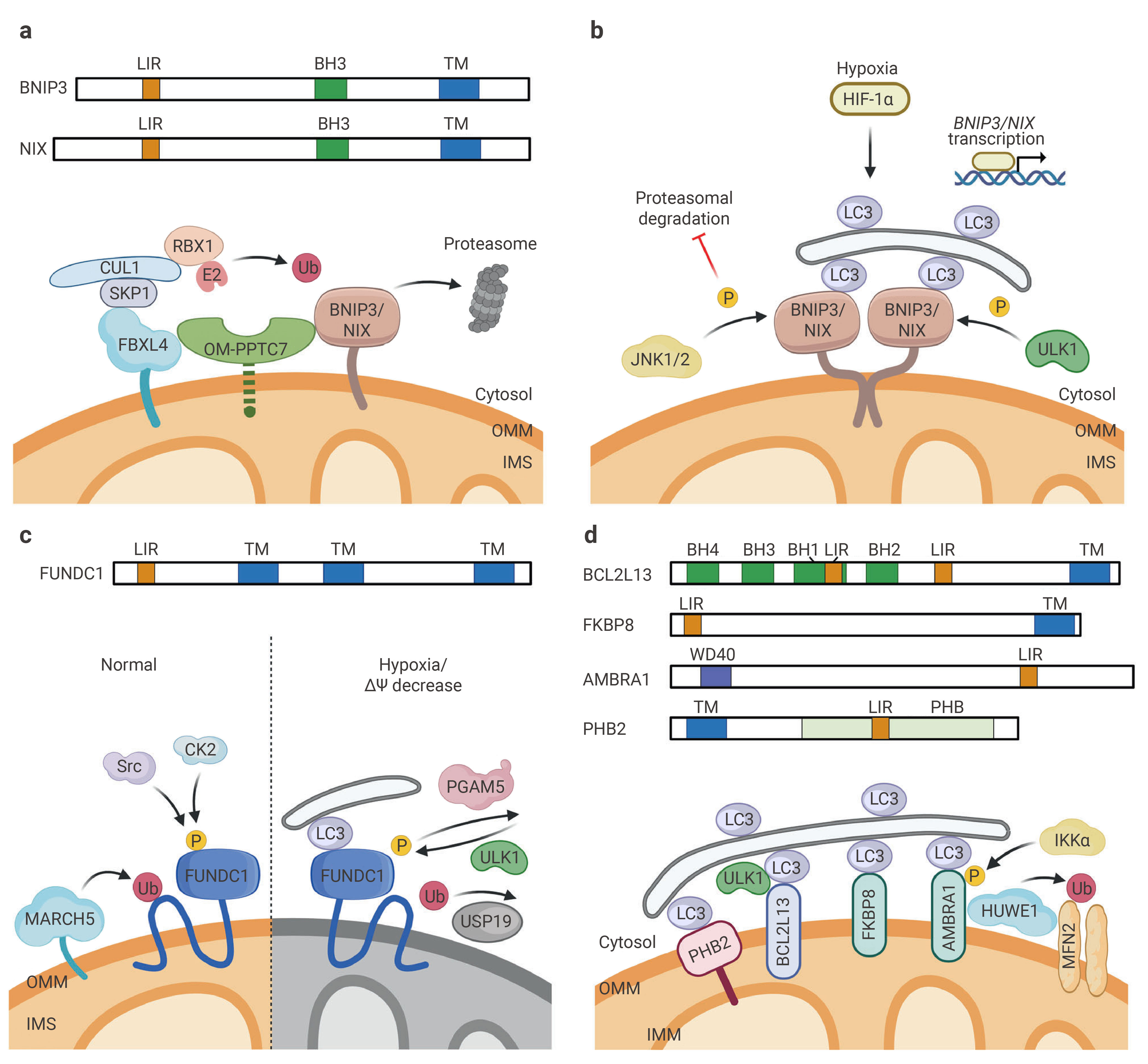
Figure 3. The main processes of mitophagy[2].
Mitophagy can also occur independently of ubiquitin. In this process, mitochondrial membrane proteins, known as "mitophagy receptors", mediate the delivery of mitochondria to autophagosomes. These receptors interact with the autophagic machinery through their LIR domains, primarily binding to ATG8s proteins.
BNIP3 and BNIP3L/NIX, BH3-only proteins located on the outer mitochondrial membrane, are key regulators of receptor-mediated mitophagy. They bind directly to LC3/GABARAP family proteins through their LC3-interacting region (LIR), thereby mediating the recognition and encapsulation of mitochondria by autophagosomes.
Their activity is tightly regulated at multiple levels. Transcriptionally, HIF-1α, stabilized in hypoxic microenvironment, upregulates its gene expression. At the post-translational level, kinases like ULK1 and JNK1/2 phosphorylate BNIP3/NIX, preventing their proteasomal degradation and enhancing their interaction with the autophagy machinery. Simultaneously, to prevent excessive autophagy, mitochondrial E3 ubiquitin ligase complexes (such as SCF-FBXL4) and phosphatase PPTC7 co-assemble to ubiquitinate and degrade BNIP3/NIX on the OMM, acting as a negative feedback "braking" mechanism under stress conditions like starvation.
BNIP3/NIX can also bind to Bcl-2/Bcl-xL, releasing Beclin-1 to promote autophagy initiation. While this pathway helps clear damaged mitochondria and maintain cell survival, overactivation can induce mitochondrial membrane potential loss and ROS accumulation, triggering non-canonical cell death. Overall, BNIP3/NIX represents a core non-ubiquitin-dependent mitophagy pathway that plays a dual role in cell fate regulation[5].
FUNDC1 is a key receptor on the OMM that primarily mediates hypoxia-induced mitophagy. Its activity is dynamically regulated by post-translational modifications: under normal conditions, FUNDC1 is phosphorylated by CK2 and Src kinases, preventing its interaction with LC3. Under hypoxic or mitochondrial stress conditions, the phosphatase PGAM5 dephosphorylates FUNDC1, while ULK1 activates it through phosphorylation, enhancing its binding to LC3 and promoting mitophagy.
In addition to the LIR-dependent mechanism, FUNDC1 also regulates mitochondrial dynamics, particularly in coordination with DRP1-mediated mitochondrial fission, suggesting it has both structural and signaling roles in mitophagy initiation. This pathway plays a crucial role in myocardial ischemia-reperfusion injury and tumor microenvironment adaptation.
In addition to BNIP3/NIX and FUNDC1, other receptor proteins involved in mitophagy have been identified in recent years, such as BCL2L13, FKBP8, and PHB2. These proteins also interact with LC3 through their LIR domains, but their functions exhibit tissue specificity and stress dependence.
For example, BCL2L13 acts as a receptor in mammals similar to yeast Atg32, independently inducing mitophagy. PHB2, located on the inner mitochondrial membrane, is exposed when the outer membrane ruptures and binds to LC3, indicating that mitophagy regulation extends beyond the OMM. Additionally, FKBP8 is involved in basal mitochondrial turnover, highlighting the importance of this pathway in maintaining mitochondrial homeostasis under normal conditions.
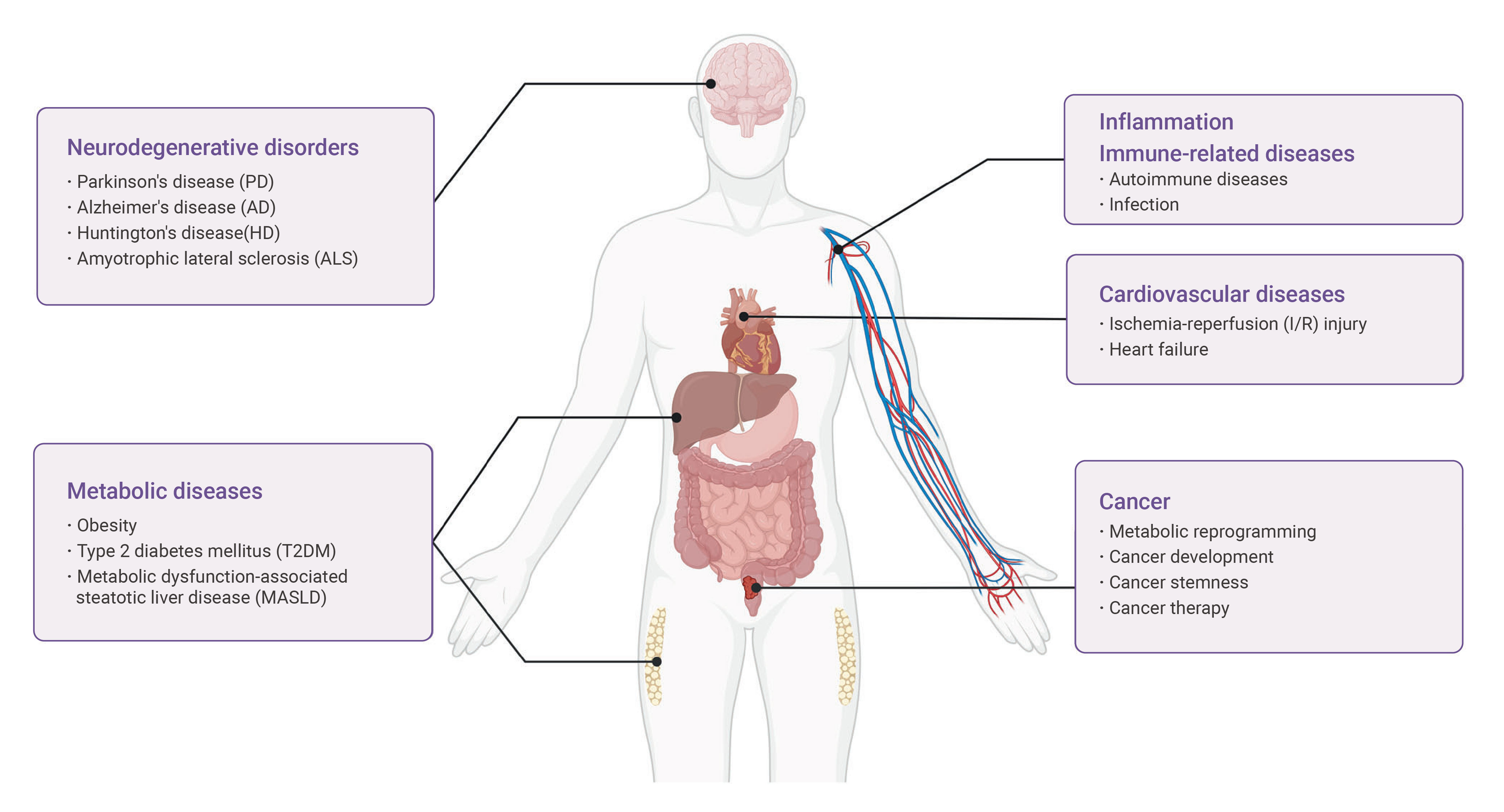
Figure 4. Summary of mitophagy-associated human diseases[2].
Abnormal mitochondrial morphology and function are closely associated with a wide range of diseases. Dysfunctional mitophagy contributes to the pathogenesis of numerous clinical conditions, including neurodegenerative disorders, cardiovascular diseases, cancer, metabolic diseases and immune-related diseases.
Neurons are highly dependent on mitochondrial energy supply and, as terminally differentiated cells, are particularly sensitive to mitochondrial quality control. Accumulating evidence indicates that defective mitophagy leads to the accumulation of damaged mitochondria, triggering oxidative stress, calcium dysregulation, and synaptic dysfunction—key pathological features of many neurodegenerative diseases.
In Parkinson's disease, mutations in PINK1 or Parkin impair ubiquitin-dependent mitophagy, resulting in mitochondrial dysfunction and dopaminergic neuron loss. In Alzheimer's disease, Aβ and tau protein disrupt mitophagy and exacerbate mitochondrial damage, forming a vicious cycle. Similarly, in Huntington's disease and amyotrophic lateral sclerosis (ALS), aberrant protein aggregation interferes with mitophagy.
Overall, insufficient mitophagy and the consequent buildup of mitochondrial toxicity are common hallmarks leading to neuronal death[6].
Diseases | Mitophagy defects under pathological conditions |
|---|---|
| AD | 1. Altered mitophagy initiation. 2. Ongoing vicious cycle of impaired mitophagy by Tau and Aβ. 3. Impaired undegraded cargo clearance within lysosomes. |
| PD | 1. Impaired PINK1-Parkin-mediated mitophagy. 2. Increased cardiolipin-mediated mitophagy in the presence of α-synuclein. |
| HD | 1. Impaired PINK1-Parkin-mediated mitophagy. 2. Affected mitophagy initiation and recruitment of mitophagy receptors. 3. Impaired GAPDH-mediated mitophagy. 4. Impaired autophagic recruitment of damaged mitochondria. 5. Affected transportation of autophagosome towards the lysosomes. |
| ALS | 1. Enhanced PINK1-Parkin-mediated mitophagy. |
Mitophagy exerts a dual role in immune regulation, both limiting excessive inflammation and, under certain conditions, promoting immune activation. Its primary function is to remove damaged mitochondria, thereby reducing the release of mitochondrial DNA (mtDNA) and ROS, key drivers of inflammatory signaling.
Mitophagy deficiency can lead to mtDNA leakage and activation of the NLRP3 inflammasome, promoting chronic inflammationn. Conversely, enhanced mitophagy suppresses the inflammatory cascade and maintains immune homeostasis. In innate immune cells such as macrophages, mitophagy also regulate metabolic reprogramming, thereby shaping inflammatory phenotypes.
In autoimmune diseases, impaired mitophagy may promote autoantigen release and loss of immune tolerance; for instance, in systemic lupus erythematosus, defective mitochondrial clearance enhances immune activation.
As central regulators of cellular metabolism, mitochondria make mitophagy essential in metabolic homeostasis. Mitophagy influences lipid metabolism, glucose balance, and insulin sensitivity by controlling mitochondrial quantity and quality.
In type 2 diabetes, impaired mitophagy leads to mitochondrial dysfunction in β-cells, compromising insulin secretion. In nonalcoholic fatty liver disease, defective mitophagy can promote lipid accumulation and oxidative stress, exacerbating disease progression.
Furthermore, in obesity-related disorders, mitophagy contributes to metabolic reprogramming by regulating mitochondrial function in adipose tissue.
Cardiomyocytes are highly dependent on mitochondrial function, making mitophagy crucial in cardiovascular disease. Studies have shown that mitophagy has a dual role in ischemia-reperfusion (I/R) injury: moderate activation clears damaged mitochondria and protects the myocardium, while excessive or insufficient activation exacerbates the injury.
For example, FUNDC1-mediated mitophagy is activated under hypoxia, helping maintain mitochondrial homeostasis and reduce cell death. Mitophagy also contributes to heart failure and cardiomyopathy by regulating mitochondrial function and myocardial energy metabolism.
Mitophagy plays a stage-dependent and dual role in tumorigenesis. In the early stages of development, mitophagy exerts anti-tumor effects by eliminating damaged mitochondria, reducing ROS levels, and maintaining genomic stability. However, during tumor progression, cancer cells can utilize mitophagy to adapt to hypoxic conditions, reprogram cellular metabolism, and enhance resistance to therapy.
Furthermore, mitophagy is involved in regulating cancer stem cell maintenance, immune evasion, and metastasis. Notably, mitophagy levels vary significantly across different tumor types, exhibiting high heterogeneity. This suggests that its role in cancer is highly dependent on the specific biological context[7].
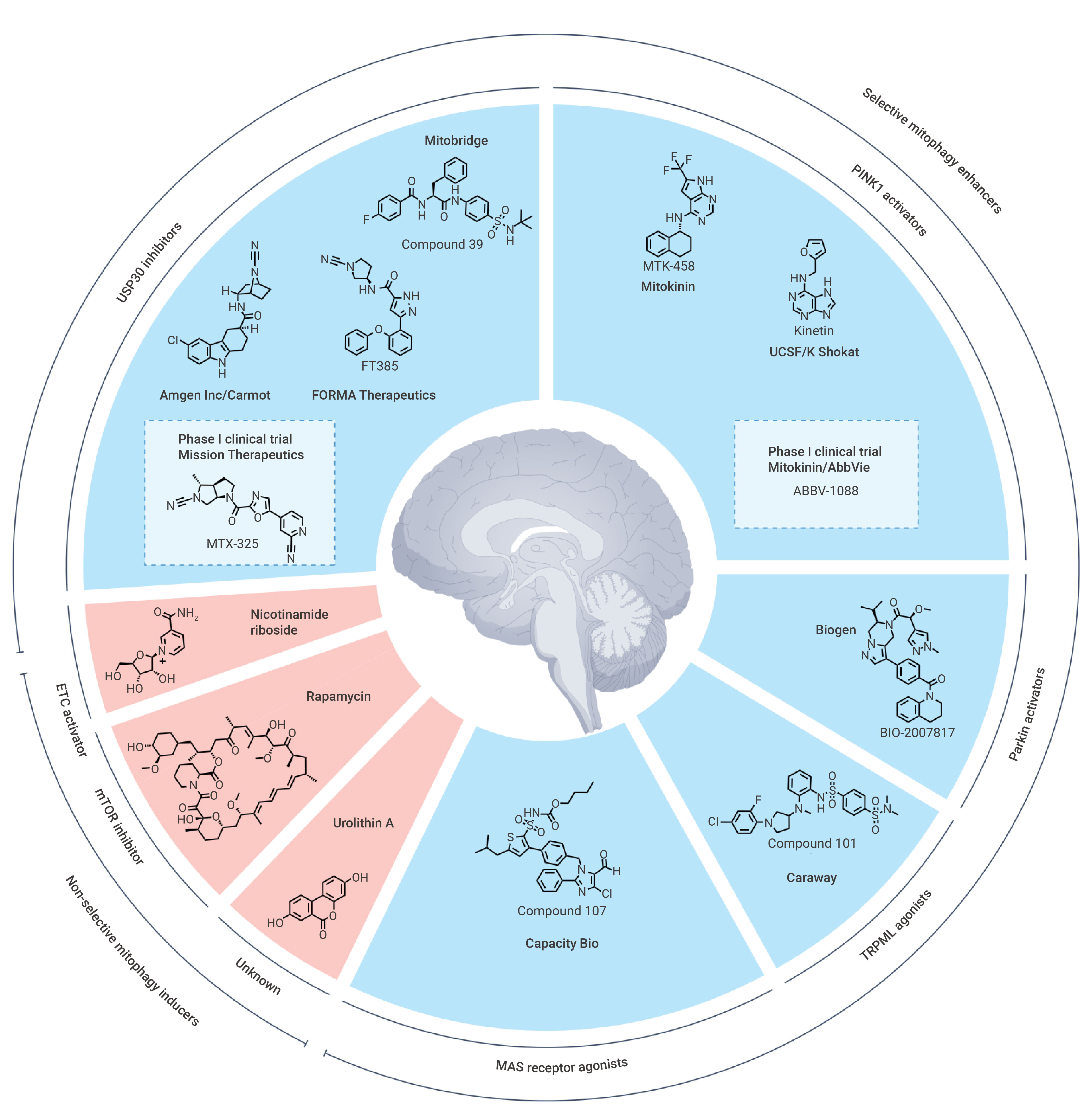
Figure 5. Selective and non-selective mitophagy activators[8].
Given the central role of mitochondrial quality control dysregulation in aging and numerous major diseases, targeted modulation of the mitophagy network has emerged as a promising avenue in translational medicine. Current mainstream clinical strategies primarily focus on "enhancing/activating mitophagy", aiming to reverse degenerative changes and restore cellular homeostasis by improving impaired autophagic flux.
The PINK1/Parkin pathway represents a major target in drug development. Activation of PINK1 or enhancement of Parkin activity promotes ubiquitination cascades, thereby accelerating the clearance of damaged mitochondria. In neurodegenerative diseases, particularly Parkinson's disease, this approach is considered to have strong disease-modifying potential.
Urolithin A, a natural metabolite produced by gut microbiota, is widely recognized as a representative mitophagy activator. It promotes mitochondrial renewal by activating the PINK1/Parkin pathway, thereby improving muscle function and delaying age-related mitochondrial decline.
Nicotinamide riboside chloride, an NAD+ precursor, enhances mitophagy and mitochondrial biogenesis through activation of the SIRT1/PGC-1α axis, demonstrating protective effects in neurodegenerative and metabolic diseases.
MTK458 is an orally active, brain-penetrant PINK1 activator. It binds to and stabilizes the active PINK1 heterocomplex, thereby increasing mitophagy and shows potential for application in Parkinson's disease research.
Several metabolic regulators (such as SIRT1 activators and AMPK agonists) have been shown to indirectly promote mitophagy. These compounds enhance mitochondrial turnover by regulating energy metabolism and autophagy pathways.
Metformin induces autophagy and mitophagy by activating the AMPK signaling pathway, and has broad therapeutic effects in metabolic diseases and cancer[9].
Resveratrol exerts protective effects in neurodegenerative and cardiovascular diseases by activating SIRT1 to induce mitophagy and reducing ROS levels.
In recent years, a novel class of molecules—autophagy-targeting chimeras (AUTACs)—has been developed to directly induce selective autophagic degradation of specific organelles, including mitochondria. These molecules activate mitophagy by linking target mitochondria to the autophagy machinery, thereby bypassing the classic PINK1/Parkin pathway.
AUTAC4 is a bifunctional molecule that induces K63-linked ubiquitination to label specific mitochondria, thereby promoting their recognition and degradation by the autophagy system and achieving selective mitophagy.
A common strategy to inhibit mitophagy involves blocking the completion of the autophagic process, particularly the fusion of autophagosomes with lysosomes.
Chloroquine inhibits mitophagy by suppressing lysosomal acidification and preventing the fusion of autophagosomes with lysosomes. It is widely used in combination cancer therapies.
Bafilomycin A1 suppresses lysosomal function by inhibiting V-ATPase, thereby blocking late-stage autophagy, preventing the fusion of autophagosomes and lysosomes, and inhibiting acidification and protein degradation in cultured cells.
Mdivi-1, a DRP1 inhibitor, indirectly affects mitophagy by modulating mitochondrial fission. In dopaminergic neuronal cell lines, Mdivi-1 counteracts the global mitotic effect induced by mutant human PINK1. It has also been shown to inhibit BCL2L13-mediated mitophagy, thereby reducing the migration and invasion in glioblastoma cells.
Mitophagy is a central mechanism of mitochondrial quality control with context-dependent roles across diverse diseases. Dysregulation of mitophagy contributes to the pathogenesis of neurodegenerative, metabolic, cardiovascular, and malignant disorders, underscoring its dual function as both a protective and pathogenic process. Advances in understanding ubiquitin-dependent and receptor-mediated pathways have facilitated the development of targeted therapeutic strategies, including small-molecule modulators and emerging technologies such as AUTACs. However, challenges remain, particularly regarding specificity and the context-dependent effects of mitophagy. These limitations highlight the need for more precise, disease-specific interventions in future translational research.
Product Name | Cat. No. | Target | The Role of Targeted Mitophagy Therapy |
|---|---|---|---|
Urolithin A | Autophagy/Mitophagy | Induced autophagy | |
Nicotinamide riboside chloride | Sirtuin | Increased NAD+ levels and activates SIRT1 and SIRT3 | |
MTK458 | HY-152943 | PINK1/Parkin | Bound to PINK1 and stabilizes an active heterocomplex, thereby increasing mitophagy |
Metformin | HY-B0627 | AMPK/Autophagy/Mitophagy/Apoptosis/mTOR | Regulated the expression of autophagy-related proteins by activating AMPK and inhibiting the mTOR signaling pathway |
Resveratrol | HY-16561 | Autophagy/Mitophagy/ Sirtuin | SIRT1 activator |
AUTAC4 | HY-134640 | AUTACs/Mitophagy | Induced mitophagy |
Chloroquine | HY-17589A | Autophagy | Autophagy inhibitor |
Bafilomycin A1 | HY-100558 | Autophagy | Autophagy inhibitor |
Mdivi-1 | HY-15886 | Mitophagy/Autophagy | Mitophagy inhibitor |
BIO-2007817 | HY-169329 | PINK1/Parkin | Enhanced the activity of wildtype Parkin |
Product Name | Cat. No. | Application | The Role of Detecting and Monitoring Mitophagy/Reactivity |
|---|---|---|---|
JC-1 | HY-15534 | Fluorescent Dye | Detection of early mitochondrial depolarization events |
MitoSOX Red | HY-D1055 | Fluorescent Dye/Reactive Oxygen Species (ROS)/ Mitochondrial Metabolism | Detection of mitochondrial ROS (mitophagy trigger signal) |
MitoTracker Green FM | HY-135056 | Fluorescent Dye | Mitochondrial localization/co-localization analysis with LC3 |
LC3A/B Antibody | HY-P80741 | WB | Human, Mouse, Rat |
SQSTM1/p62 Antibody (YA062) | HY-P80899 | WB, IHC-F, IHC-P, ICC/IF, IP | Human |
TOMM20 Antibody (YA1380) | HY-P81635 | WB, IHC-F, IHC-P, ICC/IF | Human, Hamster |
COX IV Antibody (YA5187) | HY-P85495 | WB, FC, ICC/IF, IP, IHC-P | Human, Mouse, Rat, Hamster, Goat, Monkey |
Product Name | Cat. No. | Target | The Role of Detecting and Monitoring Mitophagy |
|---|---|---|---|
CCCP | HY-100941 | OXPHOS/PINK1/Parkin/ STING | Induced activation of PINK1 leading to Parkin Ser65 phosphorylation |
FCCP | HY-100410 | PINK1/Parkin | Induced activation of PINK1 leading to Parkin Ser65 phosphorylation |
Note: MCE can provide products for research use only. We do not sell to patients.
 LinkedIn
 Researchgate
 Instagram
 X
X Facebook
 WhatsApp
 Reddit
 Mail
Mail- [1]. Onishi M, et al. EMBO J. 2021 Feb 1;40(3):e104705. [Content Brief]
- [2]. Wang Q, et al. Cell Res. 2026 Jan;36(1):11-37. [Content Brief]
- [3]. Lu Y, et al. Theranostics. 2023 Jan 1;13(2):736-766. [Content Brief]
- [4]. Clague MJ, et al. Trends Cell Biol. 2025 Jun;35(6):527-538. [Content Brief]
- [5]. Li Y, et al. Cell Death Dis. 2021 Dec 20;13(1):14. [Content Brief]
- [6]. Li X, et al. Pharmacol Res. 2021 Jan;163:105240. [Content Brief]
- [7]. Huang J, et al. J Hematol Oncol. 2025 Aug 1;18(1):78. [Content Brief]
- [8]. Antico O, et al. Nat Rev Drug Discov. 2025 Apr;24(4):276-299. [Content Brief]
- [9]. Bharath LP, et al. Cell Metab. 2020 Jul 7;32(1):44-55.e6. [Content Brief]