
M-525
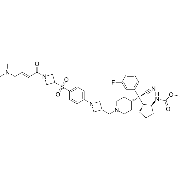
M-525 is a first-in-class, highly potent, irreversible and covalent menin-MLL protein-protein interaction inhibitor. M-525 binds to menin with an IC50 of 3 nM and achieves low nanomolar potencies in cell growth inhibition and in suppression of MLL regulated gene expression in MLL leukemia cells. Anti-leukemia activity.
For research use only. We do not sell to patients.
- CAS No.: 2173582-08-4
- Formula: C39H51FN6O5S
- Molecular Weight:734.92
-
Storage:
Please store the product under the recommended conditions in the Certificate of Analysis.
Biological Activity
Description
Cellular Effect
|
Cell Line
|
Type | Value | Description | References |
|---|---|---|---|---|
| HL-60 | GI50 |
0.9 μM
Compound: 9; M-525
|
Anticancer activity against human HL-60 cells assessed as cell growth inhibition measured after 7 days by WST-8 assay
|
[PMID: 36528996] |
| HL-60 | IC50 |
1500 nM
Compound: M-525
|
Growth inhibition of human HL60 cells after 7 days by WST-8 assay
|
[PMID: 32338903] |
| K562 | GI50 |
0.9 μM
Compound: 9; M-525
|
Anticancer activity against human K562 cells assessed as cell growth inhibition measured after 7 days by WST-8 assay
|
[PMID: 36528996] |
| MOLM-13 | GI50 |
15.4 nM
Compound: 15; M-525
|
Antiproliferative activity against human MOLM-13 cells
|
[PMID: 34726905] |
| MOLM-13 | GI50 |
2.3 nM
Compound: 9; M-525
|
Anticancer activity against human MOLM-13 cells harboring MLL-AF9 fusion protein assessed as cell growth inhibition measured after 7 days by WST-8 assay
|
[PMID: 36528996] |
| MOLM-13 | IC50 |
19 nM
Compound: M-525
|
Growth inhibition of human MOLM13 cells after 7 days by WST-8 assay
|
[PMID: 32338903] |
| MOLM-14 | GI50 |
2.3 nM
Compound: 9; M-525
|
Anticancer activity against human MOLM-14 cells harboring MLL-AF9 fusion protein assessed as cell growth inhibition measured after 7 days by WST-8 assay
|
[PMID: 36528996] |
| MOLM-16 | GI50 |
0.9 μM
Compound: 9; M-525
|
Anticancer activity against human MOLM16 cells assessed as cell growth inhibition measured after 7 days by WST-8 assay
|
[PMID: 36528996] |
| MONO-MAC-6 | GI50 |
2.3 nM
Compound: 9; M-525
|
Anticancer activity against human MONO-MAC-6 cells harboring MLL-AF9 fusion protein assessed as cell growth inhibition measured after 7 days by WST-8 assay
|
[PMID: 36528996] |
| MV4-11 | GI50 |
2.3 nM
Compound: 9; M-525
|
Anticancer activity against human MV4-11 cells harboring MLL-AF4 fusion protein assessed as cell growth inhibition measured after 7 days by WST-8 assay
|
[PMID: 36528996] |
| MV4-11 | GI50 |
2.7 nM
Compound: 15; M-525
|
Antiproliferative activity against human MV4-11 cells
|
[PMID: 34726905] |
| MV4-11 | IC50 |
4 nM
Compound: M-525
|
Growth inhibition of human MV4-11 cells after 7 days by WST-8 assay
|
[PMID: 32338903] |
| RS4-11 | GI50 |
2.3 nM
Compound: 9; M-525
|
Anticancer activity against human RS4-11 cells harboring MLL-AF4 fusion protein assessed as cell growth inhibition measured after 7 days by WST-8 assay
|
[PMID: 36528996] |
| SEM | GI50 |
2.3 nM
Compound: 9; M-525
|
Anticancer activity against human SEM cells harboring MLL-AF4 fusion protein assessed as cell growth inhibition measured after 7 days by WST-8 assay
|
[PMID: 36528996] |
| SKM-1 | GI50 |
0.9 μM
Compound: 9; M-525
|
Anticancer activity against human SKM-1 cells assessed as cell growth inhibition measured after 7 days by WST-8 assay
|
[PMID: 36528996] |
In Vitro
M-525 achieves an IC50 of 3 nM in the MV4;11 cell line and has an IC50 of 2 µM in the HL-60 cell line. M-525 (3-300 nM; 24 h) is potent and effective in suppressing the expression of MEIS1 and HOX genes in MV4;11 cells carrying the MLL-AF4 fusion and in MOLM-13 cells carrying the MLL-AF9 fusion. It demonstrates high cellular specificity over non-MLL leukemia cells[1].
MedChemExpress (MCE) has not independently confirmed the accuracy of these methods. They are for reference only. Further protocols information, click here.
Chemical Information
-
CAS No. 2173582-08-4
-
Molecular Weight 734.92
-
Formula C39H51FN6O5S
-
SMILES
O=C(OC)N[C@@H]1[C@@H]([C@](C2CCN(CC3CN(C4=CC=C(S(=O)(C5CN(C(/C=C/CN(C)C)=O)C5)=O)C=C4)C3)CC2)(C#N)C6=CC=CC(F)=C6)CCC1
-
Shipping
Room temperature in continental US; may vary elsewhere.
-
Storage
Please store the product under the recommended conditions in the Certificate of Analysis.
Protocols
-
RT-PCR
Reverse transcription technology uses RNA as a template to synthesize DNA. RT-PCR is simple, specific and sensitive, and can be used to detect gene expression levels and expression differences in cells; detect RNA virus content; clone cDNA sequences of specific genes.
-
RNA extraction experimental
By lysing cells, releasing RNA, and removing impurities such as proteins and DNA, high-purity RNA products are finally obtained. The commonly used traditional method is the guanidine isothiocyanate/phenol/chloroform method (Trizol), which is suitable for a variety of animal materials including animal tissues, microorganisms, cultured cells, etc., and most plant materials.
-
Immunoprecipitation
Immunoprecipitation (IP) is an experimental method that uses the principle of antibody specific binding to purify and enrich target proteins.
-
Somatic Cell Culture
A method of simulating the in vivo environment in vitro to maintain the cell growth, differentation and main functions.
-
Protocol for Bimolecular Fluorescence Complementation (BiFC) Assay
Bimolecular fluorescence complementation detects protein-protein proximity in living or fixed cells by fusing two candidate interaction partners to nonfluorescent N- and C-terminal fragments of a fluorescent protein; when the partners interact or remain close enough, the fluorescent fragments complement, mature, and generate a fluorescent signal at the site of the protein complex. The BiFC readout is fluorescence intensity and subcellular localization of the reconstituted fluorophore, which reflects formation or stabilization of a protein complex rather than direct biochemical binding kinetics; BiFC is therefore useful for mapping where interactions occur in cancer cells, neurons, macrophages, organoid-derived cells, or drug-screening systems, but results should be validated by independent assays such as co-IP or Western blot. BiFC signal formation is delayed by fluorophore maturation and can stabilize otherwise transient complexes, so it is not a real-time reversible interaction assay
-
CCK-8/WST-8 Cell Proliferation Assay
The CCK-8/WST-8 assay is based on the reduction of the water-soluble tetrazolium salt WST-8 to a water-soluble formazan product by cellular dehydrogenases in metabolically active cells, where the generated formazan amount is proportional to the number of living cells and is quantified by measuring absorbance in the visible range, providing a colorimetric readout for cell viability and proliferation assessment. This class of tetrazolium-based assays improves upon earlier MTT-based systems by producing a water-soluble formazan, eliminating the need for organic solubilization steps and enabling direct spectrophotometric measurement in culture medium.
-
Cell Counting-Based Growth Curve Assay
Cell counting-based growth curve assays quantify cell proliferation by directly measuring changes in viable cell number over time using manual or automated counting methods such as hemocytometer-based counting or instrument-assisted cell enumeration, enabling construction of growth curves that reflect population expansion dynamics in response to culture conditions. A widely used approach is trypan blue exclusion with hemocytometer counting, where membrane-compromised (non-viable) cells take up the dye, allowing discrimination between viable and non-viable cells while simultaneously enabling total cell number quantification. Repeated sampling across time points allows estimation of proliferation rate, growth phases, and comparative growth kinetics between experimental conditions.
-
Real Time qPCR (Q-PCR)
Real-time quantitative PCR (qPCR) quantifies an amplifiable nucleic-acid target by monitoring fluorescence during PCR cycling rather than measuring product only after amplification. The increase in fluorescence tracks accumulation of PCR product, and the quantification cycle (Cq; historically also Ct/CP) is related to the initial amount of target: samples containing more starting target generally reach the defined fluorescence threshold in fewer cycles.
-
Directly Induced Neuron Culture
Directly induced neuron culture converts somatic cells, most commonly fibroblasts, into induced neurons without passing through a pluripotent or neural progenitor stage; classic evidence shows that mouse fibroblasts can be converted by Ascl1, Brn2/Pou3f2, and Myt1l, human fibroblasts can be converted by defined neuronal transcription factors, and human fibroblasts can also be converted by miR-9/9-124 with neurogenic or subtype-specifying transcription factors. The readout is acquisition of neuronal identity and function, assessed by neuronal morphology, neuronal markers such as Tuj1/βIII-tubulin, MAP2, synapsin, and subtype markers when relevant, together with functional assays such as action-potential firing, synaptic activity, and electrophysiology.
-
Cell Viability Determination by MTT Colorimetric Assay
The following protocol uses the MTT colorimetric assay as a classic literature-established method for assessing cell viability/metabolic activity in cultured mammalian cells. MTT[3-(4,5-dimethylthiazol-2-yl)-2,5-diphenyltetrazolium bromide] is reduced by metabolically active cells to a colored formazan product; the amount of formazan is quantified spectrophotometrically and provides an indirect measure of metabolically active viable cells. Importantly, MTT reduction reflects cellular oxidoreductase/metabolic activity rather than an absolute direct count of living cells, so changes in cellular metabolism can alter the signal independently of cell number.
-
Cell Cytotoxicity Assay
Cytotoxicity assays are usually based on the assessment of cell membrane damage, which can also be indirectly detected by measuring cell viability. Detection methods include MTT assay, CKK-8 assay, LDH assay and ATP assay, etc.
-
Pull-down
The pull-down assay is an in vitro technique used to detect physical interactions between two or more proteins and an invaluable tool for confirming a predicted protein-protein interaction or identifying novel interacting partners. This method typically involves the use of affinity purification with various wash and elution steps.
-
Protocol for Cell Counting and Cell Density Analysis
Cell counting and cell-density analysis estimate the number of cells in a known volume or field area. Manual hemocytometer counting uses a chamber of defined geometry to convert counted cells into cells/mL, while automated counters and image-analysis workflows detect cell objects from optical, brightfield, fluorescence, impedance, or digital-image features. Trypan blue viability counting is based on dye exclusion: viable cells with intact membranes exclude dye, while non-viable cells with compromised membranes stain blue. The readout is total cell density, viable-cell density, dead-cell density, and percent viability. Cell density can also be estimated from microscopy images by counting objects per image area, from flow cytometry using calibrated volume or reference particles, or from in situ microscopy in bioreactors after calibration against reference methods such as hemocytometer or flow cytometry.
-
MTT Cell Proliferation Assay
The MTT assay is a colorimetric endpoint assay for estimating viable cell number, cell growth, cytotoxicity, or cell activation in cultured mammalian cells. Living cells reduce the yellow tetrazolium salt MTT into purple/blue formazan, while dead cells do not generate the same signal; the resulting color can be quantified with a multiwell spectrophotometer. MTT reduction is commonly interpreted as a readout of metabolic activity that often correlates with viable cell number, but it should not be treated as a direct cell-counting method unless the assay is optimized for the cell type and experimental condition. Studies show that MTT reduction can involve mitochondrial and non-mitochondrial reducing systems, and formazan may accumulate in intracellular lipid droplets rather than simply marking mitochondria.
-
Co-Immunoprecipitation
Co-immunoprecipitation technology can verify protein interaction based on the specific immune reaction between antibodies and antigens.
-
Protocol for Yeast Two-Hybrid (Y2H) Assay
The yeast two-hybrid assay detects binary protein-protein interactions by separating a transcription factor into a DNA-binding domain fused to a "bait" protein and a transcriptional activation domain fused to a "prey" protein; if bait and prey interact in yeast, the transcription factor is reconstituted and activates reporter genes such as HIS3, ADE2, lacZ, MEL1, or other selectable/readable reporters. The readout is yeast growth on selective medium and/or reporter activity, which reflects proximity-dependent transcriptional activation in the yeast nucleus rather than direct biochemical binding in the original mammalian, tumor, neuronal, macrophage, or organoid context. Because yeast two-hybrid can generate false positives and false negatives, interaction claims should be validated using independent assays such as co-immunoprecipitation, Western blot, immunofluorescence colocalization, BiFC, pull-down, or mammalian two-hybrid assays.
Purity & Documentation
References
Calculators
Concentration (start) × Volume (start) = Concentration (final) × Volume (final)

