



N1-Acetyl-5-methoxykynuramine hydrochloride

Based on 1 Customer Validation
N1-Acetyl-5-methoxykynuramine hydrochloride (AMK hydrochloride) is an active metabolite of Melatonin (HY-B0075) with antioxidant activity and blood-brain barrier permeability. N1-Acetyl-5-methoxykynuramine hydrochloride inhibits LPS-induced activation of inducible nitric oxide synthase and COX-2, scavenges ROS, hydroxyl radicals, carbonate radicals and ABTS cation radicals, and protects bovine serum albumin from peroxyl radical-mediated damage. N1-Acetyl-5-methoxykynuramine hydrochloride reduces the increase in cytoplasmic and mitochondrial lipid peroxidation in the substantia nigra and striatum in MPTP (HY-15608)-induced mouse models of Parkinson's disease. N1-Acetyl-5-methoxykynuramine hydrochloride can be used in studies related to Parkinson's disease, metabolism-related diseases and oxidative stress.
For research use only. We do not sell to patients.
- CAS No.: 1215711-91-3
- Formula: C12H17ClN2O3
- Molecular Weight:272.73
-
Storage:
Please store the product under the recommended conditions in the Certificate of Analysis.


To place orders, for customer services and technical support, please contact: MedChemExpress USA
Tel: 609-228-6898 E-mail: [email protected] [email protected]


-
 Biological Activity
-
Chemical Information
- Protocols
- Purity & Documentation
- References
-
 Help & FAQs


-
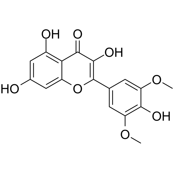
HY-N8920
SyringetinOsteoblast Differentiation Stimulator -
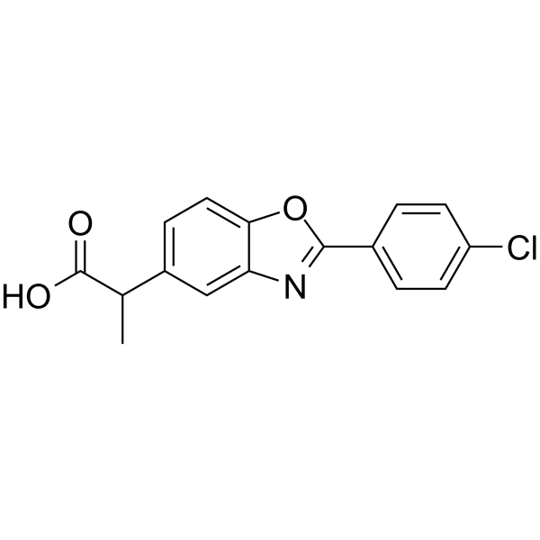
HY-13568
BenoxaprofenNonsteroidal Anti-inflammatory Agent -
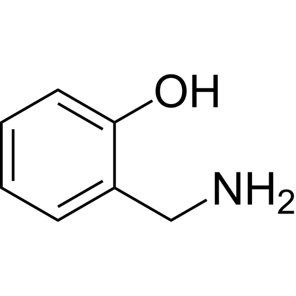
HY-34350
2-(Aminomethyl)phenolAntioxidant -
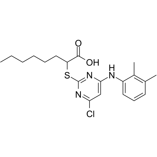
HY-111140
YS121mPGES-1/5-LOX Inhibitor -

HY-136855
MitoPBNAMPK/SIRT3/PGC-1α Modulator -

HY-13568R
Benoxaprofen (Standard)Anti-inflammatory/Antipyretic Compound -

HY-130743
Dieugenol -
HY-120255AS
17(S)-HDHA-d5Stable Isotope


Biological Activity
Description
|
COX-2 |
iNOS |
nNOS |
In Vitro
N1-Acetyl-5-methoxykynuramine hydrochloride potently scavenges hydroxyl radicals and ABTS cation radicals in cell-free systems at pH 5.0 and 7.5, but only weakly scavenges superoxide anions. It significantly protects bovine serum albumin from damage by peroxyl radicals, yet is rapidly oxidized and degraded by hydroxyl and carbonate radicals in a heme-catalyzed system at pH 8.0[1].
N1-acetyl-5-methoxykynuramine (AMK) hydrochloride spontaneously oxidizes to form MQA, AMMC, and AMNK on silica gel thin-layer plates; it traps NO in a concentration-dependent manner to generate AMMC; it reacts with peroxynitrite to produce AMNK in the presence of pH 7.4 bicarbonate; and it scavenges ABTS cations and hydroxyl radicals in a concentration-dependent manner[3].
N1-Acetyl-5-methoxykynuramine (1-1000 μM; 18 h) hydrochloride specifically inhibits lipopolysaccharide-induced expression of cyclooxygenase-2 protein in RAW 264.7 mouse macrophages without affecting the level of cyclooxygenase-1[2].
N1-acetyl-5-methoxykynuramine (AMK) hydrochloride is converted from AFMK via an enzymatic reaction catalyzed by catalase, while AFMK is generated from the reaction of melatonin with singlet oxygen and hydrogen peroxide[4].
MedChemExpress (MCE) has not independently confirmed the accuracy of these methods. They are for reference only. Further protocols information, click here.
-
Cell Line:RAW 264.7 murine macrophages
-
Concentration:1 μM, 10 μM, 100 μM, 500 μM, 1 mM
-
Incubation Time:18 h
-
Result:Prevented LPS-induced increases in COX-2 protein levels in a concentration-dependent manner.
Reduced LPS-induced COX-2 expression only at the 500 μM concentration; concentrations under 500 μM did not inhibit COX-2 upregulation.
Did not significantly alter constitutive COX-1 isoform expression at any tested concentration.
Showed comparable efficacy to melatonin and AFMK at preventing LPS-induced COX-2 upregulation at 1 mM.
In Vivo
MedChemExpress (MCE) has not independently confirmed the accuracy of these methods. They are for reference only.


Chemical Information
-
CAS No. 1215711-91-3
-
Molecular Weight 272.73
-
Formula C12H17ClN2O3
-
SMILES
O=C(C)NCCC(C1=CC(OC)=CC=C1N)=O.Cl
-
Synonyms
AMK hydrochloride
-
Shipping
Room temperature in continental US; may vary elsewhere.
-
Storage
Please store the product under the recommended conditions in the Certificate of Analysis.

Protocols
-
ROS/oxidative-stress fluorescent staining
ROS/oxidative-stress fluorescent staining uses cell-permeant fluorogenic probes that become fluorescent after oxidation inside cells or tissues; commonly used examples include DCFH-DA/DCFDA for broad cellular oxidant detection, DHE for superoxide-related signal detection, MitoSOX for mitochondrial superoxide-related signal detection, and CellROX probes for oxidative-stress-associated fluorescence readouts. The assay detects probe oxidation rather than a single ROS species unless the probe and analysis method have been chemically validated for that species. DCFH-DA enters cells, is deacetylated by intracellular esterases to DCFH, and produces fluorescent DCF after oxidation, so the readout is used as an operational measure of total cellular oxidative stress rather than a species-specific ROS measurement. DHE and MitoSOX can report superoxide-related oxidation, but red fluorescence alone can include non-specific ethidium-like oxidation products; HPLC or optimized spectral approaches are
-
Human pluripotent stem cell midbrain dopaminergic neuron differentiation
Human pluripotent stem cells are directed toward midbrain dopaminergic neurons by first inducing a neural floor-plate-like progenitor state, then patterning cells with ventralizing SHH signaling and midbrain/WNT-FGF cues, and finally maturing progenitors into neurons expressing dopaminergic markers such as TH, NURR1/NR4A2, PITX3, DAT/SLC6A3, VMAT2/SLC18A2, GIRK2/KCNJ6, FOXA2, LMX1A, and EN1. The main readouts are loss of pluripotency, acquisition of FOXA2+/LMX1A+ midbrain floor-plate progenitors, emergence of βIII-tubulin+/MAP2+ neurons, and production of TH+ dopaminergic neurons with molecular, dopamine-release, and electrophysiological features of midbrain dopaminergic identity.
-
Transepithelial/transendothelial electrical resistance assay
TEER measures electrical resistance across epithelial or endothelial monolayers cultured on permeable supports, and the readout reflects ionic conductance through the cell barrier, especially the paracellular pathway regulated by junctional integrity. TEER can be measured without destroying the monolayer and is commonly used before or during transport, permeability, barrier-disruption, and barrier-maturation experiments. TEER values are influenced by biological maturation and technical conditions; reported factors include temperature, medium formulation, passage number, electrode geometry, membrane properties, and junctional length during early monolayer maturation. Therefore, TEER should be interpreted with blank-insert subtraction, area normalization, repeated readings, and, when possible, orthogonal barrier readouts such as FITC-dextran flux or tight-junction staining.
-
Ferroptosis Solutions
Ferroptosis is an iron-dependent, non-apoptotic form of regulated cell death characterized by lethal lipid peroxidation and sensitivity to suppression by iron chelators or lipophilic radical-trapping antioxidants. The core pathway links cystine uptake through system Xc−, glutathione availability, GPX4-dependent detoxification of phospholipid hydroperoxides, iron-dependent oxidative reactions, and polyunsaturated-phospholipid metabolism into a cell-death program that is biochemically and morphologically distinct from apoptosis, necrosis, and autophagy. The ferroptosis pathway is experimentally linked to phenotype through chemical and genetic perturbation. Erastin induces ferroptosis by inhibiting cystine uptake through system Xc− and weakening antioxidant defenses, while GPX4 inhibition or depletion causes lipid peroxide accumulation and ferroptotic cancer-cell death. ACSL4 and oxidizable arachidonoyl- or adrenoyl-containing phosphatidylethanolamines shape ferroptosis sensitivity by con


Purity & Documentation
References
[1]. Ressmeyer AR, et al. Antioxidant properties of the melatonin metabolite N1-acetyl-5-methoxykynuramine (AMK): scavenging of free radicals and prevention of protein destruction. Redox report : communications in free radical research. 2003;8(4):205-13. [Content Brief]
[2]. Mayo JC, et al. Anti-inflammatory actions of melatonin and its metabolites, N1-acetyl-N2-formyl-5-methoxykynuramine (AFMK) and N1-acetyl-5-methoxykynuramine (AMK), in macrophages. Journal of neuroimmunology. 2005 Aug;165(1-2):139-49. [Content Brief]
[3]. Guenther AL, et al. Reactions of the melatonin metabolite AMK (N1-acetyl-5-methoxykynuramine) with reactive nitrogen species: formation of novel compounds, 3-acetamidomethyl-6-methoxycinnolinone and 3-nitro-AMK. Journal of pineal research. 2005 Oct;39(3):251-60. [Content Brief]
[4]. Manda K, et al. AFMK, a melatonin metabolite, attenuates X-ray-induced oxidative damage to DNA, proteins and lipids in mice. Journal of pineal research. 2007 Apr;42(4):386-93. [Content Brief]
[5]. Tapias V, et al. Melatonin and its brain metabolite N(1)-acetyl-5-methoxykynuramine prevent mitochondrial nitric oxide synthase induction in parkinsonian mice. J Neurosci Res. 2009 Oct;87(13):3002-10. [Content Brief]

Calculators
Concentration (start) × Volume (start) = Concentration (final) × Volume (final)


N1-Acetyl-5-methoxykynuramine hydrochloride Related Classifications
HY-W587743 Related Classifications
 Powered by Bioz
Keywords
- N1-Acetyl-5-methoxykynuramine
- 1215711-91-3
- AMK
- Drug Metabolite
- NO Synthase
- COX
- Reactive Oxygen Species (ROS)
- cyclooxygenase
- cyclooxygenase-2
- RAW 264.7 murine macrophages
- inducible nitric oxide synthase
- peroxynitrite
- free radical scavenger
- Lingulodinium polyedrum
- bovine serum albumin
- antioxidant
- cyclooxygenase-1
- Inhibitor
- inhibitor
- inhibit