



Denikitug

Based on 1 Customer Validation
Denikitug (GS-1811; JTX-1811) is a humanized monoclonal antibody against CCR8 receptor with a KD of 16.8 pM. Denikitug specifically binds to human CCR8, inhibits CCL1-induced downstream CCR8 signaling. Denikitug selectively depletes cells expressing CCR8 via antibody-dependent cellular cytotoxicity (ADCC). Denikitug promotes anti-tumor immunity and can be used for the research of cancer and immunology.
For research use only. We do not sell to patients.
- Purity : 99.00%
- CAS No.: 2865822-82-6
- Molecular Weight:147.2 kDa
-
Storage:
Please store the product under the recommended conditions in the Certificate of Analysis.


To place orders, for customer services and technical support, please contact: MedChemExpress USA
Tel: 609-228-6898 E-mail: [email protected] [email protected]


-
 Biological Activity
-
Chemical Information
- Protocols
- Purity & Documentation
- References
-
 Help & FAQs





Biological Activity
Description
Isotype
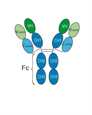
Human IgG1 kappa
Recommend Isotype Controls
Species Reactivity
Human
IC50 & Target
[1]|
CCR8 16.8 pM (Kd) |
In Vitro
Denikitug exhibits high on-cell affinity for human CCR8, with an average KD of 16.8 pM[1].
Denikitug (0.0001-100 µg/mL) specifically binds to human CCR8-expressing CHO-S cells and does not bind to human CCR4-expressing or parental CHO-S cells[1].
Denikitug (0.001-100 µg/mL) potently antagonizes CCL1-induced signaling downstream of human CCR8[1].
Denikitug (10-4-100 µg/mL, overnight) mediates potent ADCC of cells expressing human CCR8, with an average EC50 of 0.0067 µg/mL[1].
Denikitug (10-6-1000 µg/mL, overnight) potently mediates ADCC of Hut78 cells endogenously expressing human CCR8 at tumor-infiltrating Treg levels, with an average EC50 of 0.0017 µg/mL and maximum killing of 67.5%[1].
Denikitug (1 µg/mL) mediates specific depletion of tumor-infiltrating Tregs ex vivo from human ovarian, lung, and colon tumors[1].
MedChemExpress (MCE) has not independently confirmed the accuracy of these methods. They are for reference only.
-
Cell Line:CHO-S cells expressing 2500 copies of human CCR8 per cell, human NK cells isolated from healthy donors
-
Concentration:10-8-100 µg/mL
-
Incubation Time:overnight
-
Result:Exhibited potent ADCC activity, with an average EC50 of 0.0067 µg/mL (range: 0.00094-0.023 µg/mL) across 10 independent NK cell donors.
-
Cell Line:Hut78 cells (endogenously expressing human CCR8 at tumor-infiltrating Treg levels), human NK cells isolated from healthy donors
-
Concentration:10-6-1000 µg/mL
-
Incubation Time:overnight
-
Result:Had an average EC50 of 0.0017 µg/mL and an average maximum killing of 67.5% across three NK cell donors.
In Vivo
MedChemExpress (MCE) has not independently confirmed the accuracy of these methods. They are for reference only.
Clinical Trial
| NCT Number | Sponsor | Condition | Start Date |
Phase

|
|---|---|---|---|---|
| NCT01329991 | Plexxikon| | 2011-05 | PHASE1 |
Gene ID
Accession
Conjugated
Unconjugated
Reconsititution
The product can be reconstituted/diluted with sterile PBS or saline.
Format
-
IgG1-kappa
Application
ELISA, FACS, Functional assay
Verified Bioactivity
-
 Flow Cytometry analysis of Raji cells labelling CCR8 (red) with Denikitug (HY-P990914). Cells were fixed with 4% paraformaldehyde and permeabilised with 90% methanol. Then cells were stained with the primary antibody at 1/200 dilution for an hour at 4℃. Goat Anti-Human IgG H&L (AF488) (HY-P83776) was used as the secondary antibody at 1/1,000 dilution for 30 minutes at 4℃. Human IgG1 kappa (HY-P99001, blue) was used as the isotype control, cells without incubation with primary antibody were used as the unlabeled control (black).


Chemical Information
-
CAS No. 2865822-82-6
-
Appearance Liquid
-
Molecular Weight 147.2 kDa
-
Color Colorless to light yellow
-
SMILES
[Denikitug]
-
Synonyms
GS-1811; JTX-1811
-
Shipping
Shipping with dry ice.
-
Formulation
Please refer to the lot-specific COA for specific buffer information.
-
Storage
Please store the product under the recommended conditions in the Certificate of Analysis.


Purity & Documentation
-
Data Sheet (261 KB)
-
SDS (251 KB)
- English - EN (251 KB)
- Français - FR (251 KB)
- Deutsch - DE (251 KB)
- Norwegian - NO (251 KB)
- Español - ES (251 KB)
- Swedish - SV (251 KB)
- Italian - IT (251 KB)
- Korean - KR (251 KB)
- Portuguese - PT (251 KB)
-
Inhibitory Antibodies User Guide (603 KB)
References

Calculators
Concentration (start) × Volume (start) = Concentration (final) × Volume (final)


Denikitug Related Classifications
HY-P990914 Related Classifications
 Powered by Bioz