



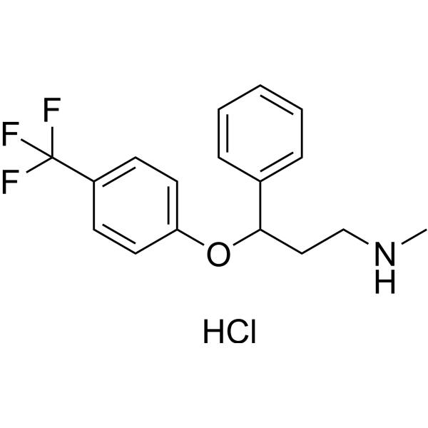
Norfluoxetine

Based on 1 Customer Validation
Norfluoxetine is the active metabolite of the antidepressant Fluoxetine (HY-B0102); it is also a TREK-2 K2P potassium channel inhibitor. Norfluoxetine exhibits neuroimmunological activity, induces apoptosis in primary microglial cells, and inhibits the release of pro-inflammatory factors. Norfluoxetine inhibits high-threshold voltage-gated Ca2+ currents in neurons with an EC50 of 20.4 μM. Norfluoxetine can be used in research related to depression, ischemic stroke, and epilepsy.
Nur für Forschungszwecke. Wir verkaufen nicht an Patienten.
- Reinheit : 98.71%
- CAS. Nr.: 83891-03-6
- Formel: C16H16F3NO
- Molecular Weight:295.30
-
Speicherung:
Store at room temperature, keep dry and cool.
In solvent -80°C, 1 year , -20°C, 6 months


To place orders, for customer services and technical support, please contact: MedChemExpress USA
Tel: 609-228-6898 E-mail: [email protected] [email protected]


-
 Biologische Aktivität
-
Chemical Information
-
Lösungsmittel & Löslichkeit
- Protokoll
- Reinheit & Dokumentation
- Verweise
-
 Help & FAQs


Alle Calcium Channel Isoform-spezifische Produkte anzeigen
More

Biologische Aktivität
Beschreibung
Cellular Effect
|
Cell Line

|
Type | Value | Description | References |
|---|---|---|---|---|
| CHO | IC50 |
0.8 μM
Compound: 40
|
Inhibition of rat voltage-gated K channel 3.1 expressed in CHO cells by patch clamp assay
Inhibition of rat voltage-gated K channel 3.1 expressed in CHO cells by patch clamp assay
|
[PMID: 23121096] |
In Vitro
Norfluoxetine (10 μM) significantly increases the percentage of non-activated primary rat microglia expressing the apoptotic marker cleaved-caspase 3[2].
Norfluoxetine (10-20 μM; 24 h) induces apoptosis in non-activated primary rat microglia by increasing cleaved-caspase 3 and decreasing full-length caspase 3, without altering caspase 8 levels[2].
Norfluoxetine (10-30 μM) reduces protein levels in non-activated primary rat microglia in a dose-dependent manner[2].
Norfluoxetine (10-30 μM) reduces viability of non-activated primary rat microglia in a dose-dependent manner[2].
Norfluoxetine (10-100 μM) reduces single-channel open probability, current amplitude, mean open time, and mean short closed time of truncated human TREK-2 channels reconstituted in lipid bilayers, with increased inhibitory efficacy at voltages above +60 mV, and exhibits similar inhibitory efficacy in high-Po and standard gating modes[1].
Norfluoxetine (1-100 μM; 30 s) concentration-dependently inhibits voltage-gated Ca2+ channel-mediated Ba2+ currents in isolated rat cochlear neurons with an EC50 of 20.4 μM[3].
MedChemExpress (MCE) has not independently confirmed the accuracy of these methods. They are for reference only.
-
Cell Line:non-activated primary rat microglia
-
Concentration:10 and 20 μM
-
Incubation Time:24 h
-
Result:Elevated Cleaved-Caspase 3.
In Vivo
MedChemExpress (MCE) has not independently confirmed the accuracy of these methods. They are for reference only.
-
Animal Model:CFLP (male, 23 g)[3]
-
Dosage:5 mg/kg; 10 mg/kg; 20 mg/kg
-
Administration:s.c.; single dose
-
Result:Increased survival rate, survival duration, and seizure latency significantly at 20 mg/kg compared to pentylenetetrazol-only controls.
Increased combined protection score significantly at 10 mg/kg and 20 mg/kg compared to controls.
Showed no significant differences in anticonvulsant effects compared to fluoxetine at matching doses.


Chemical Information
-
CAS. Nr. 83891-03-6
-
Appearance Liquid (Density: 1.205±0.06 g/cm3)
-
Molecular Weight 295.30
-
Formel C16H16F3NO
-
Color Colorless to light yellow
-
SMILES
FC(F)(F)C(C=C1)=CC=C1OC(C2=CC=CC=C2)CCN
-
Structure Classification
-
Initial Source
-
Versand
Room temperature in continental US; may vary elsewhere.
-
Speicherung
Store at room temperature, keep dry and cool
In solvent -80°C 1 year -20°C 6 months


Lösungsmittel & Löslichkeit
In Vitro:
DMSO : 100 mg/mL (338.64 mM; Need ultrasonic; Hygroscopic DMSO has a significant impact on the solubility of product, please use newly opened DMSO)
Please refer to the solubility information to select the appropriate solvent. Once prepared, please aliquot and store the solution to prevent product inactivation from repeated freeze-thaw cycles.
Storage method and period of stock solution: -80°C, 1 year; -20°C, 6 months. When stored at -80°C, please use it within 1 year. When stored at -20°C, please use it within 6 months.
Please refer to the solubility information to select the appropriate solvent. Once prepared, please aliquot and store the solution to prevent product inactivation from repeated freeze-thaw cycles.
Storage method and period of stock solution: -80°C, 1 year; -20°C, 6 months. When stored at -80°C, please use it within 1 year. When stored at -20°C, please use it within 6 months.
Konzentration (Stammlösung) × Volumen (Stammlösung) = Konzentration (Ziellösung) × Volumen (Ziellösung)
In Vivo:
Select the appropriate dissolution method based on your experimental animal and administration route.
- For the following dissolution methods, please ensure to first prepare a clear stock solution using an In Vitro approach and then sequentially add co-solvents:
- To ensure reliable experimental results, the clarified stock solution can be appropriately stored based on storage conditions. As for the working solution for In Vivo experiments, it is recommended to prepare freshly and use it on the same day.
- The percentages shown for the solvents indicate their volumetric ratio in the final prepared solution. If precipitation or phase separation occurs during preparation, heat and/or sonication can be used to aid dissolution.
Add each solvent one by one: 10% DMSO 40% PEG300 5% Tween-80 45% Saline
Solubility: ≥ 2.5 mg/mL (8.47 mM); Clear solution
This protocol yields a clear solution of ≥ 2.5 mg/mL (saturation unknown).
Taking 1 mL working solution as an example, add 100 μL DMSO stock solution (25.0 mg/mL) to 400 μL PEG300, and mix evenly; then add 50 μL Tween-80 and mix evenly; then add 450 μL Saline to adjust the volume to 1 mL.
Preparation of Saline: Dissolve 0.9 g sodium chloride in ddH₂O and dilute to 100 mL to obtain a clear Saline solution.
Add each solvent one by one: 10% DMSO 90% (20% SBE-β-CD in Saline)
Solubility: 2.5 mg/mL (8.47 mM); Suspended solution; Need ultrasonic
This protocol yields a suspended solution of 2.5 mg/mL. Suspended solution can be used for oral and intraperitoneal injection.
Taking 1 mL working solution as an example, add 100 μL DMSO stock solution (25.0 mg/mL) to 900 μL 20% SBE-β-CD in Saline, and mix evenly.
Preparation of 20% SBE-β-CD in Saline (4°C, storage for one week): 2 g SBE-β-CD powder is dissolved in 10 mL Saline, completely dissolve until clear.
In Vivo Dissolution Calculator
Please enter the basic information of animal experiments:
-
-
-
-
Recommended: Prepare an additional quantity of animals to account for potential losses during experiments.
Please enter your animal formula composition:
-
%DMSO +
Recommended: Keep the proportion of DMSO in working solution below 2% if your animal is weak.
-
%+
-
+%Tween-80 + +
-
%Saline +
The co-solvents required include: DMSO, . All of co-solvents are available by MedChemExpress (MCE). , Tween 80. All of co-solvents are available by MedChemExpress (MCE).
Working solution concentration: 0.22 mg/mL
Method for preparing stock solution: mg drug dissolved in μL DMSO. Stock solution concentration: mg/mL.
1. Take μL DMSO stock solution;
2. Add μL .
μL , mix evenly;
3. Then add μL Tween 80, mix evenly;
4. Then add μL
Please ensure that the stock solution in the first step is dissolved to a clear state, and add co-solvents in sequence. You can use ultrasonic heating (ultrasonic cleaner, recommended frequency 20-40 kHz), vortexing, etc. to assist dissolution.


Reinheit & Dokumentation
-
Data Sheet (286 KB)
-
SDS (252 KB)
- English - EN (252 KB)
- Français - FR (252 KB)
- Deutsch - DE (252 KB)
- Norwegian - NO (252 KB)
- Español - ES (252 KB)
- Swedish - SV (252 KB)
- Italian - IT (252 KB)
- Korean - KR (252 KB)
- Portuguese - PT (252 KB)
-
Handling Instructions (2659 KB)
Verweise
[1]. Proks P, et al. Norfluoxetine inhibits TREK-2 K2P channels by multiple mechanisms including state-independent effects on the selectivity filter gate. The Journal of general physiology. 2021 Aug 02;153(8):e202012812. [Content Brief]
[2]. Dhami KS, et al. Fluoxetine and its metabolite norfluoxetine induce microglial apoptosis. Journal of neurochemistry. 2019 Mar;148(6):761-778. [Content Brief]
[3]. Kecskeméti V, et al. Norfluoxetine and fluoxetine have similar anticonvulsant and Ca2+ channel blocking potencies. Brain research bulletin. 2005 Sep 30;67(1-2):126-32. [Content Brief]


Complete Stock Solution Preparation Table
Please refer to the solubility information to select the appropriate solvent. Once prepared, please aliquot and store the solution to prevent product inactivation from repeated freeze-thaw cycles.
Storage method and period of stock solution: -80°C, 1 year; -20°C, 6 months. When stored at -80°C, please use it within 1 year. When stored at -20°C, please use it within 6 months.
| Optional Solvent | Concentration Solvent Mass | 1 mg | 5 mg | 10 mg | 25 mg |
|---|---|---|---|---|---|
| DMSO | 1 mM | 3.3864 mL | 16.9319 mL | 33.8639 mL | 84.6597 mL |
| 5 mM | 0.6773 mL | 3.3864 mL | 6.7728 mL | 16.9319 mL | |
| 10 mM | 0.3386 mL | 1.6932 mL | 3.3864 mL | 8.4660 mL | |
| 15 mM | 0.2258 mL | 1.1288 mL | 2.2576 mL | 5.6440 mL | |
| 20 mM | 0.1693 mL | 0.8466 mL | 1.6932 mL | 4.2330 mL | |
| 25 mM | 0.1355 mL | 0.6773 mL | 1.3546 mL | 3.3864 mL | |
| 30 mM | 0.1129 mL | 0.5644 mL | 1.1288 mL | 2.8220 mL | |
| 40 mM | 0.0847 mL | 0.4233 mL | 0.8466 mL | 2.1165 mL | |
| 50 mM | 0.0677 mL | 0.3386 mL | 0.6773 mL | 1.6932 mL | |
| 60 mM | 0.0564 mL | 0.2822 mL | 0.5644 mL | 1.4110 mL | |
| 80 mM | 0.0423 mL | 0.2116 mL | 0.4233 mL | 1.0582 mL | |
| 100 mM | 0.0339 mL | 0.1693 mL | 0.3386 mL | 0.8466 mL |


Norfluoxetine Related Classifications
HY-135556 Related Classifications
 Powered by Bioz