



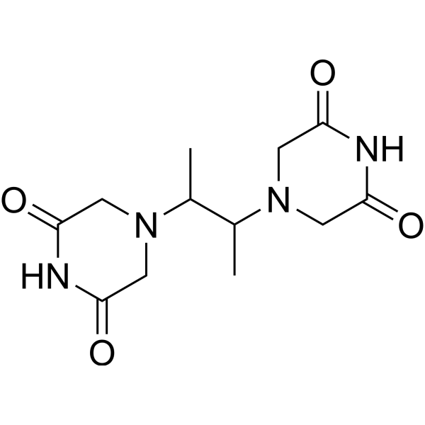
ICRF-196
ICRF-196 is an racemic mixture of the (S,S)- and (R,R)-isomers of ICRF-193 (HY-118590). ICRF-193 is a DNA Topoisomerase II inhibitor. ICRF-193 can inhibit DNA syntheses and induces apoptosis. ICRF-193 exhibits anti-cancer and anti-inflammation effects. ICRF-193 shows cardioprotective effect against anthracycline toxicity to cardiomyocytes. ICRF-193 can be used for the researches of cancer, infection, inflammation and cardiovascular disease, such as acute promyelocytic leukemia.
For research use only. We do not sell to patients.
- CAS No.: 21416-68-2
- Formula: C12H18N4O4
- Molecular Weight:282.30
-
Storage:
Please store the product under the recommended conditions in the Certificate of Analysis.

To place orders, for customer services and technical support, please contact: MedChemExpress USA
Tel: 609-228-6898 E-mail: [email protected] [email protected]


-
 Biological Activity
-
Chemical Information
- Protocols
- Purity & Documentation
- References
-
 Help & FAQs




Biological Activity
Description
IC50 & Target
[1]|
Topoisomerase II |
In Vitro
ICRF-193 (10-100 μM, 2 h) induces mitotic defects in fission yeast cells expressing histone H2A-RFP, α-tubulin Atb2-GFP and spindle pole body protein Sid4-GFP[1].
ICRF-193 (100 μM. 2 h) inhibits its own induced snapped spindles and induces unequal chromosome segregation and unequal chromosome segregation in fission yeast cells[1].
ICRF-193 (100 μM, 4 h) alters nuclear morphology and DNA content in cdc11-123 fission yeast mutant[1].
ICRF-193 (100 μM, 8 h) reduces cell viability in wild-type fission yeast[1].
ICRF-193 (5 days) inhibits the growth of human acute promyelocytic leukemia (APL) cell lines (NB4, HT-93) and other myeloid leukemia cell lines (HL-60, U937) with IC50 of 0.21-0.26 μM[2].
ICRF-193 (0.1-0.2 μM, 5 days) induces granulocytic differentiation of NB4, HT-93, HL-60, and U937 cells[2].
ICRF-193 (0.2 μM, 48 h) significantly downregulates the level of PML-RAR fusion protein and upregulates p21 and RARβ levels in NB4 cells[2].
ICRF-193 (10 μM) inhibits cells arrested at the quiescent (G0) phase reentry into the S phase and inhibits DNA syntheses in murine spleen cells[3].
ICRF-193 (150 nM, 72 h) inhibits LPS (HY-D1056)-induced IL-1β secretion in human macrophage[4].
ICRF-193 decreases DNA fragmentation induced by Etoposide (HY-13629) and induces apoptosis in murine thymocytes[6].
MedChemExpress (MCE) has not independently confirmed the accuracy of these methods. They are for reference only. Further protocols information, click here.


Chemical Information
-
CAS No. 21416-68-2
-
Molecular Weight 282.30
-
Formula C12H18N4O4
-
SMILES
CC(N(C1)CC(NC1=O)=O)C(N(C2)CC(NC2=O)=O)C
-
Shipping
Room temperature in continental US; may vary elsewhere.
-
Storage
Please store the product under the recommended conditions in the Certificate of Analysis.

Protocols
-
Research Protocol for Infectious Diseases
Infectious-disease experiments test how pathogens interact with host barriers, innate immune receptors, inflammatory signaling, pathogen replication, and tissue injury; pattern-recognition receptors such as TLRs, RIG-I-like receptors, NOD-like receptors, and inflammasomes detect microbial molecules and activate NF-κB, interferon, and cytokine responses. The central hypothesis is that infection severity reflects the balance between pathogen burden and host response: protective inflammation restricts pathogen growth, whereas excessive or mislocalized inflammation contributes to tissue damage and disease phenotype. Unresolved questions include which host pathways are protective versus pathogenic, why some infection models fail to translate to human disease, and which combined readouts best predict clinically relevant infection outcomes.
-
Apoptosis
Apoptosis, also called programmed cell death, is generally characterized by distinct morphological characteristics.
-
TUNEL staining for apoptotic DNA fragmentation
TUNEL staining detects DNA strand breaks by using terminal deoxynucleotidyl transferase to add labeled nucleotides to exposed 3′-OH DNA termini, generating either microscopic staining in fixed cells or tissue sections, or fluorescence/cytometric signal in cell suspensions. TUNEL positivity reflects DNA fragmentation but should not be interpreted alone as definitive apoptosis, because TUNEL can also label necrotic, autolytic, mechanically damaged, or DNA-repair-associated DNA breaks.
-
Annexin V plus membrane-impermeant dye apoptosis staining
Annexin V-based apoptosis assays rely on the detection of phosphatidylserine (PS) externalization from the inner leaflet of the plasma membrane to the outer leaflet, an early biochemical hallmark of apoptosis. Fluorescently labeled Annexin V binds PS in a calcium-dependent manner, enabling identification of early apoptotic cells by flow cytometry or fluorescence microscopy. When combined with a membrane-impermeant DNA-binding dye (e. g. , propidium iodide), this approach allows discrimination between viable (Annexin V−/dye−), early apoptotic (Annexin V+/dye−), and late apoptotic or necrotic (Annexin V+/dye+) cell populations by assessing membrane integrity and PS exposure.
-
Apoptosis Solutions
Apoptosis is a regulated, generally non-lytic cell-death pathway that removes unwanted, damaged, infected, or abnormal cells through coordinated morphological changes, caspase activation, DNA fragmentation, and membrane remodeling. The intrinsic apoptosis pathway is controlled mainly by mitochondrial outer membrane permeabilization, BCL-2 family proteins, cytochrome c release, apoptosome formation, caspase-9 activation, and downstream executioner caspase-3/7 activation. The extrinsic apoptosis pathway is initiated by death receptors such as Fas, TNFR, and TRAIL receptors, which recruit adaptor proteins and activate caspase-8 before engaging executioner caspases or mitochondrial amplification through BID cleavage. Apoptosis is linked to many phenotypes, including cancer cell killing, tissue homeostasis, immune regulation, neurodegeneration, infection response, and treatment-induced cytotoxicity; unresolved questions include how apoptosis interacts with necroptosis, pyroptosis, ferroptos
-
Research Protocol for Cardiovascular Diseases
Cardiovascular disease can be modeled as maladaptive cardiac remodeling, where ischemic injury or pressure overload activates inflammatory signaling, fibroblast activation, extracellular-matrix deposition, cardiomyocyte hypertrophy, vascular remodeling, and progressive ventricular dysfunction. The TGF-β/SMAD axis is a central profibrotic pathway after myocardial injury and pressure overload, while innate immune and cytokine pathways regulate leukocyte recruitment, scar formation, and adverse remodeling. Key unresolved questions include which inflammatory signals are reparative versus harmful, when fibrosis is protective versus maladaptive, and whether pathway inhibition improves function without weakening necessary infarct healing or compensatory remodeling.
-
Research Protocol for Inflammation-related Diseases
The NLRP3 inflammasome is a cytosolic innate immune signaling platform that integrates priming signals and danger-signal activation to promote caspase-1 activation, maturation of IL-1β and IL-18, and gasdermin D-mediated pyroptotic cell death. The core experimental logic is to determine whether inflammatory disease phenotypes are driven by increased NLRP3 expression, ASC-containing inflammasome assembly, caspase-1 cleavage, GSDMD cleavage, and extracellular release of IL-1β/IL-18 rather than by nonspecific cell injury alone. The pathway is strongly linked to inflammation-related disease phenotypes because monosodium urate crystals activate NALP3/NLRP3 inflammasome signaling in gout-like crystal inflammation, cholesterol crystals activate NLRP3 inflammasomes in atherogenesis models, and DSS-induced intestinal inflammation has been reported to involve NLRP3 inflammasome activity. However, experimental colitis studies also show context-dependent protective effects of NLRP3 inflammasome co


Purity & Documentation
References
[1]. Nakazawa N, et al. ICRF-193, an anticancer topoisomerase II inhibitor, induces arched telophase spindles that snap, leading to a ploidy increase in fission yeast. Genes Cells. 2016 Sep;21(9):978-93. [Content Brief]
[2]. Niitsu N, et al. The catalytic DNA topoisomerase II inhibitor ICRF-193 and all-trans retinoic acid cooperatively induce granulocytic differentiation of acute promyelocytic leukemia cells: candidate drugs for chemo-differentiation therapy against acute promyelocytic leukemia. Exp Hematol. 2002 Nov;30(11):1273-82. [Content Brief]
[3]. Hossain MS, et al. ICRF-193, a catalytic inhibitor of DNA topoisomerase II, inhibits re-entry into the cell division cycle from quiescent state in mammalian cells. Genes Cells. 2002 Mar;7(3):285-94 [Content Brief]
[4]. Brindle A, et al. The Bisdioxopiperazine ICRF-193 Attenuates LPS-induced IL-1β Secretion by Macrophages. Inflammation. 2024 Feb;47(1):84-98. [Content Brief]
[5]. Jirkovská A, et al. Structure-Activity Relationship Study of Dexrazoxane Analogues Reveals ICRF-193 as the Most Potent Bisdioxopiperazine against Anthracycline Toxicity to Cardiomyocytes Due to Its Strong Topoisomerase IIβ Interactions. J Med Chem. 2021 Apr 8;64(7):3997-4019. [Content Brief]
[6]. Tanimoto C, et al. ICRF-193 modifies etoposide-induced apoptosis in thymocytes. Acta Med Okayama. 1995 Dec;49(6):281-6. [Content Brief]

Calculators
Concentration (start) × Volume (start) = Concentration (final) × Volume (final)


ICRF-196 Related Classifications
HY-118590A Related Classifications
 Powered by Bioz