



PD173952
Based on 1 publication(s) in Google Scholar
PD173952 is a tyrosine kinases inhibitor with IC50s of 0.3, 1.7 and 6.6 nM against Lyn, Abl and Csk, respectively. PD173952 is also a potent Myt1 kinase inhibitor with a Ki of 8.1 nM. PD173952 induces apoptosis.
For research use only. We do not sell to patients.
- Purity : 99.58%
- CAS No.: 305820-75-1
- Formula: C24H21Cl2N5O2
- Molecular Weight:482.36
-
Storage:Powder -20°C, 3 years ; In solvent -80°C, 6 months , -20°C, 1 month

To place orders, for customer services and technical support, please contact: MedChemExpress USA
Tel: 609-228-6898 E-mail: [email protected] [email protected]


-
 Biological Activity
-
Chemical Information
-
Solvent & Solubility
- Protocols
- Purity & Documentation
- References
-
 Help & FAQs


-
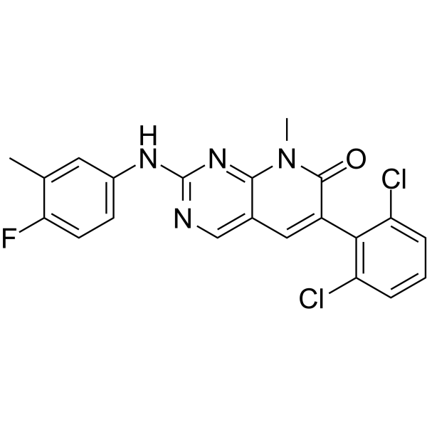
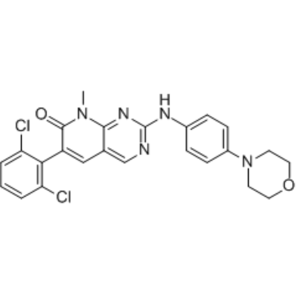
HY-103274
PD180970p210Bcr-Abl Kinase Inhibitor -
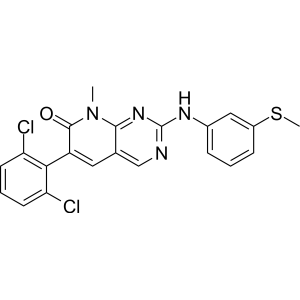
HY-10395
PD173955Src Inhibitor -
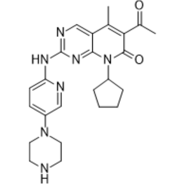
HY-50767
PalbociclibCDK4/6 Inhibitor -
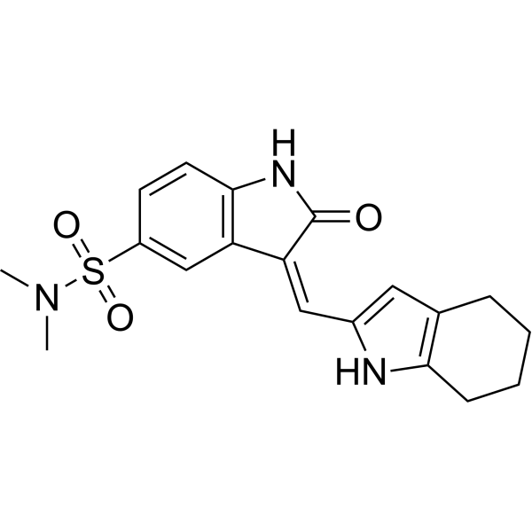
HY-B0789
SU6656Src Inhibitor -
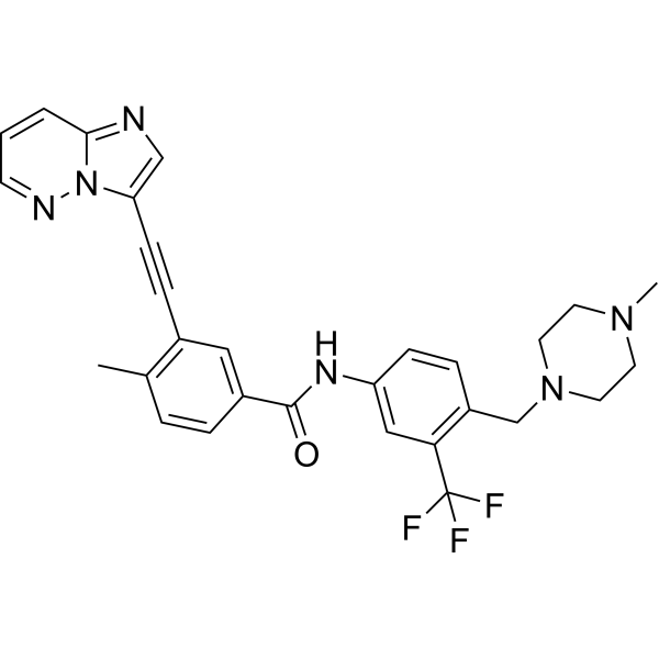
HY-12047
PonatinibMulti-targeted Kinase Inhibitor -
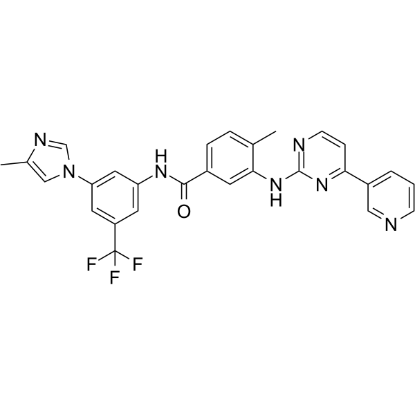
HY-10159
NilotinibBcr-Abl Inhibitor -
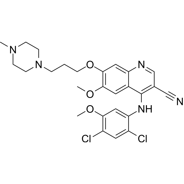
HY-10158
BosutinibSrc/Abl Inhibitor -
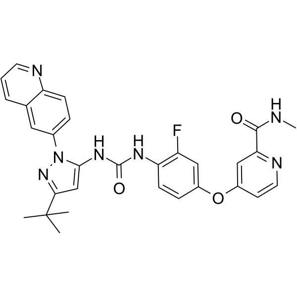
HY-13024
RebastinibKinase Inhibitor -
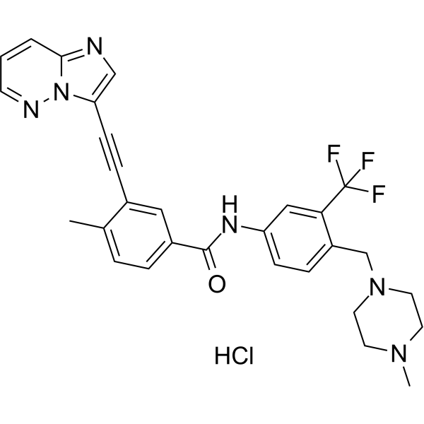
HY-108766
Ponatinib hydrochlorideMulti-targeted Kinase Inhibitor -
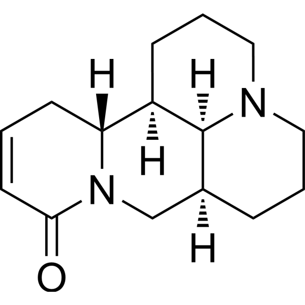
HY-N0103
SophocarpineAutophagy Inducer -
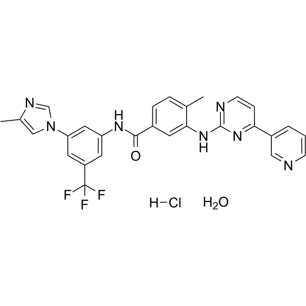
HY-10159A
Nilotinib monohydrochloride monohydrateTyrosine Kinase Inhibitor -
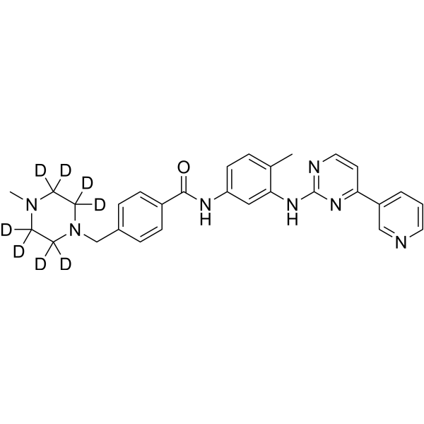
HY-15463S
Imatinib-d8c-Kit Inhibitor
Publications Citing Use of MedChemExpress (MCE) PD173952
More

Biological Activity
Description
|
Lyn 0.3 nM (IC50) |
Abl 1.7 nM (IC50) |
Csk 6.6 nM (IC50) |
Myt1 8.1 nM (Ki) |
PKMYT1 |
Cellular Effect
|
Cell Line

|
Type | Value | Description | References |
|---|---|---|---|---|
| A-431 | IC50 |
0.245 μM
Compound: PD-0173952
|
Inhibition of 3H-thymidine incorporation into A-431 cells
Inhibition of 3H-thymidine incorporation into A-431 cells
|
[PMID: 10974196] |
| HCT-8 | IC50 |
1.26 μM
Compound: PD-0173952
|
Inhibition of HCT-8 clonogenic growth in soft agar without pretreatment
Inhibition of HCT-8 clonogenic growth in soft agar without pretreatment
|
[PMID: 10974196] |
| HCT-8 | IC50 |
1.38 μM
Compound: PD-0173952
|
Inhibition of HCT-8 clonogenic growth in soft agar with pretreatment
Inhibition of HCT-8 clonogenic growth in soft agar with pretreatment
|
[PMID: 10974196] |
| HCT-8 | IC50 |
1.5 μM
Compound: 6v
|
Inhibitory activity against HCT-8 human colon adenocarcinoma cells was determined.
Inhibitory activity against HCT-8 human colon adenocarcinoma cells was determined.
|
[PMID: 10956222] |
| HCT-8 | IC50 |
1.53 μM
Compound: PD-0173952
|
Inhibition of HCT-8 cell proliferation
Inhibition of HCT-8 cell proliferation
|
[PMID: 10974196] |
| HT-29 | IC50 |
0.2 μM
Compound: 6v
|
Inhibitory activity against HT-29 human colon adenocarcinoma cells was determined.
Inhibitory activity against HT-29 human colon adenocarcinoma cells was determined.
|
[PMID: 10956222] |
| HT-29 | IC50 |
0.2 μM
Compound: PD-0173952
|
Inhibition of HT-29 cell proliferation
Inhibition of HT-29 cell proliferation
|
[PMID: 10974196] |
| HT-29 | IC50 |
1.21 μM
Compound: PD-0173952
|
Inhibition of HT-29 clonogenic growth in soft agar with pretreatment
Inhibition of HT-29 clonogenic growth in soft agar with pretreatment
|
[PMID: 10974196] |
| HT-29 | IC50 |
1.37 μM
Compound: PD-0173952
|
Inhibition of HT-29 clonogenic growth in soft agar without pretreatment
Inhibition of HT-29 clonogenic growth in soft agar without pretreatment
|
[PMID: 10974196] |
| NIH3T3 | IC50 |
0.17 μM
Compound: PD-0173952
|
Inhibition of 3H-thymidine incorporation into NIH3T3 cells
Inhibition of 3H-thymidine incorporation into NIH3T3 cells
|
[PMID: 10974196] |
| SW-620 | IC50 |
0.17 μM
Compound: 6v
|
Inhibitory activity against SW-620 human colon adenocarcinoma cells was determined.
Inhibitory activity against SW-620 human colon adenocarcinoma cells was determined.
|
[PMID: 10956222] |
| SW-620 | IC50 |
0.174 μM
Compound: PD-0173952
|
Inhibition of SW-620 cell proliferation
Inhibition of SW-620 cell proliferation
|
[PMID: 10974196] |
| SW-620 | IC50 |
0.47 μM
Compound: PD-0173952
|
Inhibition of SW-620 clonogenic growth in soft agar without pretreatment
Inhibition of SW-620 clonogenic growth in soft agar without pretreatment
|
[PMID: 10974196] |
| SW-620 | IC50 |
0.53 μM
Compound: PD-0173952
|
Inhibition of SW-620 clonogenic growth in soft agar with pretreatment
Inhibition of SW-620 clonogenic growth in soft agar with pretreatment
|
[PMID: 10974196] |
In Vitro
PD173952 (0-1000 nM; 12 h) inhibits tyrosine phosphorylation of p210Bcr-Abl and CrkL in K562 cells in a concentration-dependent manner[1].
PD173952 (0.5 μM; 1-4 days) inhibits K562 cell viability[1].
PD173952 (0.5 μM; 24 and 48 h) induces apoptosis of K562 and MEG-01 cells[1].
MedChemExpress (MCE) has not independently confirmed the accuracy of these methods. They are for reference only. Further protocols information, click here.
-
Cell Line:K562 cells
-
Concentration:0, 25, 50, 100, 200, 500 and 1000 nM
-
Incubation Time:12 h
-
Result:Inhibited tyrosine phosphorylation of p210Bcr-Abl and CrkL.
-
Cell Line:K562 cells
-
Concentration:0.5 μM
-
Incubation Time:1-4 days
-
Result:Caused cell death in a time-dependent manner.
-
Cell Line:K562 and MEG-01 cells
-
Concentration:0.5 μM
-
Incubation Time:24 and 48 h
-
Result:85-kDa PARP fragment was detected.


Chemical Information
-
CAS No. 305820-75-1
-
Appearance Solid
-
Molecular Weight 482.36
-
Formula C24H21Cl2N5O2
-
Color Light yellow to yellow
-
SMILES
O=C1C(C2=C(Cl)C=CC=C2Cl)=CC3=CN=C(NC4=CC=C(N5CCOCC5)C=C4)N=C3N1C
-
Shipping
Room temperature in continental US; may vary elsewhere.
-
Storage
Powder -20°C 3 years In solvent -80°C 6 months -20°C 1 month


Publications (1)
-
Journal Impact Factor
-
Most Recent
-
iScience
Decoding sunitinib resistance in ccRCC: Metabolic-reprogramming-induced ABAT and GABAergic system shifts. [Abstract]2024 Jun 28;27(7):110415. PMID: 39100925


Solvent & Solubility
In Vitro:
DMSO : 16.67 mg/mL (34.56 mM; ultrasonic and warming and heat to 60°C; Hygroscopic DMSO has a significant impact on the solubility of product, please use newly opened DMSO)
Please refer to the solubility information to select the appropriate solvent. Once prepared, please aliquot and store the solution to prevent product inactivation from repeated freeze-thaw cycles.
Storage method and period of stock solution: -80°C, 6 months; -20°C, 1 month. When stored at -80°C, please use it within 6 months. When stored at -20°C, please use it within 1 month.
Please refer to the solubility information to select the appropriate solvent. Once prepared, please aliquot and store the solution to prevent product inactivation from repeated freeze-thaw cycles.
Storage method and period of stock solution: -80°C, 6 months; -20°C, 1 month. When stored at -80°C, please use it within 6 months. When stored at -20°C, please use it within 1 month.
Concentration (start) × Volume (start) = Concentration (final) × Volume (final)

Protocols
-
Apoptosis
Apoptosis, also called programmed cell death, is generally characterized by distinct morphological characteristics.
-
TUNEL staining for apoptotic DNA fragmentation
TUNEL staining detects DNA strand breaks by using terminal deoxynucleotidyl transferase to add labeled nucleotides to exposed 3′-OH DNA termini, generating either microscopic staining in fixed cells or tissue sections, or fluorescence/cytometric signal in cell suspensions. TUNEL positivity reflects DNA fragmentation but should not be interpreted alone as definitive apoptosis, because TUNEL can also label necrotic, autolytic, mechanically damaged, or DNA-repair-associated DNA breaks.
-
Annexin V plus membrane-impermeant dye apoptosis staining
Annexin V-based apoptosis assays rely on the detection of phosphatidylserine (PS) externalization from the inner leaflet of the plasma membrane to the outer leaflet, an early biochemical hallmark of apoptosis. Fluorescently labeled Annexin V binds PS in a calcium-dependent manner, enabling identification of early apoptotic cells by flow cytometry or fluorescence microscopy. When combined with a membrane-impermeant DNA-binding dye (e. g. , propidium iodide), this approach allows discrimination between viable (Annexin V−/dye−), early apoptotic (Annexin V+/dye−), and late apoptotic or necrotic (Annexin V+/dye+) cell populations by assessing membrane integrity and PS exposure.
-
Apoptosis Solutions
Apoptosis is a regulated, generally non-lytic cell-death pathway that removes unwanted, damaged, infected, or abnormal cells through coordinated morphological changes, caspase activation, DNA fragmentation, and membrane remodeling. The intrinsic apoptosis pathway is controlled mainly by mitochondrial outer membrane permeabilization, BCL-2 family proteins, cytochrome c release, apoptosome formation, caspase-9 activation, and downstream executioner caspase-3/7 activation. The extrinsic apoptosis pathway is initiated by death receptors such as Fas, TNFR, and TRAIL receptors, which recruit adaptor proteins and activate caspase-8 before engaging executioner caspases or mitochondrial amplification through BID cleavage. Apoptosis is linked to many phenotypes, including cancer cell killing, tissue homeostasis, immune regulation, neurodegeneration, infection response, and treatment-induced cytotoxicity; unresolved questions include how apoptosis interacts with necroptosis, pyroptosis, ferroptos


Purity & Documentation
-
Data Sheet (275 KB)
-
SDS (252 KB)
- English - EN (252 KB)
- Français - FR (252 KB)
- Deutsch - DE (252 KB)
- Norwegian - NO (252 KB)
- Español - ES (252 KB)
- Swedish - SV (252 KB)
- Italian - IT (252 KB)
- Korean - KR (252 KB)
- Portuguese - PT (252 KB)
-
Handling Instructions (2659 KB)
References
[1]. Dorsey JF, et al. Interleukin-3 protects Bcr-Abl-transformed hematopoietic progenitor cells from apoptosis induced by Bcr-Abl tyrosine kinase inhibitors. Leukemia. 2002 Sep;16(9):1589-95. [Content Brief]
[2]. Wichapong K, et al. Application of docking and QM/MM-GBSA rescoring to screen for novel Myt1 kinase inhibitors. J Chem Inf Model. 2014 Mar 24;54(3):881-93. [Content Brief]


Complete Stock Solution Preparation Table
Please refer to the solubility information to select the appropriate solvent. Once prepared, please aliquot and store the solution to prevent product inactivation from repeated freeze-thaw cycles.
Storage method and period of stock solution: -80°C, 6 months; -20°C, 1 month. When stored at -80°C, please use it within 6 months. When stored at -20°C, please use it within 1 month.
| Optional Solvent | Concentration Solvent Mass | 1 mg | 5 mg | 10 mg | 25 mg |
|---|---|---|---|---|---|
| DMSO | 1 mM | 2.0731 mL | 10.3657 mL | 20.7314 mL | 51.8285 mL |
| 5 mM | 0.4146 mL | 2.0731 mL | 4.1463 mL | 10.3657 mL | |
| 10 mM | 0.2073 mL | 1.0366 mL | 2.0731 mL | 5.1829 mL | |
| 15 mM | 0.1382 mL | 0.6910 mL | 1.3821 mL | 3.4552 mL | |
| 20 mM | 0.1037 mL | 0.5183 mL | 1.0366 mL | 2.5914 mL | |
| 25 mM | 0.0829 mL | 0.4146 mL | 0.8293 mL | 2.0731 mL | |
| 30 mM | 0.0691 mL | 0.3455 mL | 0.6910 mL | 1.7276 mL |


PD173952 Related Classifications
HY-122113 Related Classifications
 Powered by Bioz