



Phosphatidylserine
Based on 2 publication(s) in Google Scholar
Phosphatidylserine (Phospholipids) is a well-conserved anti-inflammatory and immunosuppressive signal. Phosphatidylserine is involved in membrane translocation and the activation of protein kinase C, participating in Akt signaling through its interaction with PIP3. The local exposure of Phosphatidylserine can interact with complement and other proteins, promoting microglial phagocytosis during critical periods of synaptic refinement. Phosphatidylserine can promote blood coagulation in the extracellular environment and acts as a "eat me" signal to clear out apoptotic cells. Phosphatidylserine can suppress inflammation in tissues by inducing TGF-β secretion and inhibiting immune responses.
For research use only. We do not sell to patients.
- Purity : 98%
- CAS No.: 1446756-47-3
- Molecular Weight:788.10
-
Storage:Powder -20°C, 3 years ; In solvent -80°C, 6 months , -20°C, 1 month

To place orders, for customer services and technical support, please contact: MedChemExpress USA
Tel: 609-228-6898 E-mail: [email protected] [email protected]


-
 Biological Activity
-
Chemical Information
-
Solvent & Solubility
- Protocols
- Purity & Documentation
- References
-
 Help & FAQs


-

HY-W250118
Cephalin form bovine brainBiochemical Assay Reagent -

HY-139533
Phosphatidylinositols, soya, sodium saltsPhosphatidylinositols Mixture -

HY-125853
Phosphatidylcholines,soyaPhosphatidylcholine -

HY-156041
Lysophosphatidylethanolamines, eggEndogenous Metabolite -

HY-18739
Phorbol 12-myristate 13-acetatePKC Activator -

HY-139414
LysophosphatidylcholinesHemolytic Agent -

HY-125854
Phosphatidylcholines, eggPhosphatidylcholine -

HY-W040255
1-Palmitoyl-2-glutaryl-sn-glycero-3-phosphocholineOxidized Phospholipid -
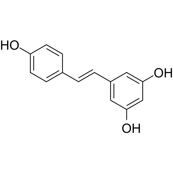
HY-16561
ResveratrolPolyphenolic Phytoalexin -
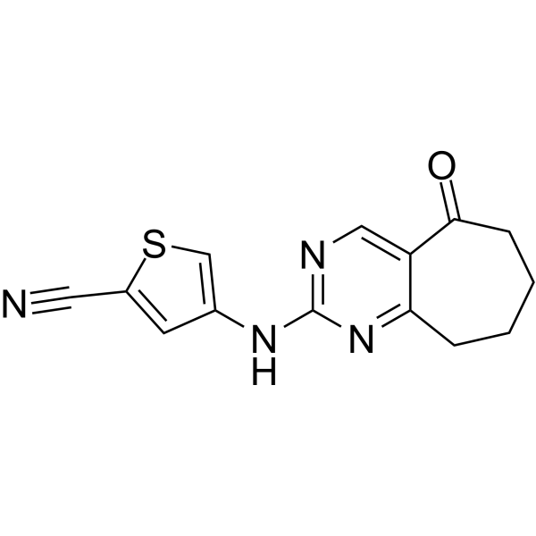
HY-W107464
G6PDi-1G6PD Inhibitor -
HY-W251428
Phosphatidylglycerols (egg) sodium saltBiochemical Assay Reagent -
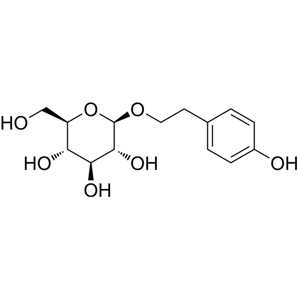
HY-N0109
SalidrosideProlyl Endopeptidase Inhibitor
Publications Citing Use of MedChemExpress (MCE) Phosphatidylserine
More

Biological Activity
Description
In Vitro
Phosphatidylserine can inhibit inflammation in tissues by inducing TGF-β secretion, thereby avoiding activation of immune responses through the production of regulatory T cells[2]. Phosphatidylserine can promote neuronal survival, differentiation, and synaptic neurotransmission[3]. Phosphatidylserine mediates the pruning of microglia developmental synapses when undergoing local externalization[4].
MedChemExpress (MCE) has not independently confirmed the accuracy of these methods. They are for reference only. Further protocols information, click here.
In Vivo
MedChemExpress (MCE) has not independently confirmed the accuracy of these methods. They are for reference only.
-
Animal Model:BALB/cAnN and C57BL/6 male mice[2].
-
Dosage:Phosphatidylserine (PS) liposomes (0.5 mg of total lipid, PS-containing liposomes consisted of 30:30:40 molar ratio PS to PC to cholesterol)
-
Administration:Subcutaneous injection (s.c.); single dose
-
Result:Inhibited responses to Ags as determined by decreased draining lymph node tissue mass, with reduced numbers of total leukocytes and Ag-specific CD4+ T cells.
Clinical Trial
| NCT Number | Sponsor | Condition | Start Date |
Phase

|
|---|---|---|---|---|
| NCT01329991 | Plexxikon| | 2011-05 | PHASE1 |


Chemical Information
-
CAS No. 1446756-47-3
-
Appearance Solid
-
Molecular Weight 788.10
-
Color Light yellow to yellow
-
SMILES
[Phosphatidylserine]
-
Synonyms
Phospholipids, phosphatidylserines; Serine glycerophosphatides
-
Initial Source
Most organisms
-
Shipping
Room temperature in continental US; may vary elsewhere.
-
Storage
Powder -20°C 3 years In solvent -80°C 6 months -20°C 1 month


Publications (2)
-
Journal Impact Factor
-
Most Recent
-
Nat Commun
Transferrin-phosphatidylserine liposomes target TDP-43 and neuroinflammation in male mice with neuropathic pain. [Abstract]2025 Dec 12;16(1):11314. PMID: 41387431 -
Adv Sci (Weinh)
2026 Jul;13(39):e20095. PMID: 41972457


Solvent & Solubility
In Vitro:
DMSO : < 1 mg/mL (insoluble or slightly soluble)

Protocols
-
Phagocytosis Functional Assay
A phagocytosis functional assay measures the ability of phagocytic cells, such as neutrophils, macrophages, monocytes, or microglia/macrophages, to bind and internalize particulate targets including bacteria, yeast particles, beads, or myelin particles. Fluorescent flow-cytometry assays detect target uptake as fluorescence associated with gated phagocytes, while pH-sensitive dyes such as pHrodo increase signal in acidic phagosomal compartments and therefore preferentially report internalized particles rather than particles remaining outside the cell. Microscopy or high-content imaging can be used to confirm intracellular localization and, in some protocols, to follow uptake kinetics.
-
Apoptosis
Apoptosis, also called programmed cell death, is generally characterized by distinct morphological characteristics.
-
TUNEL staining for apoptotic DNA fragmentation
TUNEL staining detects DNA strand breaks by using terminal deoxynucleotidyl transferase to add labeled nucleotides to exposed 3′-OH DNA termini, generating either microscopic staining in fixed cells or tissue sections, or fluorescence/cytometric signal in cell suspensions. TUNEL positivity reflects DNA fragmentation but should not be interpreted alone as definitive apoptosis, because TUNEL can also label necrotic, autolytic, mechanically damaged, or DNA-repair-associated DNA breaks.
-
Annexin V plus membrane-impermeant dye apoptosis staining
Annexin V-based apoptosis assays rely on the detection of phosphatidylserine (PS) externalization from the inner leaflet of the plasma membrane to the outer leaflet, an early biochemical hallmark of apoptosis. Fluorescently labeled Annexin V binds PS in a calcium-dependent manner, enabling identification of early apoptotic cells by flow cytometry or fluorescence microscopy. When combined with a membrane-impermeant DNA-binding dye (e. g. , propidium iodide), this approach allows discrimination between viable (Annexin V−/dye−), early apoptotic (Annexin V+/dye−), and late apoptotic or necrotic (Annexin V+/dye+) cell populations by assessing membrane integrity and PS exposure.
-
Apoptosis Solutions
Apoptosis is a regulated, generally non-lytic cell-death pathway that removes unwanted, damaged, infected, or abnormal cells through coordinated morphological changes, caspase activation, DNA fragmentation, and membrane remodeling. The intrinsic apoptosis pathway is controlled mainly by mitochondrial outer membrane permeabilization, BCL-2 family proteins, cytochrome c release, apoptosome formation, caspase-9 activation, and downstream executioner caspase-3/7 activation. The extrinsic apoptosis pathway is initiated by death receptors such as Fas, TNFR, and TRAIL receptors, which recruit adaptor proteins and activate caspase-8 before engaging executioner caspases or mitochondrial amplification through BID cleavage. Apoptosis is linked to many phenotypes, including cancer cell killing, tissue homeostasis, immune regulation, neurodegeneration, infection response, and treatment-induced cytotoxicity; unresolved questions include how apoptosis interacts with necroptosis, pyroptosis, ferroptos
-
Research Protocol for Inflammation-related Diseases
The NLRP3 inflammasome is a cytosolic innate immune signaling platform that integrates priming signals and danger-signal activation to promote caspase-1 activation, maturation of IL-1β and IL-18, and gasdermin D-mediated pyroptotic cell death. The core experimental logic is to determine whether inflammatory disease phenotypes are driven by increased NLRP3 expression, ASC-containing inflammasome assembly, caspase-1 cleavage, GSDMD cleavage, and extracellular release of IL-1β/IL-18 rather than by nonspecific cell injury alone. The pathway is strongly linked to inflammation-related disease phenotypes because monosodium urate crystals activate NALP3/NLRP3 inflammasome signaling in gout-like crystal inflammation, cholesterol crystals activate NLRP3 inflammasomes in atherogenesis models, and DSS-induced intestinal inflammation has been reported to involve NLRP3 inflammasome activity. However, experimental colitis studies also show context-dependent protective effects of NLRP3 inflammasome co


Purity & Documentation
-
Data Sheet (271 KB)
-
SDS (252 KB)
- English - EN (252 KB)
- Français - FR (252 KB)
- Deutsch - DE (252 KB)
- Norwegian - NO (252 KB)
- Español - ES (252 KB)
- Swedish - SV (252 KB)
- Italian - IT (252 KB)
- Korean - KR (252 KB)
- Portuguese - PT (252 KB)
-
Handling Instructions (2659 KB)
References
[1]. Kay JG, et al. Phosphatidylserine-mediated cellular signaling. Adv Exp Med Biol. 2013;991:177-93. [Content Brief]
[2]. Hoffmann PR, et al. Interaction between phosphatidylserine and the phosphatidylserine receptor inhibits immune responses in vivo. J Immunol. 2005 Feb 1;174(3):1393-404. [Content Brief]
[3]. Kim HY, et al. Phosphatidylserine in the brain: metabolism and function. Prog Lipid Res. 2014 Oct;56:1-18. [Content Brief]
[4]. Scott-Hewitt N, et al. Local externalization of phosphatidylserine mediates developmental synaptic pruning by microglia. EMBO J. 2020 Aug 17;39(16):e105380. [Content Brief]

Calculators
Concentration (start) × Volume (start) = Concentration (final) × Volume (final)


Phosphatidylserine Related Classifications
HY-A0183 Related Classifications
 Powered by Bioz