



PROTAC HPK1 Degrader-1

Based on 1 Customer Validation
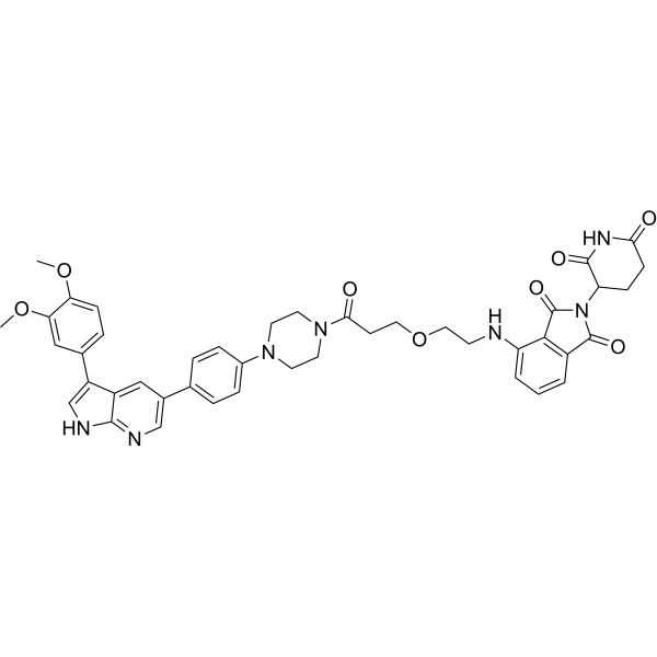
PROTAC HPK1 Degrader-1 (compound B1) is a PROTAC degrader that recruits cereblon to induce HPK1 degradation, with a DC50 of 1.8 nM. PROTAC HPK1 Degrader-1 inhibits HPK1 kinase activity, with an IC50 of 140.5 nM. PROTAC HPK1 Degrader-1 suppresses the phosphorylation of SLP76 protein, with an IC50 of 496.1 nM. PROTAC HPK1 Degrader-1 can be used to investigate non-kinase-dependent signaling of HPK1 in the TCR pathway and cancer-related research.
(Pink: MAP4K Target protein ligand; Blue: Cereblon ligand (HY-41547); Black: linker (HY-W067489)).
For research use only. We do not sell to patients.
- Purity : 99.70%
- CAS No.: 3034182-97-0
- Formula: C43H43N7O8
- Molecular Weight:785.84
-
Storage:
4°C, protect from light, stored under nitrogen
* In solvent : -80°C, 6 months; -20°C, 1 month (protect from light, stored under nitrogen)

To place orders, for customer services and technical support, please contact: MedChemExpress USA
Tel: 609-228-6898 E-mail: [email protected] [email protected]


-
 Biological Activity
-
Chemical Information
-
Solvent & Solubility
- Protocols
- Purity & Documentation
- References
-
 Help & FAQs




Biological Activity
Description
IC50 & Target
[1]|
HPK1 1.8 nM (DC50) |
HPK1 140.5 nM (IC50) |
SLP-76 496.1 nM (IC50) |
In Vitro
PROTAC HPK1 Degrader-1 (compound B1) inhibits purified HPK1 kinase activity with an IC50 of 140.5 nM[1].
PROTAC HPK1 Degrader-1 (compound B1) (0.68-500 nM; 24 h) induces dose-dependent HPK1 degradation in Jurkat cells with a DC50 of 1.8 nM, time-dependent degradation with maximum effect at 24 h, and acts via a ubiquitin-proteasome system-dependent mechanism requiring CRBN and HPK1 binding[1].
PROTAC HPK1 Degrader-1 (compound B1) (0.5-2 μM; 24 h) inhibits SLP76 phosphorylation in Jurkat cells with an IC50 of 496.1 nM[1].
MedChemExpress (MCE) has not independently confirmed the accuracy of these methods. They are for reference only. Further protocols information, click here.
-
Cell Line:Jurkat cells
-
Concentration:0.68, 2.07, 6.17, 18.5, 55, 166, 500 nM
-
Incubation Time:24 h
-
Result:Dose-dependently degraded HPK1 with a DC50 of 1.8 nM.
Complete degradation was observed at 100 nM.
-
Cell Line:Jurkat cells
-
Concentration:0.5, 1, 2 μM
-
Incubation Time:24 h
-
Result:Inhibited SLP76 phosphorylation in a dose-dependent manner, with an IC50 value of 496.1 nM as measured by ELISA.
Showed reduced p-SLP76 levels with increasing concentrations of the reagent via Western blot analysis.
-
Cell Line:Jurkat cells
-
Concentration:50 nM
-
Incubation Time:0, 8, 12, 24, 48 h
-
Result:HPK1 degradation became evident at 12 h, was nearly complete at 24 h and partially recovered at 48 h.


Chemical Information
-
CAS No. 3034182-97-0
-
Appearance Solid
-
Molecular Weight 785.84
-
Formula C43H43N7O8
-
Color Light yellow to yellow
-
SMILES
O=C1N(C(CC2)C(NC2=O)=O)C(C3=C1C=CC=C3NCCOCCC(N4CCN(C5=CC=C(C6=CN=C(NC=C7C8=CC=C(OC)C(OC)=C8)C7=C6)C=C5)CC4)=O)=O
-
Shipping
Room temperature in continental US; may vary elsewhere.
-
Storage
4°C, protect from light, stored under nitrogen
* In solvent : -80°C, 6 months; -20°C, 1 month (protect from light, stored under nitrogen)


Solvent & Solubility
In Vitro:
DMSO : 100 mg/mL (127.25 mM; Need ultrasonic; Hygroscopic DMSO has a significant impact on the solubility of product, please use newly opened DMSO)
Please refer to the solubility information to select the appropriate solvent. Once prepared, please aliquot and store the solution to prevent product inactivation from repeated freeze-thaw cycles.
Storage method and period of stock solution: -80°C, 6 months; -20°C, 1 month (protect from light, stored under nitrogen). When stored at -80°C, please use it within 6 months. When stored at -20°C, please use it within 1 month.
Please refer to the solubility information to select the appropriate solvent. Once prepared, please aliquot and store the solution to prevent product inactivation from repeated freeze-thaw cycles.
Storage method and period of stock solution: -80°C, 6 months; -20°C, 1 month (protect from light, stored under nitrogen). When stored at -80°C, please use it within 6 months. When stored at -20°C, please use it within 1 month.
Concentration (start) × Volume (start) = Concentration (final) × Volume (final)

Protocols
-
Kinase activity and phosphorylation assays
Kinase activity assays measure the ability of kinases to transfer phosphate groups from ATP to specific substrates, while phosphorylation assays detect the presence and levels of phosphorylated proteins. Common methods include radiolabeled ATP incorporation (e. g. ,), ADP release detection via bioluminescence (e. g. ,[3]), enzyme-linked immunosorbent assays (ELISA) for phospho-specific epitopes (e. g. ,[6]), and microtiter-based formats for high-throughput screening (e. g. ,[8]). The ADP-Glo assay quantifies kinase activity by measuring ADP produced during phosphorylation using a luciferase-based system. Radiometric assays involve autoradiography or scintillation counting after incorporation of 32P-labeled ATP into substrate proteins. ELISA-based approaches rely on phospho-specific antibodies to detect activated kinases in cell lysates or purified samples.
-
Western Blot
Western blotting (WB) is a commonly used experimental method in molecular biology, biochemistry, and immunogenetics for identifying and quantifying target proteins. It combines gel electrophoresis with immunoassay, enabling researchers to analyze protein expression, post-translational modifications, and molecular weight.
-
Cell Cytotoxicity Assay
Cytotoxicity assays are usually based on the assessment of cell membrane damage, which can also be indirectly detected by measuring cell viability. Detection methods include MTT assay, CKK-8 assay, LDH assay and ATP assay, etc.
-
Protocol for Kinase activity and phosphorylation assays
Kinase activity assays measure transfer of phosphate from ATP to a protein or peptide substrate, generating phosphorylated substrate, ADP, or incorporated radiolabeled phosphate as the readout; phosphorylation assays measure site-specific phosphorylation in cells or tissues as a proxy for kinase-pathway activation, inhibition, or substrate regulation. Phosphorylation can be detected by phospho-specific Western blot, immunoprecipitation kinase assay, phospho-immunofluorescence, phospho-flow cytometry, luminescent ADP detection, radiolabeled ATP incorporation, or reporter-based pathway assays, and these readouts can be applied to cancer cells, primary neurons, mouse tumors, organoids, inflammatory macrophages, ferroptosis studies, and mitophagy studies when the kinase target is biologically relevant.


Purity & Documentation
-
Data Sheet (273 KB)
-
SDS (252 KB)
- English - EN (252 KB)
- Français - FR (252 KB)
- Deutsch - DE (252 KB)
- Norwegian - NO (252 KB)
- Español - ES (252 KB)
- Swedish - SV (252 KB)
- Italian - IT (252 KB)
- Korean - KR (252 KB)
- Portuguese - PT (252 KB)
-
Handling Instructions (2659 KB)
References


Complete Stock Solution Preparation Table
Please refer to the solubility information to select the appropriate solvent. Once prepared, please aliquot and store the solution to prevent product inactivation from repeated freeze-thaw cycles.
Storage method and period of stock solution: -80°C, 6 months; -20°C, 1 month (protect from light, stored under nitrogen). When stored at -80°C, please use it within 6 months. When stored at -20°C, please use it within 1 month.
| Optional Solvent | Concentration Solvent Mass | 1 mg | 5 mg | 10 mg | 25 mg |
|---|---|---|---|---|---|
| DMSO | 1 mM | 1.2725 mL | 6.3626 mL | 12.7252 mL | 31.8131 mL |
| 5 mM | 0.2545 mL | 1.2725 mL | 2.5450 mL | 6.3626 mL | |
| 10 mM | 0.1273 mL | 0.6363 mL | 1.2725 mL | 3.1813 mL | |
| 15 mM | 0.0848 mL | 0.4242 mL | 0.8483 mL | 2.1209 mL | |
| 20 mM | 0.0636 mL | 0.3181 mL | 0.6363 mL | 1.5907 mL | |
| 25 mM | 0.0509 mL | 0.2545 mL | 0.5090 mL | 1.2725 mL | |
| 30 mM | 0.0424 mL | 0.2121 mL | 0.4242 mL | 1.0604 mL | |
| 40 mM | 0.0318 mL | 0.1591 mL | 0.3181 mL | 0.7953 mL | |
| 50 mM | 0.0255 mL | 0.1273 mL | 0.2545 mL | 0.6363 mL | |
| 60 mM | 0.0212 mL | 0.1060 mL | 0.2121 mL | 0.5302 mL | |
| 80 mM | 0.0159 mL | 0.0795 mL | 0.1591 mL | 0.3977 mL | |
| 100 mM | 0.0127 mL | 0.0636 mL | 0.1273 mL | 0.3181 mL |


PROTAC HPK1 Degrader-1 Related Classifications
HY-163372 Related Classifications
 Powered by Bioz