



PSMA-Val-Cit-PAB-MMAE

Based on 1 Customer Validation
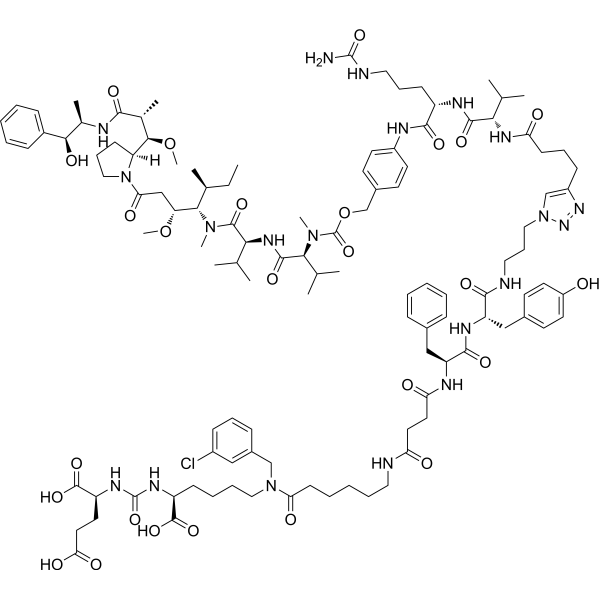
PSMA-Val-Cit-PAB-MMAE is a small-molecule conjugate targeting PSMA, with Monomethyl auristatin E (MMAE) (HY-15162) as its cytotoxic payload. PSMA-Val-Cit-PAB-MMAE binds to PSMA, thereby being delivered into PSMA-expressing prostate cancer cells. Subsequently, the Val-Cit linker is cleaved under the mediation of cathepsin B, releasing active MMAE. PSMA-Val-Cit-PAB-MMAE inhibits CYP3A4 activity (IC50 = 11.2 μM), induces intracellular ROS production and oxidative stress, disrupts the cytoskeleton through microtubule destabilization, and induces prostate cancer cell death. PSMA-Val-Cit-PAB-MMAE can be used in research related to prostate cancer.
For research use only. We do not sell to patients.
- Purity : 99.52%
- CAS No.: 2748039-79-2
- Formula: C114H165ClN20O26
- Molecular Weight:2267.10
-
Storage:Powder -20°C, 3 years , 4°C, 2 years ; In solvent -80°C, 6 months , -20°C, 1 month

To place orders, for customer services and technical support, please contact: MedChemExpress USA
Tel: 609-228-6898 E-mail: [email protected] [email protected]


-
 Biological Activity
-
Chemical Information
-
Solvent & Solubility
- Protocols
- Purity & Documentation
- References
-
 Help & FAQs




Biological Activity
Description
IC50 & Target
|
Auristatin |
Cellular Effect
|
Cell Line

|
Type | Value | Description | References |
|---|---|---|---|---|
| CWR22R | CC50 |
29 nM
Compound: PSMA-Val-Cit-PAB-MMAE
|
Cytotoxicity against human 22Rv1 cells expressing PMSA measured using MTT assay
Cytotoxicity against human 22Rv1 cells expressing PMSA measured using MTT assay
|
[PMID: 34797052] |
| PC-3 | CC50 |
27 nM
Compound: PSMA-Val-Cit-PAB-MMAE
|
Cytotoxicity against human PC-3 cells not expressing PMSA measured using MTT assay
Cytotoxicity against human PC-3 cells not expressing PMSA measured using MTT assay
|
[PMID: 34797052] |
In Vitro
PSMA-Val-Cit-PAB-MMAE exhibits nanomolar cytotoxicity in both PSMA-expressing 22Rv1 and PSMA-null PC-3 human prostate cancer cell lines, with CC50 values of 29 nM and 27 nM, respectively[1].
PSMA-Val-Cit-PAB-MMAE (27-29 nM; 1 h) induces significant oxidative stress in 22Rv1 and PC-3 human prostate cancer cells, increasing intracellular hydrogen peroxide levels to twice and three times the control levels, respectively, after 1 h incubation at its CC50 concentration[1].
PSMA-Val-Cit-PAB-MMAE (27-29 nM; up to 40 min) significantly reduces the stiffness of 22Rv1 human prostate cancer cells after incubation at its CC50 concentration, but does not alter the stiffness of PC-3 human prostate cancer cells over 40 minutes[1].
PSMA-Val-Cit-PAB-MMAE (40 μM; up to 8 h) is efficiently cleaved by recombinant cathepsin B in a pH-dependent manner, with the fastest hydrolysis at pH 3.6, but the highest release of free MMAE occurs at pH 5.6[1].
PSMA-Val-Cit-PAB-MMAE (1-10000 μg/mL) does not induce gene mutations in Salmonella typhimurium strains TA98, TA97, or TA100 at concentrations up to 10000 μg/mL, with or without metabolic activation[1].
MedChemExpress (MCE) has not independently confirmed the accuracy of these methods. They are for reference only. Further protocols information, click here.
In Vivo
PSMA-Val-Cit-PAB-MMAE (0.3 mg/kg; i.v.; three times at five-day intervals) shows weak antitumor activity against low-PSMA PC-3 prostate cancer xenografts in nude mice, achieving a maximum 37.7% tumor growth inhibition[1].
PSMA-Val-Cit-PAB-MMAE (0.3 mg/kg; i.p.; daily; 5 days) exhibits potent antitumor activity against PSMA-positive 22Rv1 prostate cancer xenografts in nude mice, achieving 70−85% tumor growth inhibition with 100% survival[1].
PSMA-Val-Cit-PAB-MMAE (0.2-0.4 mg/kg; i.p.; daily; 5 days) inhibits tumor growth in DU145 prostate cancer xenografts in nude mice[1].
PSMA-Val-Cit-PAB-MMAE (2-60 mg/kg; i.v.; single dose) has a median lethal dose of 6.3 mg/kg in healthy male ICR mice following a single intravenous injection, resulting in a therapeutic index of 21[1].
PSMA-Val-Cit-PAB-MMAE (1-30 mg/kg; i.v.; single dose) has a median lethal dose of 4.9 mg/kg in healthy male Wistar rats following a single intravenous injection[1].
PSMA-Val-Cit-PAB-MMAE (150-225 μg/kg; i.v.; once every three weeks; three months) exhibits moderate chronic toxicity in healthy male Wistar rats, with dose-dependent testicular pathology and transient gastrointestinal effects at the highest tested dose of 225 μg/kg, and no mortality observed[1].
PSMA-Val-Cit-PAB-MMAE (81-122 μg/kg; i.v.; once every three weeks; three months) exhibits moderate chronic toxicity in healthy male Soviet Chinchilla rabbits, with one death in the highest dose group, immunological and pancreatic effects, and testicular pathology observed in some animals[1].
PSMA-Val-Cit-PAB-MMAE (up to 14 mg/kg cumulative; i.v.; daily) does not exhibit significant cumulative toxicity in healthy male Wistar rats with daily repeated intravenous injections, as indicated by a cumulation index of 1.45, despite increased mortality with higher cumulative doses[1].
MedChemExpress (MCE) has not independently confirmed the accuracy of these methods. They are for reference only.
-
Animal Model:BALB/c nu/nu (male, 8−10 weeks old, 21−25 g, subcutaneous xenograft of 22Rv1 PSMA-positive human prostate carcinoma cells)[1]
-
Dosage:0.3 mg/kg
-
Administration:i.v.; three times at five-day intervals
-
Result:Achieved tumor growth inhibition (TGI) ranging from 77.4−84.5% relative to the control group.
Had no effect on the general condition or body weight of the mice.
-
Animal Model:BALB/c nu/nu (male, 8−10 weeks old, 21−25 g, subcutaneous xenograft of PC-3 low-PSMA human prostate adenocarcinoma cells)[1]
-
Dosage:0.3 mg/kg
-
Administration:i.v.; three times at five-day intervals
-
Result:Achieved tumor growth inhibition (TGI) not exceeding 37.7% relative to the control group.
Had no effect on the general condition or body weight of the mice.
-
Animal Model:Nude mice (male, subcutaneous xenograft of 22Rv1 PSMA-positive human prostate carcinoma cells)[1]
-
Dosage:0.3 mg/kg
-
Administration:i.p.; daily; 5 days
-
Result:Maintained 100% mice survival during the experiment.
Achieved tumor growth inhibition (TGI) ranging from 70−85% relative to the control group.
-
Animal Model:Nude mice (male, subcutaneous xenograft of DU145 human prostate cancer cells)[1]
-
Dosage:0.2 mg/kg; 0.4 mg/kg
-
Administration:i.p.; daily; 5 days
-
Result:Reduced average tumor volume relative to the control group at both 0.2 mg/kg and 0.4 mg/kg.
Inhibited tumor growth to levels comparable to low-dose docetaxel groups.
-
Animal Model:ICR (male, ~2 months old, 19−21 g)[1]
-
Dosage:2 mg/kg; 3 mg/kg; 5 mg/kg; 6 mg/kg; 7 mg/kg; 8 mg/kg; 9 mg/kg; 30 mg/kg; 45 mg/kg; 60 mg/kg
-
Administration:i.v.; single dose
-
Result:Resulted in a median lethal dose (LD50) of 6.3 mg/kg.
Produced a therapeutic index of 21, calculated as LD50/ED50.
-
Animal Model:Wistar (male, ~2 months old, 190−210 g)[1]
-
Dosage:1 mg/kg; 2 mg/kg; 3 mg/kg; 4 mg/kg; 4.5 mg/kg; 5 mg/kg; 5.1 mg/kg; 5.4 mg/kg; 6 mg/kg; 8 mg/kg; 9 mg/kg; 15 mg/kg; 23 mg/kg; 30 mg/kg
-
Administration:i.v.; single dose
-
Result:Resulted in a median lethal dose (LD50) of 4.9 mg/kg.
-
Animal Model:Wistar (male, ~2 months old, 190−210 g)[1]
-
Dosage:150 μg/kg; 188 μg/kg; 225 μg/kg
-
Administration:i.v.; once every three weeks; three months
-
Result:Ensured all rats survived to the end of the experiment.
Caused externally visible signs of intoxication (diarrhea resolving within seven days post-injection) only in the 225 μg/kg group.
Induced dose-dependent hypo- and aplasia of spermatogenesis in the testes.
Resulted in body weight changes significantly different from controls only at the highest dose, with changes not constant.
-
Animal Model:Soviet Chinchilla (male, ~2 months old, 2.0−2.2 kg)[1]
-
Dosage:81 μg/kg; 102 μg/kg; 122 μg/kg
-
Administration:i.v.; once every three weeks; three months
-
Result:Caused one death in the 122 μg/kg group after the second injection; all other rabbits survived.
Induced decreased blood lymphocyte counts and increased glucose levels.
Caused hypo- and aplasia of spermatogenesis in some animals, with no clear dose-dependence.
Resulted in body weight changes significantly different from controls only at the highest dose, with changes not constant.


Chemical Information
-
CAS No. 2748039-79-2
-
Appearance Solid
-
Molecular Weight 2267.10
-
Formula C114H165ClN20O26
-
Color White to off-white
-
SMILES
OC(CC[C@@H](C(O)=O)NC(N[C@@H](CCCCN(CC1=CC(Cl)=CC=C1)C(CCCCCNC(CCC(N[C@H](C(N[C@H](C(NCCCN2N=NC(CCCC(N[C@H](C(N[C@H](C(NC3=CC=C(C=C3)COC(N([C@H](C(N[C@@H](C(C)C)C(N([C@H]([C@@H](CC(N4CCC[C@]4([C@@H]([C@@H](C)C(N[C@@H]([C@H](C5=CC=CC=C5)O)C)=O)OC)[H])=O)OC)[C@@H](C)CC)C)=O)=O)C(C)C)C)=O)=O)CCCNC(N)=O)=O)C(C)C)=O)=C2)=O)CC6=CC=C(C=C6)O)=O)CC7=CC=CC=C7)=O)=O)=O)C(O)=O)=O)=O
-
Shipping
Room temperature in continental US; may vary elsewhere.
-
Storage
Powder -20°C 3 years 4°C 2 years In solvent -80°C 6 months -20°C 1 month


Solvent & Solubility
In Vitro:
DMSO : 100 mg/mL (44.11 mM; Need ultrasonic; Hygroscopic DMSO has a significant impact on the solubility of product, please use newly opened DMSO)
Please refer to the solubility information to select the appropriate solvent. Once prepared, please aliquot and store the solution to prevent product inactivation from repeated freeze-thaw cycles.
Storage method and period of stock solution: -80°C, 6 months; -20°C, 1 month. When stored at -80°C, please use it within 6 months. When stored at -20°C, please use it within 1 month.
Please refer to the solubility information to select the appropriate solvent. Once prepared, please aliquot and store the solution to prevent product inactivation from repeated freeze-thaw cycles.
Storage method and period of stock solution: -80°C, 6 months; -20°C, 1 month. When stored at -80°C, please use it within 6 months. When stored at -20°C, please use it within 1 month.
Concentration (start) × Volume (start) = Concentration (final) × Volume (final)

Protocols
-
Cell Cytotoxicity Assay
Cytotoxicity assays are usually based on the assessment of cell membrane damage, which can also be indirectly detected by measuring cell viability. Detection methods include MTT assay, CKK-8 assay, LDH assay and ATP assay, etc.
-
Mammalian live/dead viability and cytotoxicity staining
Live/dead viability and cytotoxicity staining assays are based on the simultaneous detection of intracellular esterase activity in metabolically active (viable) cells and membrane integrity loss in non-viable cells. In commonly used dual-staining approaches, membrane-permeant fluorogenic substrates are converted by intracellular esterases into fluorescent products in live cells, while impermeant DNA-binding dyes selectively enter cells with compromised plasma membranes and label nucleic acids in dead or dying cells, enabling discrimination between viable and non-viable populations by fluorescence microscopy or flow cytometry.
-
ROS/oxidative-stress fluorescent staining
ROS/oxidative-stress fluorescent staining uses cell-permeant fluorogenic probes that become fluorescent after oxidation inside cells or tissues; commonly used examples include DCFH-DA/DCFDA for broad cellular oxidant detection, DHE for superoxide-related signal detection, MitoSOX for mitochondrial superoxide-related signal detection, and CellROX probes for oxidative-stress-associated fluorescence readouts. The assay detects probe oxidation rather than a single ROS species unless the probe and analysis method have been chemically validated for that species. DCFH-DA enters cells, is deacetylated by intracellular esterases to DCFH, and produces fluorescent DCF after oxidation, so the readout is used as an operational measure of total cellular oxidative stress rather than a species-specific ROS measurement. DHE and MitoSOX can report superoxide-related oxidation, but red fluorescence alone can include non-specific ethidium-like oxidation products; HPLC or optimized spectral approaches are
-
Apoptosis Solutions
Apoptosis is a regulated, generally non-lytic cell-death pathway that removes unwanted, damaged, infected, or abnormal cells through coordinated morphological changes, caspase activation, DNA fragmentation, and membrane remodeling. The intrinsic apoptosis pathway is controlled mainly by mitochondrial outer membrane permeabilization, BCL-2 family proteins, cytochrome c release, apoptosome formation, caspase-9 activation, and downstream executioner caspase-3/7 activation. The extrinsic apoptosis pathway is initiated by death receptors such as Fas, TNFR, and TRAIL receptors, which recruit adaptor proteins and activate caspase-8 before engaging executioner caspases or mitochondrial amplification through BID cleavage. Apoptosis is linked to many phenotypes, including cancer cell killing, tissue homeostasis, immune regulation, neurodegeneration, infection response, and treatment-induced cytotoxicity; unresolved questions include how apoptosis interacts with necroptosis, pyroptosis, ferroptos
-
Cell Viability Determination by MTT Colorimetric Assay
The following protocol uses the MTT colorimetric assay as a classic literature-established method for assessing cell viability/metabolic activity in cultured mammalian cells. MTT[3-(4,5-dimethylthiazol-2-yl)-2,5-diphenyltetrazolium bromide] is reduced by metabolically active cells to a colored formazan product; the amount of formazan is quantified spectrophotometrically and provides an indirect measure of metabolically active viable cells. Importantly, MTT reduction reflects cellular oxidoreductase/metabolic activity rather than an absolute direct count of living cells, so changes in cellular metabolism can alter the signal independently of cell number.
-
Phalloidin F-actin cytoskeleton staining
Phalloidin F-actin staining detects polymerized filamentous actin in fixed and permeabilized specimens by using fluorescent phalloidin or phalloidin-derived phallotoxins that bind actin filaments and generate a fluorescence microscopy readout corresponding to F-actin organization, including stress fibers, cortical actin, filament bundles, and tissue-specific actin networks. Phalloidin stabilizes F-actin by reducing actin subunit dissociation from filament ends, and fluorescent phallotoxins were established as tools for visualizing actin-containing structures in eukaryotic cells.


Purity & Documentation
-
Data Sheet (293 KB)
-
SDS (254 KB)
- English - EN (254 KB)
- Français - FR (254 KB)
- Deutsch - DE (254 KB)
- Norwegian - NO (254 KB)
- Español - ES (254 KB)
- Swedish - SV (254 KB)
- Italian - IT (254 KB)
- Korean - KR (254 KB)
- Portuguese - PT (254 KB)
-
Handling Instructions (2659 KB)
References


Complete Stock Solution Preparation Table
Please refer to the solubility information to select the appropriate solvent. Once prepared, please aliquot and store the solution to prevent product inactivation from repeated freeze-thaw cycles.
Storage method and period of stock solution: -80°C, 6 months; -20°C, 1 month. When stored at -80°C, please use it within 6 months. When stored at -20°C, please use it within 1 month.
| Optional Solvent | Concentration Solvent Mass | 1 mg | 5 mg | 10 mg | 25 mg |
|---|---|---|---|---|---|
| DMSO | 1 mM | 0.4411 mL | 2.2055 mL | 4.4109 mL | 11.0273 mL |
| 5 mM | 0.0882 mL | 0.4411 mL | 0.8822 mL | 2.2055 mL | |
| 10 mM | 0.0441 mL | 0.2205 mL | 0.4411 mL | 1.1027 mL | |
| 15 mM | 0.0294 mL | 0.1470 mL | 0.2941 mL | 0.7352 mL | |
| 20 mM | 0.0221 mL | 0.1103 mL | 0.2205 mL | 0.5514 mL | |
| 25 mM | 0.0176 mL | 0.0882 mL | 0.1764 mL | 0.4411 mL | |
| 30 mM | 0.0147 mL | 0.0735 mL | 0.1470 mL | 0.3676 mL | |
| 40 mM | 0.0110 mL | 0.0551 mL | 0.1103 mL | 0.2757 mL |


PSMA-Val-Cit-PAB-MMAE Related Classifications
HY-141860 Related Classifications
 Powered by Bioz