



Ru(acac)3

Based on 1 Customer Validation
Ru (acac) 3 (Tris (acetylacetonato) ruthenium (III)) is a caspase-3 activator and Apoptosis inducer. Ru (acac) 3 exerts growth inhibitory effects on various cell lines in vitro by inhibiting DNA/RNA synthesis and inducing mild reversible S-phase cell cycle arrest. Ru (acac) 3 is commonly used in research related to ovarian cancer, osteosarcoma, cervical cancer, melanoma, and other fields.
Nur für Forschungszwecke. Wir verkaufen nicht an Patienten.
- Reinheit : 98.48%
- CAS. Nr.: 14284-93-6
- Formel: C15H21O6Ru
- Molecular Weight:398.39
-
Speicherung:
RT, protect from light, stored under nitrogen.
In solvent -80°C, 1 year , -20°C, 6 months


To place orders, for customer services and technical support, please contact: MedChemExpress USA
Tel: 609-228-6898 E-mail: [email protected] [email protected]


-
 Biologische Aktivität
-
Chemical Information
-
Lösungsmittel & Löslichkeit
- Protokoll
- Reinheit & Dokumentation
- Verweise
-
 Help & FAQs


-
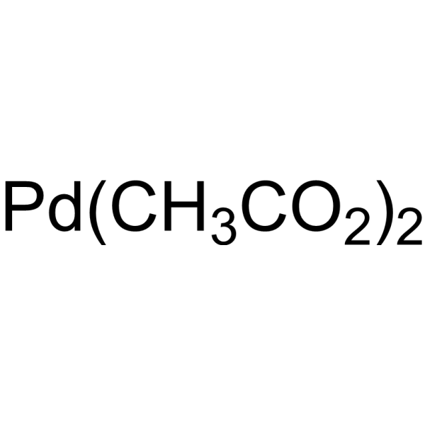
HY-W002072
Palladium (II) acetateCatalyst -

HY-W020952
Palladium(Ⅱ) acetylacetonateAnticancer Agent -
![[Ir(cod)Cl]2](https://file.medchemexpress.com/product_pic/hy-w008260.gif)
HY-W008260
[Ir(cod)Cl]2Biochemical Reagent -
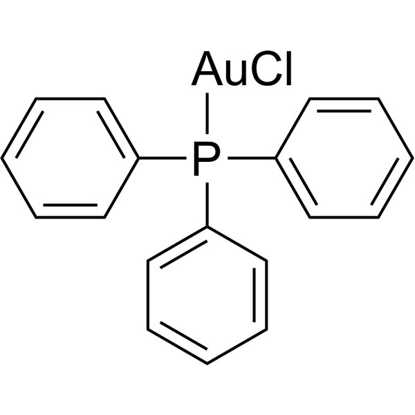
HY-W020955
TriphenylphosphinechlorogoldApoptosis Inducer -
![Dichloro[1,1'-bis(diphenylphosphino)ferrocene]palladium(II) dichloromethane adduct](https://file.medchemexpress.com/product_pic/hy-21623.gif)
HY-21623
Dichloro[1,1'-bis(diphenylphosphino)ferrocene]palladium(II) dichloromethane adductCatalyst -

HY-Y1109
Dichloro(p-cymene)ruthenium(II) dimerPrecursor of Metal Complexes -
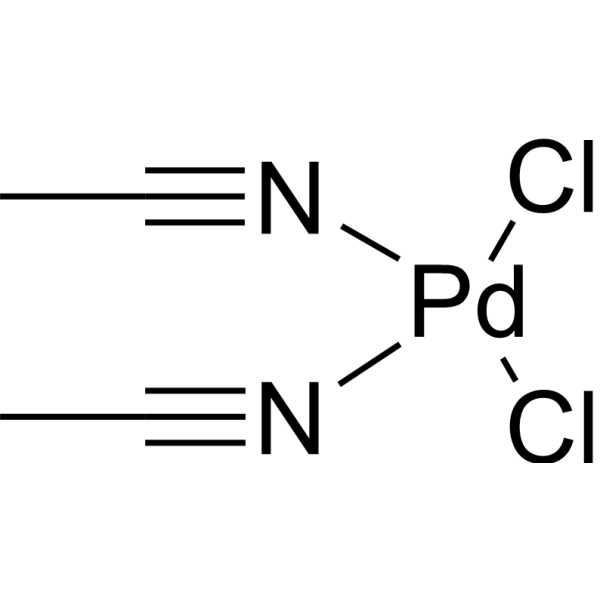
HY-W002087
Bis(acetonitrile)palladium(II) dichlorideBiochemical Reagent -
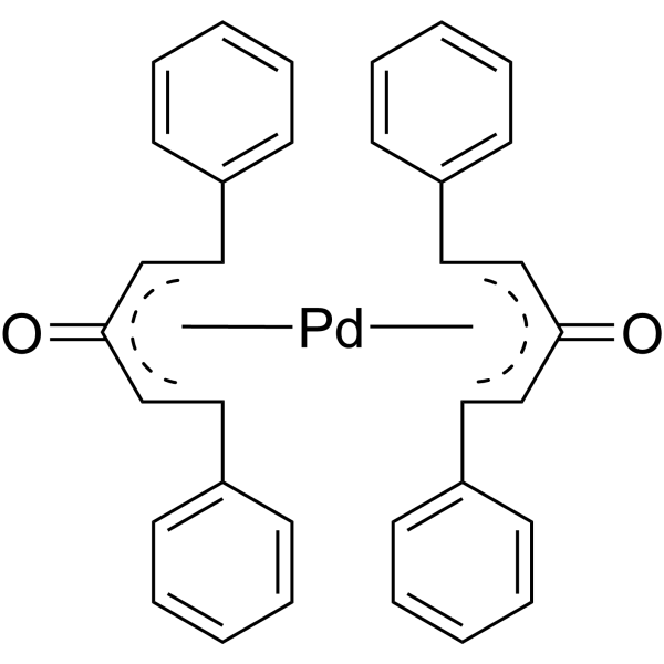
HY-W001137
Bis(dibenzylideneacetone)palladiumBiochemical Reagent -
![[Ir(Cp-)Cl2]2](https://file.medchemexpress.com/product_pic/hy-w018845.gif)
HY-W018845
[Ir(Cp-)Cl2]2Catalyst -

HY-W021022
Iridium(III) chloride hydrateHydrated Iridium (III) Chloride Salt -
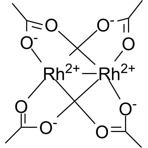
HY-W001951
Dirhodium tetraacetateAnticancer Agent -

HY-Y1237
DPPFMetal Ligand
Alle Caspase Isoform-spezifische Produkte anzeigen
More

Biologische Aktivität
Beschreibung
In Vitro
Ru (acac)3 (100 μM; 24 h) does not inhibit the growth of A2780, U2-OS or U2-OS/Pt cells at concentrations up to 100 μM, with an IC50 value greater than 100 μM[1].
Ru (acac) 3 (treated with U2-OS and U2-OS/Pt cells for 48 h, and HeLa and B16 cells for 72 h; 300 μM) inhibits the growth of HeLa, B16 and U2-OS cells, with IC50 values ranging from 114 μM to 173 μM; it shows no detectable activity against U2-OS/Pt cells at concentrations up to 300 μM[1].
Ru (acac)3 (100 μM; 24 h, 48 h) induces apoptosis in A2780 cells, with the proportion of apoptotic cells reaching 9.93% after 24 h and 14.70% after 48 h[1].
Ru (acac)3 (IC50, 1.5×IC50; 48 h, 72 h) induces a mild, time-dependent decrease in the proportion of G1- and G2-phase cells in HeLa cells, and increases the proportion of apoptotic sub-G1-phase cells to approximately 10% after 72 h[1].
Treatment with Ru (acac)3 (1.5×IC50-10×IC50; 5-48 h) for 24 h induces apoptosis in 7.48% of HeLa cells and activates caspase-3[1].
Ru (acac)3 (30-100 μM; 6-24 h) reversibly inhibits RNA synthesis and suppresses DNA synthesis in a time- and concentration-dependent manner in A2780 cells[1].
MedChemExpress (MCE) has not independently confirmed the accuracy of these methods. They are for reference only.
-
Cell Line:human cervix carcinoma HeLa cells, murine melanoma B16 cells, human osteosarcoma U2-OS cells, human osteosarcoma CDDP-resistant U2-OS/Pt cells
-
Concentration:Up to 300 μM
-
Incubation Time:48 h (U2-OS and U2-OS/Pt); 72 h (HeLa and B16)
-
Result:Exhibited IC50 values of 114 μM for HeLa cells, 148 μM for B16 cells, 173 μM for U2-OS cells, and >300 μM for U2-OS/Pt cells.
Showed that U2-OS cells had slightly higher sensitivity than U2-OS/Pt cells.
-
Cell Line:human cervix carcinoma HeLa cells
-
Concentration:IC50; 1.5×IC50
-
Incubation Time:48 h; 72 h
-
Result:Caused a time- and concentration-dependent slight decrease in the percentage of cells in G1 and G2 phases, with a corresponding increase in sub-G1 (apoptotic) cells reaching ~10% after 72 h incubation at 1.5×IC50.
-
Cell Line:human cervix carcinoma HeLa cells
-
Concentration:1.5×IC50
-
Incubation Time:24 h; 48 h
-
Result:Induced apoptosis in 7.48% of cells after 24 h exposure and 29.09% of cells after 48 h exposure.
Showed that 31.78% of cells were PI-positive after 48 h, indicating necrotic involvement in cell death.


Chemical Information
-
CAS. Nr. 14284-93-6
-
Appearance Solid
-
Molecular Weight 398.39
-
Formel C15H21O6Ru
-
Color Red to reddish brown
-
SMILES
CC1=CC(C)=O[Ru]O1.[3]
-
Synonyms
Tris(acetylacetonato)ruthenium (III)
-
Versand
Room temperature in continental US; may vary elsewhere.
-
Speicherung
RT, protect from light, stored under nitrogen
In solvent -80°C 1 year -20°C 6 months


Lösungsmittel & Löslichkeit
In Vitro:
DMSO : 50 mg/mL (125.51 mM; Need ultrasonic; Hygroscopic DMSO has a significant impact on the solubility of product, please use newly opened DMSO)
Please refer to the solubility information to select the appropriate solvent. Once prepared, please aliquot and store the solution to prevent product inactivation from repeated freeze-thaw cycles.
Storage method and period of stock solution: -80°C, 1 year; -20°C, 6 months. When stored at -80°C, please use it within 1 year. When stored at -20°C, please use it within 6 months.
Please refer to the solubility information to select the appropriate solvent. Once prepared, please aliquot and store the solution to prevent product inactivation from repeated freeze-thaw cycles.
Storage method and period of stock solution: -80°C, 1 year; -20°C, 6 months. When stored at -80°C, please use it within 1 year. When stored at -20°C, please use it within 6 months.
Konzentration (Stammlösung) × Volumen (Stammlösung) = Konzentration (Ziellösung) × Volumen (Ziellösung)


Reinheit & Dokumentation
-
Data Sheet (279 KB)
-
SDS (480 KB)
- English - EN (480 KB)
- Français - FR (480 KB)
- Deutsch - DE (480 KB)
- Norwegian - NO (480 KB)
- Español - ES (480 KB)
- Swedish - SV (480 KB)
- Italian - IT (480 KB)
- Korean - KR (480 KB)
- Portuguese - PT (480 KB)
-
Handling Instructions (2659 KB)
Verweise


Complete Stock Solution Preparation Table
Please refer to the solubility information to select the appropriate solvent. Once prepared, please aliquot and store the solution to prevent product inactivation from repeated freeze-thaw cycles.
Storage method and period of stock solution: -80°C, 1 year; -20°C, 6 months. When stored at -80°C, please use it within 1 year. When stored at -20°C, please use it within 6 months.
| Optional Solvent | Concentration Solvent Mass | 1 mg | 5 mg | 10 mg | 25 mg |
|---|---|---|---|---|---|
| DMSO | 1 mM | 2.5101 mL | 12.5505 mL | 25.1010 mL | 62.7526 mL |
| 5 mM | 0.5020 mL | 2.5101 mL | 5.0202 mL | 12.5505 mL | |
| 10 mM | 0.2510 mL | 1.2551 mL | 2.5101 mL | 6.2753 mL | |
| 15 mM | 0.1673 mL | 0.8367 mL | 1.6734 mL | 4.1835 mL | |
| 20 mM | 0.1255 mL | 0.6275 mL | 1.2551 mL | 3.1376 mL | |
| 25 mM | 0.1004 mL | 0.5020 mL | 1.0040 mL | 2.5101 mL | |
| 30 mM | 0.0837 mL | 0.4184 mL | 0.8367 mL | 2.0918 mL | |
| 40 mM | 0.0628 mL | 0.3138 mL | 0.6275 mL | 1.5688 mL | |
| 50 mM | 0.0502 mL | 0.2510 mL | 0.5020 mL | 1.2551 mL | |
| 60 mM | 0.0418 mL | 0.2092 mL | 0.4184 mL | 1.0459 mL | |
| 80 mM | 0.0314 mL | 0.1569 mL | 0.3138 mL | 0.7844 mL | |
| 100 mM | 0.0251 mL | 0.1255 mL | 0.2510 mL | 0.6275 mL |


Ru(acac)3 Related Classifications
HY-W020958 Related Classifications
 Powered by Bioz