
1017 Results for "

Detection

" in MedChemExpress (MCE) Product Catalog:
Products (1017)

1017 Results for "Detection" in MCE Product Catalog:

Cat. No.: HY-D3085
CAS No.: 2882924-23-2
SSR-LDs is a fluorescent probe for lipid droplet polarity detection and lipid droplet imaging. SSR-LDs exhibits polarity-dependent fluorescence: it produces intense short-wavelength emission in low-polarity environments, while generating weak long-wavelength emission in high-polarity environments. SSR-LDs enters cells via free diffusion, specifically targets lipid droplets, and is unaffected by intracellular viscosity or pH values ranging from 4.5 to 9.0. SSR-LDs shows variable Ex/Em wavelengths in different solvents, including 590/663 nm in THF, 600/660 nm in mouse tissue and in vivo imaging, and an excitation wavelength of 580 nm with an emission wavelength range of 610-750 nm in cell imaging. SSR-LDs can be used for research related to fatty liver, liver injury and hepatitis .
loading...
    loading...
Cat. No.: HY-D3086
CAS No.: 1668565-72-7
Target:  

Fluorescent Dye

Research Areas:  

Cancer

Mito-polarity is a mitochondria-targeted polarity probe. Mito-polarity can be used to detect mitochondrial polarity. The detection mechanism of Mito-polarity is based on an intramolecular charge transfer (ICT) system designed with a donor-π-bridge-acceptor (D-π-A) structure. Among them, the 467 nm green emission generated by the Coumarin (HY-N0709) structure is extremely sensitive to polarity changes, while the 642 nm red emission produced by the extended π-conjugation and ICT effect between the Coumarin and benzothiazine structures shows only a weak response, thus forming a ratiometric fluorescence response that is linearly correlated with solvent polarity. When Mito-polarity is excited at 405 nm in methanol, its excitation/emission wavelengths are Ex/Em = 405/467 nm and Ex/Em = 405/642 nm, with maximum absorption wavelengths of 426 nm and 561 nm .
loading...
    loading...
Cat. No.: HY-D3186
CAS No.: 2163840-29-5
HMRef-βGal is a fluorescent probe and a substrate responsive to β-galactosidase (β-galactosidase) (Ex/Em=498 nm/505-600 nm). After being cleaved by β-galactosidase, HMRef-βGal triggers significant fluorescence enhancement via intramolecular spirocyclic function regulation. HMRef-βGal generates bright fluorescence in cancer cells with elevated β-galactosidase activity, enabling visualization of tiny peritoneal metastases in mouse models. HMRef-βGal exhibits low in vitro cytotoxicity and low acute in vivo toxicity in mice. HMRef-βGal can be used for preclinical fluorescence-guided diagnosis and cytoreductive surgery of peritoneal metastases, including compatibility with real-time naked-eye detection and endoscopic imaging, as well as for studies related to peritoneal metastases of ovarian cancer .
loading...
    loading...
Cat. No.: HY-D3391
RMR-Tre is a fluorescent probe targeting the mycobacterial acyltransferase Ag85. Under the catalysis of Ag85, RMR-Tre undergoes 6-position mycoloylation and anchors to the mycobacterial membrane, while achieving fluorescence activation by inhibiting the intramolecular twisted charge transfer state transition. RMR-Tre can distinguish live mycobacteria from dead ones through metabolism-driven labeling, enabling rapid, wash-free, low-background detection of viable bacteria. RMR-Tre reports the drug resistance of Mycobacterium tuberculosis via the trehalose catalytic shift activity readout associated with TreS. In addition, RMR-Tre can be combined with flow cytometry or high-content imaging techniques to visualize and quantitatively analyze the metabolic heterogeneity of Mycobacterium tuberculosis related to persistence and drug resistance. RMR-Tre is widely used in tuberculosis-related research .
loading...
    loading...
Cat. No.: HY-P11590
Target:  

EGFR

Research Areas:  

Cancer

WGYRGFYC (WC8) is a selective HER2-targeting peptide that binds specifically to HER2 by mimicking the antigen-binding site of trastuzumab. The DOTA precursor of WGYRGFYC has a KD of 61.20 nM for HER2. WGYRGFYC enables specific and highly sensitive detection of HER2 expression in HER2-positive breast cancer cells and tumor tissues, and monitors the dynamic downregulation of HER2 expression. WGYRGFYC rapidly distributes to target tissues and is efficiently cleared from non-target tissues via the kidneys, generating an ideal tumor-to-background ratio in imaging; it is a component of the PET radiotracer Ga-DOTA-WC8. WGYRGFYC exhibits no significant cytotoxicity in breast cancer cells, and can be used for non-invasive imaging diagnosis and therapeutic efficacy evaluation of HER2-positive breast cancer .
loading...
    loading...
Cat. No.: HY-P990232

Target:  

IFNAR

Research Areas:  

Others

Anti-Mouse IFNγRα/CD119 Antibody (2E2) is an Armenian hamster-derived IgG type antibody inhibitor, targeting to mouse IFNγRα/CD119. Anti-Mouse IFNγRα/CD119 Antibody (2E2) reacts with the mouse IFNγR (interferon gamma receptor) α chain also known as CD119 and IFNγ receptor 1. Anti-Mouse IFNγRα/CD119 Antibody (2E2) is a non-neutralizing antibody and does not block the binding of IFNγ to the receptor. Anti-Mouse IFNγRα/CD119 Antibody (2E2) can be used for detections of western blot, flow cytometry and immunoprecipitation .
loading...
    loading...
Cat. No.: HY-P990301

Target:  

Amyloid-β

Research Areas:  

Neurological Disease

Anti-Human/Mouse/Rat Amyloid-beta Antibody (MOAB-2) is a mouse-derived IgG2b λ type antibody inhibitor, targeting to Amyloid-beta. Anti-Human/Mouse/Rat Amyloid-beta Antibody (MOAB-2) recognizes unaggregated, oligomeric or fibrillar forms of Aβ42 and unaggregated Aβ40. Anti-Human/Mouse/Rat Amyloid-beta Antibody (MOAB-2) is selective for human Aβ42 over Aβ40, but not amyloid precursor protein (APP). Anti-Human/Mouse/Rat Amyloid-beta Antibody (MOAB-2) can immunostain human or rat and mouse tissue. Anti-Human/Mouse/Rat Amyloid-beta Antibody (MOAB-2) can be used for detections of western blot, immunohistochemistry, immunofluorescence, immunoprecipitation and ELISA .
loading...
    loading...
Cat. No.: HY-P992275

Target:  

γ-secretase

Research Areas:  

Neurological Disease Cancer

Anti-Nicastrin Antibody (A5226A) is a monoclonal antibody against Nicastrin and an inhibitor of γ-secretase. Anti-Nicastrin Antibody (A5226A) recognizes the fully glycosylated mature presenilin enhancer in the active γ-secretase complex and inhibits its activity via competition for substrate binding. Anti-Nicastrin Antibody (A5226A) abrogates the growth of cancer cells dependent on γ-secretase activity. Anti-Nicastrin Antibody (A5226A) serves as an imaging tool to visualize the endocytic trafficking of active γ-secretase, and also acts as a detection reagent to evaluate the endocytic efficiency of γ-secretase. Anti-Nicastrin Antibody (A5226A) can be used in studies related to non-small cell lung cancer, T-cell acute lymphoblastic leukemia and Alzheimer's disease .
loading...
    loading...
Cat. No.: HY-W110910
CAS No.: 1787-61-7
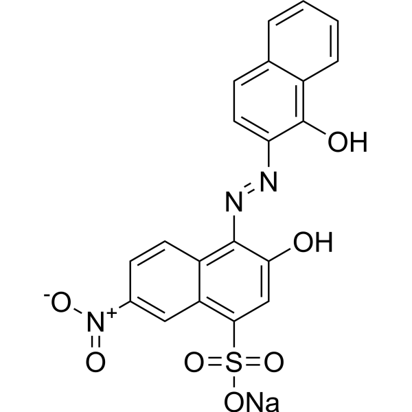
Eriochrome black T, Indicator is a complexing agent for metal ions (e.g., Ca 2+, Mg 2+) and is used as an indicator in complexometric titrations. Eriochrome black T, Indicator forms colored complexes with metal ions through covalent coordination bonds, and indicates the endpoint of the titration by color change. Eriochrome black T, Indicator can be used as an anionic azo dye in photocatalytic degradation studies to evaluate the performance of photocatalysts. The reaction solution of Eriochrome black T, Indicator combined with Mg 2+ is initially purple. During loop-mediated isothermal amplification (LAMP), the color changes from purple to sky blue due to the consumption of Mg 2+ by the formation of magnesium pyrophosphate, indicating a positive reaction. The optimal concentration of Eriochrome black T, Indicator in LAMP is 60 μM, and the detection limit for Mycobacterium tuberculosis is 1 pg DNA/reaction .
loading...
    loading...
Cat. No.: HY-W115721
CAS No.: 523-21-7
Synonyms: Sodium rhodizonate dibasic
Rhodizonic acid disodium (Sodium rhodizonate dibasic) is a transition metal-dependent pro-oxidant and lead detection agent that induces reactive oxygen species generation, DNA damage, and inhibits Aconitase activity. Rhodizonic acid disodium generates superoxide anion radicals in an iron (II)-dependent manner, leading to aconitase inactivation. Rhodizonic acid disodium also triggers hydroxyl radical-mediated DNA strand breaks and 8-OHdG formation via copper ion reduction. Rhodizonic acid disodium reacts with lead to form a scarlet precipitate, with the color intensity proportional to lead content, enabling qualitative or quantitative analysis of lead. Rhodizonic acid disodium can also be used for real-time visualization of the dynamic process of lead sequestration in the plant rhizosphere and evaluation of the effects of environmental factors such as soil type on the stability of lead-sequestering structures .
loading...
    loading...
Cat. No.: HY-L123
6,767 compounds

Human metabolism is an integral part of cellular function that reflects individual differences in health, disease, diet, and lifestyle. Many health conditions such as obesity, diabetes, hypertension, heart disease, and cancer are associated with abnormal metabolic states. In the pathological state of the human body, metabolic pathways are significantly altered, resulting in aberrant levels of intermediates or end-products that can be viewed as potential diagnostic biomarkers or even therapeutic targets. Therefore, detection, identification and quantification of human metabolites are very important for drug metabolism research in drug development.

MCE offers a unique collection of 6,767 human metabolites, including endogenous metabolites and exogenous metabolites, covering multiple structure types, such as lipids, amino acids, nucleic acids, carbohydrates, organic acids, biogenic amines, vitamins,. MCE Human Metabolites Library is a helpful tool for studying the relationship between diseases and metabolism.

Cat. No.: HY-131498
CAS No.: 140430-53-1
Purity:  99.67%
Synonyms: Mca-Pro-Leu-Gly-Leu-Dpa-Ala-Arg-NH2
Target:  

Fluorescent Dye MMP

Research Areas:  

Cancer

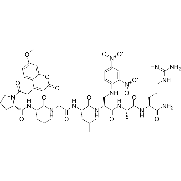
MOCAc-PLGL (Dpa) AR (Mca-Pro-Leu-Gly-Leu-Dpa-Ala-Arg-NH2) is a synthetic fluorescent substrate mainly used for in vitro enzymatic activity assays of MMP-7 (detection at 393 nm after excitation at 328 nm). MOCAc-PLGL (Dpa) AR is specifically cleaved and hydrolyzed by MMP-7 at the Gly-Leu peptide bond, allowing enzymatic activity to be monitored via fluorometry or HPLC (DMSO is usually added to ensure sufficient dissolution when determining kinetic parameters). MOCAc-PLGL (Dpa) AR is used to accurately evaluate the catalytic activity and pH dependence of recombinant mature human MMP-7 in vitro. MOCAc-PLGL (Dpa) AR is suitable for research on the mechanisms of tumor metastasis and invasion, particularly in fields such as prostate cancer, colon cancer, lung cancer and breast cancer .\n
loading...
    loading...
Cat. No.: HY-137276
CAS No.: 97753-82-7
Purity:  99.73%
Synonyms: Bluo-Gal
Research Areas:  

Others

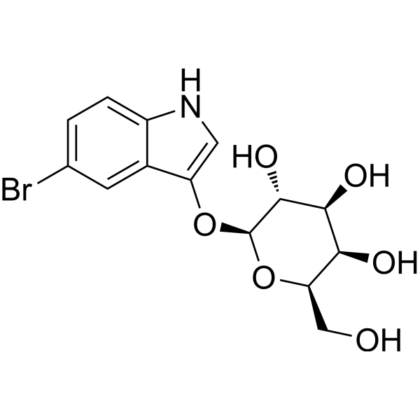
5-Bromo-3-indolyl β-D-galactopyranoside (Bluo-Gal) is a chromogenic substrate for β-galactosidase. 5-Bromo-3-indolyl β-D-galactopyranoside is hydrolyzed by the enzyme to generate a 5-bromoindole intermediate, which is further oxidized to form an insoluble blue precipitate. 5-Bromo-3-indolyl β-D-galactopyranoside can specifically recognize bacterial β-galactosidases (such as the product of the Escherichia coli lacZ gene) and reacts at pH 7.4, making it suitable for light and electron microscopic observations. 5-Bromo-3-indolyl β-D-galactopyranoside can be used in histochemical detection of reporter gene expression in transgenic organisms, such as the localization analysis of β-galactosidase activity in mouse embryos or muscle tissues .
loading...
    loading...
Cat. No.: HY-186341
Research Areas:  

Metabolic Disease

PAB-QMSF2-PEG2-Biotin is a biotin probe for the CAT-S photocatalytic proximity labeling system. PAB-QMSF2-PEG2-Biotin undergoes uncaging under the action of a mitochondria-targeted iridium photocatalyst and blue light (450 nm) to release difluorothioquinone methide (thioQM), which covalently labels nucleophilic residues (Lys/Tyr/Glu/Asp/Trp, etc.) of neighboring proteins. PAB-QMSF2-PEG2-Biotin preferentially labels mitochondrial matrix proteins in live cells. PAB-QMSF2-PEG2-Biotin is conjugated with a biotin tag, enabling detection or isolation of captured proteins. PAB-QMSF2-PEG2-Biotin can be used for research related to type 2 diabetes .
loading...
    loading...
Cat. No.: HY-D2745
CAS No.: 1638544-48-5
BP Fluor 594 NHS ester is the most popular tool for modifying proteins or antibodies through the primary amines (Lys), amine-modified oligonucleotides, and other amine-containing biomolecules with BP Fluor 594 label. The labeling occurs most efficiently at pH 7-9 and forms a stable, covalent amide bond. BP Fluor 594 dye can be used for proteins labeling at high molar ratios without significant self-quenching, enabling brighter conjugates and more sensitive detection. BP Fluor 594 is bright, water-soluble, and pH-insensitive from pH 4 to pH 10 red-fluorescent dye with absorption and emission maxima at 590 and 617 nm, respectively. It can be used with the 561 nm and 594 nm laser lines. BP Fluor 594 dye conjugated to a variety of antibodies, peptides, proteins, tracers, and amplification substrates often used for generation of stable signal in imaging and flow cytometry.
loading...
    loading...
Cat. No.: HY-D3075
CAS No.: 2490504-23-7
Target:  

Fluorescent Dye

Research Areas:  

Others

TPE-2E N-oxide is a fluorescent probe used for hypoxia detection and tumor hypoxia imaging. Due to its active intramolecular motion, TPE-2E N-oxide shows no fluorescence in aqueous solution. In hypoxic environments, its N-oxide group undergoes two-electron reduction by CYP450 reductase and other heme protein reductases expressed under hypoxia, forming hydrophobic aggregates that trigger aggregation-induced emission through the restriction of intramolecular motion. TPE-2E N-oxide exhibits selective lipid droplet localization in cells. The absorption wavelength of TPE-2E N-oxide in dichloromethane is 313 nm; in a 99% hexane/dichloromethane mixed system, its excitation wavelength is 330 nm and emission wavelength is 460 nm. For in vitro cell imaging, excitation is performed at 405 nm, with an emission wavelength range of 430-560 nm .
loading...
    loading...
Cat. No.: HY-D3080
Target:  

Fluorescent Dye

Research Areas:  

Others

CR-1 is a ratiometric photoacoustic (PA) probe for in-situ real-time imaging of Zn 2+ in deep living tissues. CR-1 is formed by coupling the near-infrared cyanine dye IR825 with the Zn 2+ ligand tris (2-pyridylmethyl) amine (TMPA). Upon binding to Zn 2+, the five N atoms on TMPA coordinate with Zn 2+, leading to double bond rearrangement of IR825 and attenuation of its π-electron conjugated system. The absorption peak of CR-1 blue-shifts from 710 nm to 532 nm, enabling quantitative detection of Zn 2+ via the PA532/PA710 ratio. Free CR-1 generates a strong PA signal at 710 nm, while CR-1 bound to Zn 2+ produces a strong PA signal at 532 nm .
loading...
    loading...
Cat. No.: HY-D3090
CAS No.: 2757682-04-3
Target:  

Fluorescent Dye

Research Areas:  

Others

NRLD is a lipid droplet-targeting solvatochromic fluorescent probe developed based on Nile Red (HY-D0718), which can target lipid droplets and sense the non-polar lipid microenvironment inside lipid droplets. NRLD exhibits superior lipid droplet-targeting selectivity compared to the parent Nile Red dye in HeLa cell imaging. NRLD achieves detection relying on the solvatochromic effect generated by excited-state charge transfer, and its emission wavelength shifts according to the local polarity changes of the microenvironment it locates in: the more compact and ordered the lipid packing and the lower the environmental polarity, the more blue-shifted the emission; the looser the lipid packing or the stronger the hydration and the higher the environmental polarity, the more red-shifted the emission. NRLD shows blue shift in the non-polar oil core of lipid droplets and red shift in high-polarity environments such as phosphate buffer .
loading...
    loading...
Cat. No.: HY-D3102
CAS No.: 1015251-87-2
Target:  

Fluorescent Dye

Research Areas:  

Others

CGTDP is a fluorescent probe used for selective labeling of active-site arginine residues, detection of local polarity, and monitoring of conformational changes in enzyme active sites. CGTDP contains a neutral red moiety serving as a polarity-sensitive fluorophore, and a phenylglyoxal unit acting as an arginine-specific labeling group; it undergoes a selective reaction via its α-dicarbonyl moiety with the guanidyl group of arginine residues in enzyme active sites to form a covalent linkage, thereby enabling the reporting of the local microenvironment at the labeled site. The excitation/emission wavelengths are Ex/Em = 398/607 nm and Ex/Em = 507/607 nm; these wavelengths blue-shift in organic solvents, such as Ex/Em = 375/569 nm and Ex/Em = 469/569 nm in THF, and Ex/Em = 375/587 nm and Ex/Em = 470/587 nm in acetonitrile. When labeled onto creatine kinase, its excitation/emission wavelengths are Ex/Em = 493/590 nm .
loading...
    loading...
Cat. No.: HY-W127719
CAS No.: 96087-38-6
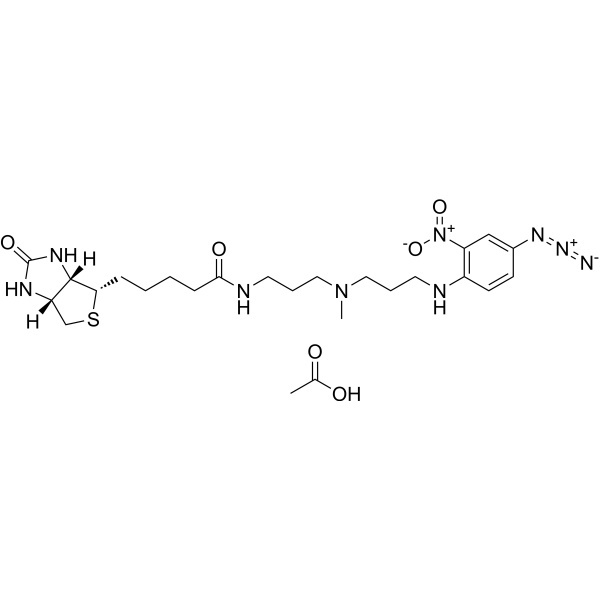
Photobiotin (acetate)It is a biological probe used to study biochemical processes such as protein interactions and enzymatic reactions. It is a molecule containing a photosensitive group, which can be combined with specific target molecules (such as proteins, nucleic acids, etc.) through photochemical cross-linking technology, so as to realize the labeling and detection of these molecules. During the photosensitive crosslinking process, Photobiotin (acetate)Can participate in the formation of covalent bonds and form stable compounds. In addition, the compound also has high biocompatibility and biological activity, so it is widely used in the field of biomedical research, such as enzymatic research, proteomics, western blotting and other aspects. Photobiotin (acetate) is a click chemistry reagent, it contains an Azide group and can undergo copper-catalyzed azide-alkyne cycloaddition reaction (CuAAc) with molecules containing Alkyne groups. It can also undergo strain-promoted alkyne-azide cycloaddition (SPAAC) reactions with molecules containing DBCO or BCN groups.
loading...
    loading...

