



FR104
Based on 2 publication(s) in Google Scholar
FR104 is a selective CD28 inhibitor (binding to CD28 with a Kd value of 4.6 nM; ED50 value of 7.5 ng/mL). FR104 blocks the binding of CD28 to CD80/CD86 and ICOSL, inhibiting conventional T cell costimulation, proliferation, IL-2 secretion, alloantigen responses, and donor-specific alloantibody production. FR104 does not affect the CTLA-4/PD-L1 pathway, preserves the suppressive function of regulatory T cells, and accumulates Helios-negative Foxp3-positive regulatory T cells. FR104 has a prolonged half-life via pegylation (PEG300) (HY-Y0873) and sustained receptor occupancy, and alleviates central nervous system inflammation, demyelinating lesions, and anti-rhMOG responses. FR104 can be used for research on graft-versus-host disease, renal allograft rejection, and experimental autoimmune encephalomyelitis.
For research use only. We do not sell to patients.
- Purity : 97.26%
- Molecular Weight:145.08 kDa
-
Storage:Shipping with dry ice
Please store the product under the recommended conditions in the Certificate of Analysis.


To place orders, for customer services and technical support, please contact: MedChemExpress USA
Tel: 609-228-6898 E-mail: [email protected] [email protected]


-
 Biological Activity
-
Chemical Information
- Protocols
- Purity & Documentation
- References
-
 Help & FAQs


-

HY-P9975
TheralizumabCD28 Superagonist -
HY-P99003
Human IgG4 (S228P) kappa, Isotype ControlIgG4κ Isotype Control -

HY-P99302
LulizumabCD28 Antagonist -
HY-P99002
Human IgG2 kappa, Isotype ControlIgG2κ Isotype Control -
HY-P99001
Human IgG1 kappa, Isotype ControlIgG1κ Isotype Control -

HY-P99315
RuplizumabAnti-CD40L Antibody


Biological Activity
Description
IC50 & Target
[1]|
IL-2 0.027 μg/mL (IC50) |
CD28 4.6 nM (Kd) |
CD28 0.32 μg/mL (ED50) |
Cellular Effect
|
Cell Line

|
Type | Value | Description | References |
|---|---|---|---|---|
| PBMC | IC50 |
0.16 μg/mL
|
Inhibition of allogeneic T cell proliferation in human PBMC mixed lymphocyte reaction assessed by 3H-thymidine incorporation after 5 days.
Inhibition of allogeneic T cell proliferation in human PBMC mixed lymphocyte reaction assessed by 3H-thymidine incorporation after 5 days.
|
22759318 |
| Jurkat | IC50 |
0.027 μg/mL
|
Inhibition of IL-2 secretion by superantigen-stimulated Jurkat T cells co-cultured with Raji B cells assessed by ELISA after 48 hrs.
Inhibition of IL-2 secretion by superantigen-stimulated Jurkat T cells co-cultured with Raji B cells assessed by ELISA after 48 hrs.
|
22759318 |
| PBMC | IC50 |
0.10 μg/mL
|
Inhibition of T-lymphocyte proliferation in baboon peripheral blood mononuclear cells assessed by allogeneic mixed lymphocyte reaction with 3H-thymidine incorporation after 5 days.
Inhibition of T-lymphocyte proliferation in baboon peripheral blood mononuclear cells assessed by allogeneic mixed lymphocyte reaction with 3H-thymidine incorporation after 5 days.
|
25488654 |
| PBMC | IC50 |
0.15 μg/mL
|
Inhibition of T-lymphocyte proliferation in human peripheral blood mononuclear cells assessed by allogeneic mixed lymphocyte reaction with 3H-thymidine incorporation after 5 days.
Inhibition of T-lymphocyte proliferation in human peripheral blood mononuclear cells assessed by allogeneic mixed lymphocyte reaction with 3H-thymidine incorporation after 5 days.
|
25488654 |
In Vitro
FR104 (5 μg/mL; 30 min at room temperature) binds CD28 on human T lymphocytes with an ED50 of 0.32 μg/mL[1].
FR104 (0.001-10 μg/mL; 5 days) inhibits the proliferation of baboon and human T lymphocytes in a dose-dependent manner in allogeneic MLR, with an IC50 of 0.10 μg/mL for baboon PBMC and an IC50 of 0.15 μg/mL for human PBMC[2].
FR104 (1-10000 ng/mL; 5 days) inhibits baboon PBMC proliferation in an allogeneic mixed lymphocyte reaction[5].
FR104 (10 mg/mL; 8 days) reduces human tonsil pre-Tfh and Tfh cell proliferation more effectively than Belatacept (HY-108813)[5].
FR104 pretreatment does not impair the suppressive effect of NHP Tregs on Tconv allogeneic proliferation in MLR[3].
FR104 (10 µg/mL; 5 days) does not interfere with the suppressive function of natural CD4+CD25+ Treg cells from rhesus macaques in vitro, while inhibiting the proliferation of CD4+CD25− effector T cells[4].
FR104 (10 µg/mL; 5 days) blocks the ex vivo rhMOG-specific recall proliferative response of rhMOG-sensitized rhesus monkey monocytes, with an effect comparable to that of CTLA4-Ig[4].
MedChemExpress (MCE) has not independently confirmed the accuracy of these methods. They are for reference only.
-
Cell Line:baboon and human T lymphocytes
-
Concentration:0.001, 0.01, 0.1, 1, 10 μg/mL
-
Incubation Time:5 days
-
Result:Dose-dependently inhibited the proliferation of baboon and human T lymphocytes in allogeneic MLR, with an IC50 of 0.10 μg/mL for baboon PBMC and an IC50 of 0.15 μg/mL for human PBMC.
-
Cell Line:CD4+CD25+ Treg cells from rhesus macaques and CD4+CD25− effector T cells
-
Concentration:10 µg/mL
-
Incubation Time:5 days
-
Result:Did not interfere with the suppressive function of natural CD4+CD25+ Treg cells from rhesus macaques in vitro, while inhibiting the proliferation of CD4+CD25− effector T cells.
In Vivo
FR104 (5 mg/kg; intravenous injection; administered on days 0, 4, and 14, then once every 2 weeks until 3 months) in combination with Rapamycin (HY-10219) without steroids prevents acute renal allograft rejection during the 3-month dosing period[2].
FR104 (1-3.5 mg/kg; intravenous injection; single injection) achieves complete CD28 saturation in cynomolgus monkeys without inducing cytokine release[1].
FR104 (5 mg/kg; intravenous injection; administered on days 0, 4, and 14, then once every 2 weeks until 3 months) combined with MMF and steroids induces delayed rejection and prevents DSA production, but the effect is suboptimal, with half of the recipients developing rejection within 3 months[2].
MedChemExpress (MCE) has not independently confirmed the accuracy of these methods. They are for reference only.
-
Animal Model:NOD/SCID (7 weeks old)[1]
-
Dosage:5 mg/kg
-
Administration:i.p.; single injection; one week after engraftment
-
Result:Did not induce human cytokine release compared with vehicle and reduced IFNγ release.
Did not elicit CD25 or CD69 activation markers on human T cells in blood or spleen.
-
Animal Model:NOD/SCID (7 weeks old)[1]
-
Dosage:5 mg/kg (monotherapy); 5 mg/kg (co-administered with 5 mg/kg anti-human CTLA-4 antibody)
-
Administration:i.p.; twice weekly; day 0 to day 25
-
Result:Prevented GVHD; mice showed no signs of disease, continued gaining weight, and survived through day 60.
No human T-cell infiltration was observed in liver, small intestine, or lung at 3 weeks after engraftment.
At 2 months after engraftment, mice had low human cell engraftment (< 10%).
Co-administration with a blocking anti-human CTLA-4 antibody completely abrogated protection against GVHD.
-
Animal Model:Cynomolgus monkeys (Macaca fascicularis)[1]
-
Dosage:1 mg/kg and 3.5 mg/kg
-
Administration:i.v.; single injection
-
Result:100% CD28 saturation was maintained as long as trough levels were above 0.5-2 µg/mL.
No cytokine release was observed after i.v. injection.
-
Animal Model:Cynomolgus monkeys (Macaca fascicularis)[2]
-
Dosage:5 mg/kg
-
Administration:i.v.; on days 0, 4, 14, then every 2 weeks until 3 months
-
Result:Mean survival time (MST) was 18.5 days; individual survival times were 14, 16, 21, and 28 days, all with acute rejection.
DSA development was prevented in all recipients.
At rejection, intragraft frequencies of Foxp3+, CTLA-4+, Foxp3+CTLA-4+, and Foxp3+Helios− T lymphocytes were significantly higher compared to controls.
Peripheral blood Tregs were > 90% Helios+ before transplantation; Helios-negative Tregs accumulated in frequency and number.
-
Animal Model:Renal allograft rejection model in baboons (Papio anubis, body weight 8-13 kg)[2]
-
Dosage:5 mg/kg (FR104); 35 mg/kg (MMF); 1 mg/kg/day (Solumedrol)
-
Administration:i.v.; on days 0, 4, 14, then every 2 weeks until 3 months; MMF p.o. twice daily from day 0 to day 90; Solumedrol i.v. daily from day 0 to day 14
-
Result:With MMF/steroids, an MST of 69.5 days was observed.
Survival times were 21, 49, 158, and 291 days; two recipients had acute rejection within 3 months (days 21 and 49), one had acute rejection after withdrawal at day 158, and one died of hypoglycemic coma at day 291.
DSA development was prevented in all recipients.
-
Animal Model:Renal allograft rejection model in baboons (Papio anubis, body weight 8-13 kg)[2]
-
Dosage:5 mg/kg (FR104); 0.05 mg/kg/day (tacrolimus)
-
Administration:i.v.; on days 0, 4, 14, then every 2 weeks until 3 months; tacrolimus i.m. daily from day 0 to day 90, adjusted to trough levels of 5-10 ng/mL
-
Result:With low-dose tacrolimus, allograft rejection was prevented over the 3-month dosing period.
Survival times were 77, 77, 84, and 112 days; three recipients were euthanized for ESBL-producing E. coli, Proteus mirabilis, and ESBL-producing E. coli, respectively; one had acute rejection after withdrawal at day 112.
No detectable DSA developed.
-
Animal Model:Renal allograft rejection model in baboons (Papio anubis, body weight 8–13 kg)[2]
-
Dosage:5 mg/kg (FR104); 1 mg/kg/day (rapamycin)
-
Administration:i.v.; on days 0, 4, 14, then every 2 weeks until 3 months; rapamycin p.o. daily from day 0 to day 90, adjusted to trough levels of 10-25 ng/mL
-
Result:With rapamycin, acute rejection was prevented over the 3-month dosing period.
Survival times were 84, 91, and 123 days; one unexplained death without rejection occurred at 84 days, one had an ESBL-producing E. coli infection at 91 days, and one had acute rejection after withdrawal at 123 days.
One recipient developed early IgG DSA without rejection.
-
Animal Model:baboons (Papio anubis, body weight 8–13 kg)[4]
-
Dosage:10 mg/kg (protein equivalent)
-
Administration:i.v.; days 0, 7, 14, and 21
-
Result:All six placebo-treated animals developed EAE and were euthanized on days 12, 13, 16, 18, 21, and 27.
None of the six animals developed EAE; they were euthanized on days 41, 41, 42, 42, 43, and 43 with EAE score 0.
Anti-rhMOG MNC proliferation in axillary lymph nodes was lower (stimulation index 2.42) than in placebo-treated animals (10.4); PBMC and spleen responses remained below the positive threshold (SI ≥ 3), except for monkey F3.
Serum anti-rhMOG IgM on day 11 was 126.1 AU and anti-rhMOG IgG was 14.02 AU; suppression of IgM and IgG production persisted throughout the observation period.
CSF anti-rhMOG IgM ranged from 0.000 to 20.90 AU, and anti-rhMOG IgG ranged from 0.000 to 2.100 AU.
No leukocytes were detected in the CSF.
Brain histology showed no inflammatory infiltrates in five of six animals; the remaining animal had only some small perivascular infiltrates.
Demyelination was observed in four of six placebo-treated animals; no demyelination was observed in the other six animals.
Brain mRNA for CD28, CTLA4, CD25, CCR7, IFN-γ, IL-6, CD63, and CXCL8 (IL-8) was lower relative to placebo; CD3 and CD19 mRNA levels were similar between groups.
CSF cytokine/chemokine medians included IL-2 0.00 pg/mL, IL-6 5.61 pg/mL, IL-12 0.00 pg/mL, IL-15 0.00 pg/mL, IFN-γ 0.00 pg/mL, IL-1RA 8.75 pg/mL, G-CSF 12.0 pg/mL, MIF 34.2 pg/mL, CCL2 123 pg/mL, CXCL10 2.79 pg/mL, CXCL11 3.36 pg/mL, IL-1β 0.56 pg/mL, IL-4 13.1 pg/mL, IL-5 5.12 pg/mL, IL-10 3.19 pg/mL, FGF-basic 0.00 pg/mL, GM-CSF 0.60 pg/mL, VEGF 0.00 pg/mL, and CXCL8 2.56 pg/mL.
At necropsy, serum IL-1β, CCL2, CCL11, and CCL22 were higher than in placebo-treated animals.
LCV DNA in blood increased after day 7, peaked at day 14, decreased at day 21, and increased again after day 21 in animals F1-F4; LCV was not detected in brain tissue.
Animal F3 had elevated CMV, LCV, and SA12 in tissues.
No marked reactivation was observed for CMV or the polyomaviruses; HEV was undetectable.
-
Animal Model:Papio anubis (8-13 kg)[5]
-
Dosage:10 mg/kg
-
Administration:i.v.; days 0, 7, and 14, then every 2 weeks; up to 1 year
-
Result:In recipients, two animals developed acute rejection on days 51 and 58; both reversed after steroid administration.
Two animals were lost to anesthetic complications or septicemia.
At 1 year, the three remaining animals had stable kidney function.
No animal had donor-specific antibodies by flow cytometry.
One-month protocol biopsies showed interstitial CD3+ T-cell infiltrate in all animals, with no differences in B-lymphocyte or major T-lymphocyte populations.
Blood CD25hi/CD127lo CD4+ Tregs and graft Foxp3 expression were not different; TSDR demethylation showed a trend toward a stronger Treg signal in animals, and TGFβ expression showed a nonsignificant increase.
No differences were found in CD28-negative CD4+ or CD8+ T cells, perforin, 2B4, PD1, or BTLA expression.
Th17-related transcripts (RORγc, IL-17, IL-23) were not different; graft IL-21 expression was lower than in belatacept group animals.
In rejection biopsies, CD4+PD1+ graft-infiltrating cells were 408/mm2 (n=2).
Parmacokinetics
Clinical Trial
| NCT Number | Sponsor | Condition | Start Date |
Phase
|
|---|---|---|---|---|
| NCT01329991 | 2011-05 | PHASE1 |
Application
ELISA, FACS, Functional assay
Verified Bioactivity
-
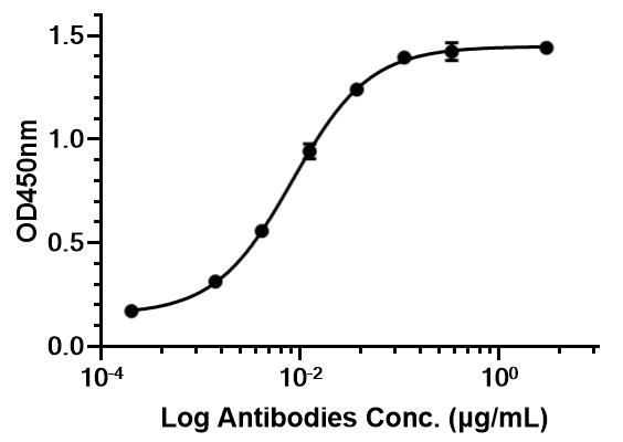 Immobilized CD28 Protein, Human can bind FR104. The EC50 for this effect is 8.381 ng/mL.
-
 Flow cytometric analysis of 106 Jurkat cells with FR104 (HY-P990587, red). Cells were fixed with 4% paraformaldehyde. Then stained with the primary antibody at 1/200 dilution for an hour at 4℃. Alexa Fluor® 488-conjugated AffiniPure Goat Anti-Human IgG H&L (HY-P83776) was used as the secondary antibody at 1/1,000 dilution for 30 minutes at 4℃. Human IgG2 kappa (HY-P99002, blue) was used as the isotype control.


Product Details
-
Accession P10747
-
Molecular Weight 145.08 kDa
-
Species Reactivity
Human
-
Conjugated
Unconjugated
-
Isotype
Human IgG2 kappa
-
Recommend Isotype Controls
Product Properties
-
Appearance
Liquid
-
Formulation
Please refer to the lot-specific COA for specific buffer information.
-
Purity
97.26%
-
Reconstitution
The product can be reconstituted/diluted with sterile PBS or saline.
-
Storage & Stability
Please store the product under the recommended conditions in the Certificate of Analysis.
-
Shipping
Shipping with dry ice.


Publications (2)
Purity & Documentation
-
Data Sheet (287 KB)
-
SDS (251 KB)
- English - EN (251 KB)
- Français - FR (251 KB)
- Deutsch - DE (251 KB)
- Norwegian - NO (251 KB)
- Español - ES (251 KB)
- Swedish - SV (251 KB)
- Italian - IT (251 KB)
- Korean - KR (251 KB)
- Portuguese - PT (251 KB)
-
Inhibitory Antibodies User Guide (603 KB)


References
[1]. Poirier N, et al. Preclinical efficacy and immunological safety of FR104, an antagonist anti-CD28 monovalent Fab' antibody. American journal of transplantation : official journal of the American Society of Transplantation and the American Society of Transplant Surgeons. 2012 Oct;12(10):2630-40. [Content Brief]

Calculators
Concentration (start) × Volume (start) = Concentration (final) × Volume (final)


FR104 Related Classifications
HY-P990587 Related Classifications
 Powered by Bioz