



Terrein

Based on 1 Customer Validation
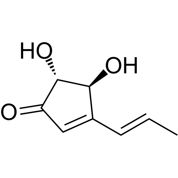
Terrein is a melanogenesis inhibitor. Terrein induces apoptosis in breast cancer cell lines . Terrein is an inhibitor of quorum sensing and c-di-GMP in Pseudomonas aeruginosa.
For research use only. We do not sell to patients.
- Purity : 99.44%
- CAS No.: 582-46-7
- Formula: C8H10O3
- Molecular Weight:154.16
-
Storage:
4°C, sealed storage, away from moisture
* In solvent : -80°C, 6 months; -20°C, 1 month (sealed storage, away from moisture)

To place orders, for customer services and technical support, please contact: MedChemExpress USA
Tel: 609-228-6898 E-mail: [email protected] [email protected]


-
 Biological Activity
-
Chemical Information
- Protocols
- Purity & Documentation
- References
-
 Help & FAQs


-
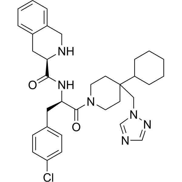
HY-10624
THIQMC4R Agonist -
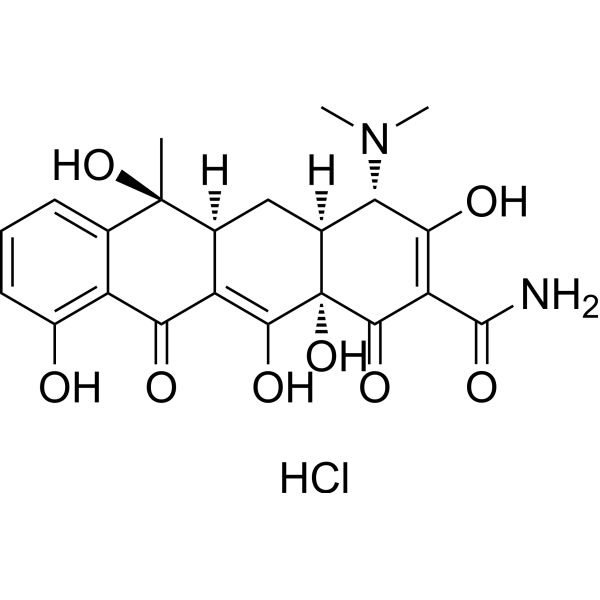
HY-B0474
Tetracycline hydrochlorideAntibiotic -
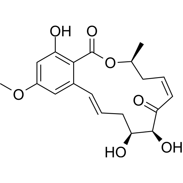
HY-12686
5Z-7-OxozeaenolTAK1 Inhibitor -
HY-B1456A
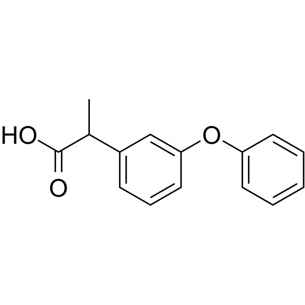
FenoprofenNSAID -

HY-19870
SetmelanotideMC4R Agonist -

HY-10624R
THIQ (Standard)Reference Standard -

HY-B0474R
Tetracycline hydrochloride (Standard)Bacterial Inhibitor -
HY-107139
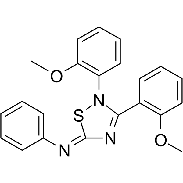
JNJ-10229570Melanocortin Receptor Antagonist -
HY-P1726
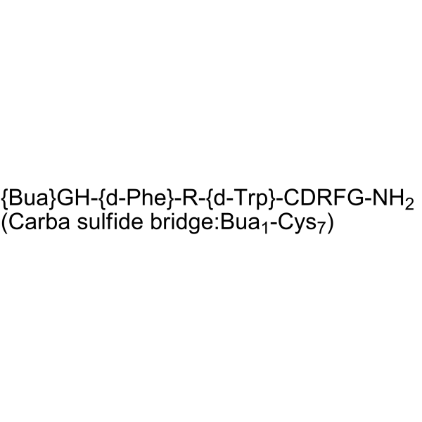
MSG606MC1R Antagonist -
HY-129337
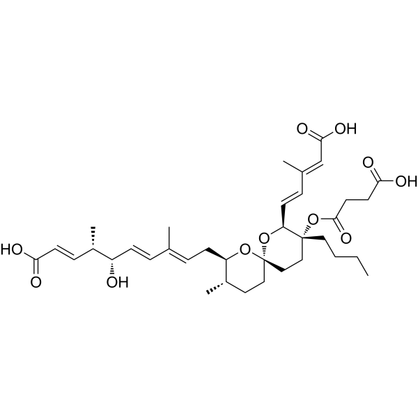
Reveromycin AAntifungal Agent -
HY-N2466A
Melanotan I acetateMelanocortin Receptor Activator -
HY-11029
SNT-207707MC-4 Receptor Antagonist
All Antibiotic Isoforms
More

Biological Activity
Description
In Vitro
Treatment of Mel-Ab cells with Terrein (10-100 μM) for 4 days significantly reduces melanin levels in a dose-dependent manner. Terrein reduces melanin synthesis by reducing tyrosinase production via ERK activation[1].
Terrein (5-500 μM; 24 and 48 hours) reduces the viability of these cells in concentration- and time-dependent manners[2].
Terrein (150, 250, 500μM; 24 hours) induces programmed cell death in both invasive and non-invasive breast cancer cell lines. Thus, the optimal non-toxic concentrations of Terrein (25 and 75 μM) were used for subsequent experiments[2].
Terrein (25 and 75 μM) significantly decrease the mRNA levels of both MMP-2 and MMP-9 in MDA-MB-231 cells. Terrein (75 μM) significantly decrease the mRNA levels of both MMP-2 and MMP-9 in MCF-7 cells[2].
Terrein (75 μM) inhibits the expression of RhoA, RhoB, p-Rac1 in MCF-7 cells. Terrein (75 μM) inhibits the expression of RhoB, p-Rac1 in MDA-MB-231 cells[2].
Terrein (10, 30, and 100 μM) inhibits the production of virulence factors such as elastase, pyocyanin, and rhamnolipid, as well as biofilm formation in P. aeruginosa PAO1 and PA14 strains[3].
MedChemExpress (MCE) has not independently confirmed the accuracy of these methods. They are for reference only. Further protocols information, click here.
-
Cell Line:MCF-7 and MDA-MB-231 cells
-
Concentration:5, 25, 50, 75, 100, 150, 250, 500 µM
-
Incubation Time:24 and 48 hours
-
Result:The IC50s for MCF-7 and MDA-MB-231 cells after 24 h of incubation were 2.34 mM and 700 µM, respectively. After 48 h of incubation, the IC50s are 244.3 µM for MCF-7 and 244.5 µM for MDA-MB-231 cells.
-
Cell Line:MCF-7 and MDA-MB-231 cells
-
Concentration:25, 75, 150, 250, 500 µM
-
Incubation Time:24 hours
-
Result:The percentages of apoptotic cells after 25 and 75 µM treatment were 7.52 and 5.82%, respectively for MCF-7 cells (9.94% for the control), and 15.77 and 15.82%, respectively for MDA-MB-231 cells (8.79% for the control).
-
Cell Line:MCF-7 and MDA-MB-231 cells
-
Concentration:25, 75 µM
-
Incubation Time:
-
Result:Decreased the mRNA levels of both MMP-2 and MMP-9 in a concentration-dependent manner.
-
Cell Line:MCF-7 and MDA-MB-231 cells
-
Concentration:75 µM
-
Incubation Time:6, 12 and 24 hours
-
Result:RhoA and RhoB protein levels were significantly decreased after 6 h of 75 µM treatment in MCF-7 cells.
Phosphorylated (p)-Rac1 was also slightly decreased at 12 h. However, RhoC and Cdc42 levels remained unchanged. RhoB protein levels and Rac1 phosphorylation were also decreased in MDA-MB-231 cells at 12 and 24 h, respectively.


Chemical Information
-
CAS No. 582-46-7
-
Appearance Solid
-
Molecular Weight 154.16
-
Formula C8H10O3
-
Color White to off-white
-
SMILES
O=C1C=C(/C=C/C)[C@H](O)[C@H]1O
-
Shipping
Room temperature in continental US; may vary elsewhere.
-
Storage
4°C, sealed storage, away from moisture
* In solvent : -80°C, 6 months; -20°C, 1 month (sealed storage, away from moisture)

Protocols
-
Apoptosis
Apoptosis, also called programmed cell death, is generally characterized by distinct morphological characteristics.
-
TUNEL staining for apoptotic DNA fragmentation
TUNEL staining detects DNA strand breaks by using terminal deoxynucleotidyl transferase to add labeled nucleotides to exposed 3′-OH DNA termini, generating either microscopic staining in fixed cells or tissue sections, or fluorescence/cytometric signal in cell suspensions. TUNEL positivity reflects DNA fragmentation but should not be interpreted alone as definitive apoptosis, because TUNEL can also label necrotic, autolytic, mechanically damaged, or DNA-repair-associated DNA breaks.
-
Annexin V plus membrane-impermeant dye apoptosis staining
Annexin V-based apoptosis assays rely on the detection of phosphatidylserine (PS) externalization from the inner leaflet of the plasma membrane to the outer leaflet, an early biochemical hallmark of apoptosis. Fluorescently labeled Annexin V binds PS in a calcium-dependent manner, enabling identification of early apoptotic cells by flow cytometry or fluorescence microscopy. When combined with a membrane-impermeant DNA-binding dye (e. g. , propidium iodide), this approach allows discrimination between viable (Annexin V−/dye−), early apoptotic (Annexin V+/dye−), and late apoptotic or necrotic (Annexin V+/dye+) cell populations by assessing membrane integrity and PS exposure.
-
Apoptosis Solutions
Apoptosis is a regulated, generally non-lytic cell-death pathway that removes unwanted, damaged, infected, or abnormal cells through coordinated morphological changes, caspase activation, DNA fragmentation, and membrane remodeling. The intrinsic apoptosis pathway is controlled mainly by mitochondrial outer membrane permeabilization, BCL-2 family proteins, cytochrome c release, apoptosome formation, caspase-9 activation, and downstream executioner caspase-3/7 activation. The extrinsic apoptosis pathway is initiated by death receptors such as Fas, TNFR, and TRAIL receptors, which recruit adaptor proteins and activate caspase-8 before engaging executioner caspases or mitochondrial amplification through BID cleavage. Apoptosis is linked to many phenotypes, including cancer cell killing, tissue homeostasis, immune regulation, neurodegeneration, infection response, and treatment-induced cytotoxicity; unresolved questions include how apoptosis interacts with necroptosis, pyroptosis, ferroptos
-
Breast Cancer Modeling
Breast cancer is a heterogeneous cancer, and it has been distinguished into four subtypes: luminal A, luminal B, HER2-positive and basal-like. Molecular mutations, epigenetic alterations, hormone exposure and immune microenvironment are related to the progression of breast cancer.


Purity & Documentation
-
Data Sheet (275 KB)
-
SDS (393 KB)
- English - EN (393 KB)
- Français - FR (393 KB)
- Deutsch - DE (393 KB)
- Norwegian - NO (393 KB)
- Español - ES (393 KB)
- Swedish - SV (393 KB)
- Italian - IT (393 KB)
- Korean - KR (393 KB)
- Portuguese - PT (393 KB)
-
Handling Instructions (2659 KB)
References
[1]. S-H Park, et al. Terrein: A New Melanogenesis Inhibitor and Its Mechanism. Cell Mol Life Sci. 2004 Nov;61(22):2878-85. [Content Brief]
[2]. Anongnard Kasorn, et al. Terrein Inhibits Migration of Human Breast Cancer Cells via Inhibition of the Rho and Rac Signaling Pathways. Oncol Rep. 2018 Mar;39(3):1378-1386. [Content Brief]
[3]. Bomin Kim, et al. Terrein Is an Inhibitor of Quorum Sensing and c-di-GMP in Pseudomonas Aeruginosa: A Connection Between Quorum Sensing and c-di-GMP. Sci Rep. 2018 Jun 5;8(1):8617. [Content Brief]

Calculators
Concentration (start) × Volume (start) = Concentration (final) × Volume (final)


Terrein Related Classifications
HY-119808 Related Classifications
 Powered by Bioz