



Ticarcillin monosodium
Based on 7 publication(s) in Google Scholar
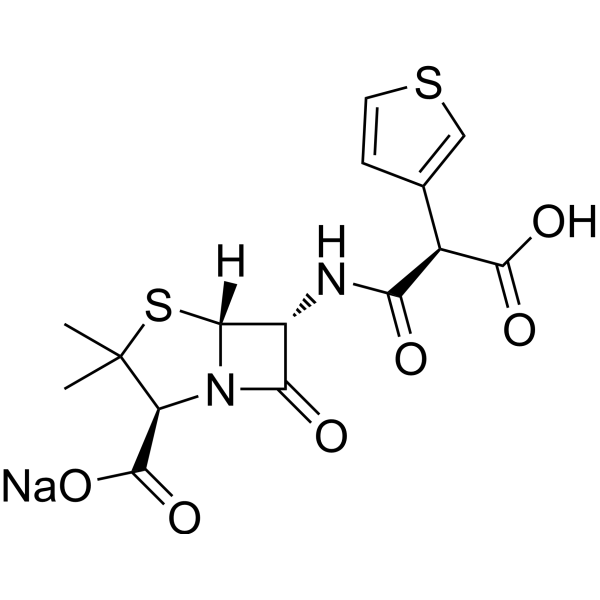
Ticarcillin monosodium is a semisynthetic, extended-spectrum, carboxypenicillin antibacterial agent, and is active against gram-positive cocci, including streptococci and staphylococci. Ticarcillin monosodium is also effective against most gram-negative organisms, including Pseudomonas aeruginosa. Ticarcillin monosodium can be used in lower respiratory tract infections, skin and skin structure infections, urinary tract infections, and intraabdominal infections research.
For research use only. We do not sell to patients.
- CAS No.: 74682-62-5
- Formula: C15H15N2NaO6S2
- Molecular Weight:406.41
-
Storage:
Please store the product under the recommended conditions in the Certificate of Analysis.

To place orders, for customer services and technical support, please contact: MedChemExpress USA
Tel: 609-228-6898 E-mail: [email protected] [email protected]


-
 Biological Activity
-
Chemical Information
- Protocols
- Purity & Documentation
- References
-
 Help & FAQs


Publications Citing Use of MedChemExpress (MCE) Ticarcillin monosodium
More- Virulence. 2026 Dec 31;17(1):2646808. [Abstract]
- Molecules. 2025 Mar 9;30(6):1224. [Abstract]
- Antimicrob Agents Chemother. 2023 Jun 15;67(6):e0160322. [Abstract]
- J Antimicrob Chemother. 2025 Nov 5:dkaf408. [Abstract]
- Microbiol Spectr. 2023 Jun 15;11(3):e0069223. [Abstract]
- Microbiol Spectr. 2023 Feb 14;11(1):e0303822. [Abstract]
- Infect Drug Resist. 2026 May 8;19.
All Antibiotic Isoforms
More

Biological Activity
Description


Chemical Information
-
CAS No. 74682-62-5
-
Molecular Weight 406.41
-
Formula C15H15N2NaO6S2
-
SMILES
OC([C@H](C1=CSC=C1)C(N[C@H]2[C@]3([H])N(C2=O)[C@H](C(C)(S3)C)C(O[Na])=O)=O)=O
-
Shipping
Room temperature in continental US; may vary elsewhere.
-
Storage
Please store the product under the recommended conditions in the Certificate of Analysis.


Publications (7)
-
Journal Impact Factor
-
Most Recent
-
Virulence
Antibacterial efficacy and mechanism of the novel antimicrobial peptide lachnospirin-1 against Acinetobacter baumannii. [Abstract]2026 Dec 31;17(1):2646808. PMID: 41838520 -
Molecules
Seeking Correlation Among Porin Permeabilities and Minimum Inhibitory Concentrations Through Machine Learning: A Promising Route to the Essential Molecular Descriptors. [Abstract]2025 Mar 9;30(6):1224. PMID: 40142001 -
Antimicrob Agents Chemother
Penicillin-Binding Protein 5/6 Acting as a Decoy Target in Pseudomonas aeruginosa Identified by Whole-Cell Receptor Binding and Quantitative Systems Pharmacology. [Abstract]2023 Jun 15;67(6):e0160322. PMID: 37199612 -
J Antimicrob Chemother
Unravelling the triad of penicillin-binding proteins, β-lactamase activity, and mRNA dynamics in Pseudomonas aeruginosa AmpC induction. [Abstract]2025 Nov 5:dkaf408. PMID: 41206063 -
Microbiol Spectr
Penicillin-Binding Protein Occupancy Dataset for 18 β-Lactams and 4 β-Lactamase Inhibitors in Neisseria gonorrhoeae. [Abstract]2023 Jun 15;11(3):e0069223. PMID: 37093051 -
Microbiol Spectr
PBP Target Profiling by β-Lactam and β-Lactamase Inhibitors in Intact Pseudomonas aeruginosa: Effects of the Intrinsic and Acquired Resistance Determinants on the Periplasmic Drug Availability. [Abstract]2023 Feb 14;11(1):e0303822. PMID: 36475840 -


Purity & Documentation
References
[1]. Cecile Formosa, et al. Nanoscale effects of antibiotics on P. aeruginosa. Nanomedicine. 2012 Jan;8(1):12-6. [Content Brief]
[2]. G B van der Voet, et al. Comparison of the antibacterial activity of azlocillin and ticarcillin in vitro and in irradiated neutropenic mice. J Antimicrob Chemother. 1985 Nov;16(5):605-13. [Content Brief]
[3]. Oriel Spierer, et al. Comparative activity of antimicrobials against Pseudomonas aeruginosa, Achromobacter xylosoxidans and Stenotrophomonas maltophilia keratitis isolates. Br J Ophthalmol. 2018 May;102(5):708-712. [Content Brief]

Calculators
Concentration (start) × Volume (start) = Concentration (final) × Volume (final)


Ticarcillin monosodium Related Classifications
HY-139805A Related Classifications
 Powered by Bioz