



TNF-α-IN-19

Based on 1 Customer Validation
TNF-α-IN-19 is an inhibitor of TNFα that can block the interaction between TNFαRI, TRADD, and RIP1, the EC50 values for TNFα, IL-1β, and IL-1β/TNFα are 2.451, 3.792 and 1.54 μM, respectively. TNF-α-IN-19 only inhibits the degradation of IκBα when cells are stimulated by TNFα and not by IL-1β.
For research use only. We do not sell to patients.
- CAS No.: 338773-13-0
- Formula: C16H8ClF3N4
- Molecular Weight:348.71
-
Storage:Powder -20°C, 3 years ; In solvent -80°C, 6 months , -20°C, 1 month


To place orders, for customer services and technical support, please contact: MedChemExpress USA
Tel: 609-228-6898 E-mail: [email protected] [email protected]


-
 Biological Activity
-
Chemical Information
- Protocols
- Purity & Documentation
- References
-
 Help & FAQs


-
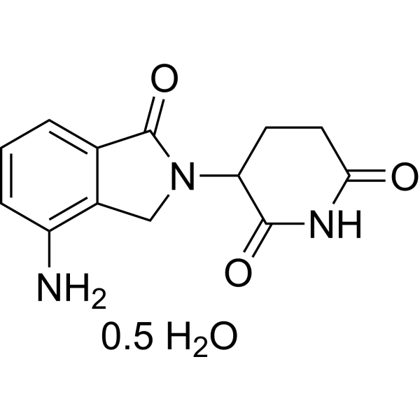
HY-A0003B
Lenalidomide hemihydrateImmunomodulator -

HY-P9908
AdalimumabTNF-α Inhibitor -

HY-108847
EtanerceptTNF Inhibitor -

HY-P9970
InfliximabTNF-α Inhibitor -
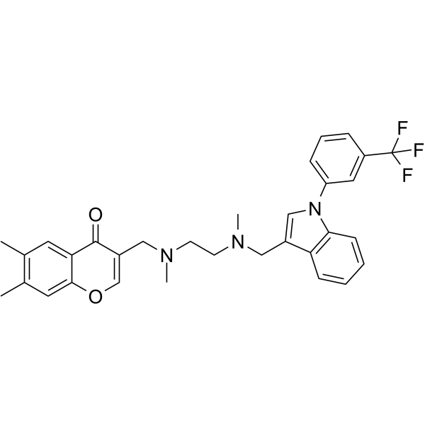
HY-111255
SPD304TNFα Inhibitor -

HY-A0003BR
Lenalidomide hemihydrate (Standard)Molecular Glues -

HY-P1875
TNF-α (46-65), humanTNF-α -
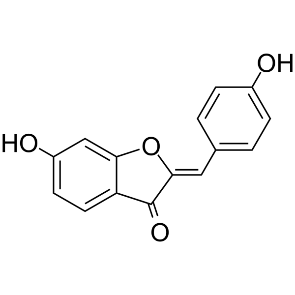
HY-102040
HispidolTNF-α Inhibitor -
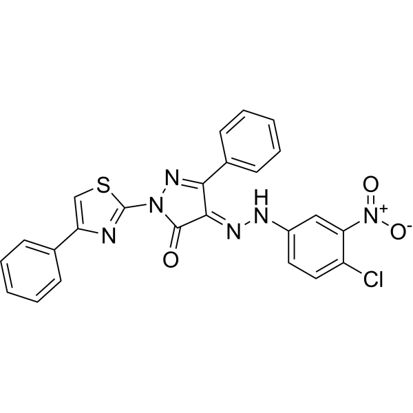
HY-100735
C 87TNFα Inhibitor -
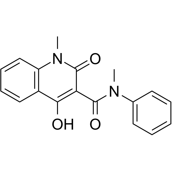
HY-13743
RoquinimexImmunomodulator -
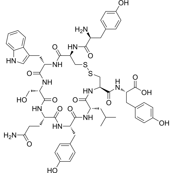
HY-P2612
WP9QYRANKL Inhibitor -
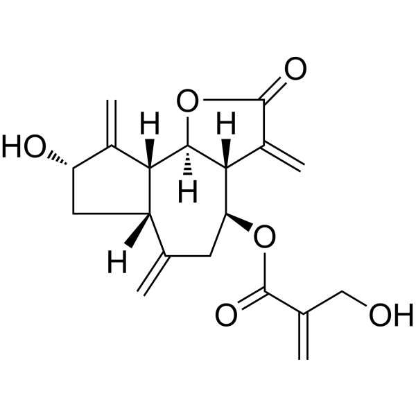
HY-N2350
CynaropicrinTNF Receptor Inhibitor


Biological Activity
Description
IC50 & Target
|
IL-1β |
Cellular Effect
|
Cell Line

|
Type | Value | Description | References |
|---|---|---|---|---|
| 2008 | IC50 |
2.5 μM
Compound: 7, LQ011
|
Cytotoxicity against human cDDP-sensitive 2008 cells after 72 hrs using crystal violet staining by spectrophotometric analysis
Cytotoxicity against human cDDP-sensitive 2008 cells after 72 hrs using crystal violet staining by spectrophotometric analysis
|
[PMID: 20951595] |
| A2780 | IC50 |
2.3 μM
Compound: 7, LQ011
|
Cytotoxicity against human cDDP-sensitive A2780 cells after 72 hrs using crystal violet staining by spectrophotometric analysis
Cytotoxicity against human cDDP-sensitive A2780 cells after 72 hrs using crystal violet staining by spectrophotometric analysis
|
[PMID: 20951595] |
| A2780 | IC50 |
4.7 μM
Compound: 7, LQ011
|
Cytotoxicity against human cDDP-resistant A2780 cells after 72 hrs using crystal violet staining by spectrophotometric analysis
Cytotoxicity against human cDDP-resistant A2780 cells after 72 hrs using crystal violet staining by spectrophotometric analysis
|
[PMID: 20951595] |
| C13 | IC50 |
3.4 μM
Compound: 7, LQ011
|
Cytotoxicity against human cDDP-resistant C13 cells after 72 hrs using crystal violet staining by spectrophotometric analysis
Cytotoxicity against human cDDP-resistant C13 cells after 72 hrs using crystal violet staining by spectrophotometric analysis
|
[PMID: 20951595] |
| Vero | IC50 |
1.2 μM
Compound: 7, LQ011
|
Cytotoxicity against african green monkey Vero cells after 72 hrs using crystal violet staining by spectrophotometric analysis
Cytotoxicity against african green monkey Vero cells after 72 hrs using crystal violet staining by spectrophotometric analysis
|
[PMID: 20951595] |


Chemical Information
-
CAS No. 338773-13-0
-
Appearance Solid
-
Molecular Weight 348.71
-
Formula C16H8ClF3N4
-
Color White to off-white
-
SMILES
ClC1=CC=C(C2=NN=C3N2C4=C(N=C3C(F)(F)F)C=CC=C4)C=C1
-
Shipping
Room temperature in continental US; may vary elsewhere.
-
Storage
Powder -20°C 3 years In solvent -80°C 6 months -20°C 1 month

Protocols
-
Pyroptosis Solutions
Pyroptosis is a lytic inflammatory cell-death pathway executed by gasdermin pores, most classically through inflammasome-mediated activation of caspase-1, cleavage of gasdermin D, membrane pore formation, LDH release, and secretion of IL-1β and IL-18. The canonical pathway is commonly modeled by priming cells with an inflammatory signal such as LPS to induce pro-IL-1β and inflammasome components, followed by an activation signal such as ATP or nigericin to activate NLRP3, ASC speck formation, caspase-1 cleavage, GSDMD cleavage, cytokine release, and pyroptotic membrane rupture. The non-canonical pathway is triggered when cytosolic LPS activates mouse caspase-11 or human caspase-4/5, leading to GSDMD cleavage and pyroptosis, and this can secondarily activate NLRP3-dependent IL-1β release. Pyroptosis is linked to inflammatory injury, infection, cancer, liver disease, ocular disease, placental inflammation, and other disease phenotypes, but unresolved questions include which gasdermin fam


Purity & Documentation
-
Data Sheet (273 KB)
-
SDS (252 KB)
- English - EN (252 KB)
- Français - FR (252 KB)
- Deutsch - DE (252 KB)
- Norwegian - NO (252 KB)
- Español - ES (252 KB)
- Swedish - SV (252 KB)
- Italian - IT (252 KB)
- Korean - KR (252 KB)
- Portuguese - PT (252 KB)
-
Handling Instructions (2659 KB)
References

Calculators
Concentration (start) × Volume (start) = Concentration (final) × Volume (final)


TNF-α-IN-19 Related Classifications
HY-114573 Related Classifications
 Powered by Bioz