



AD4
Based on 1 publication(s) in Google Scholar
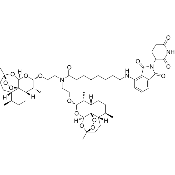
AD4 is a PROTAC that induces the degradation of PCLAF by recruiting cereblon. AD4 is also an artemisinin derivative. AD4 directly binds to PCLAF with a KD of 6.1 μM, and induces apoptosis and G1-phase cell cycle arrest. By degrading PCLAF, AD4 upregulates p21, reduces Rb phosphorylation, downregulates Bcl-2 and upregulates Bax, thereby activating the p21/Rb axis. AD4 can be used in studies related to acute B-lymphoblastic leukemia and PCLAF-dependent anticancer mechanisms .
(Pink: Caspase and Apoptosis ligand (HY-160962); Blue: Cereblon ligand (HY-14658); Black: linker (HY-W018678)).
For research use only. We do not sell to patients.
- Purity : 98.01%
- CAS No.: 2918262-09-4
- Formula: C55H78N4O15
- Molecular Weight:1035.23
-
Storage:Powder -20°C, 3 years , 4°C, 2 years ; In solvent -80°C, 6 months , -20°C, 1 month

To place orders, for customer services and technical support, please contact: MedChemExpress USA
Tel: 609-228-6898 E-mail: [email protected] [email protected]


-
 Biological Activity
-
Chemical Information
-
Solvent & Solubility
- Protocols
- Purity & Documentation
- References
-
 Help & FAQs


Publications Citing Use of MedChemExpress (MCE) AD4
MoreAll PROTACs Isoforms
More

Biological Activity
Description
IC50 & Target
[1]|
PCLAF 54.92 nM (DC50) |
Cellular Effect
|
Cell Line

|
Type | Value | Description | References |
|---|---|---|---|---|
| RS4-11 | IC50 |
50.6 nM
|
Antiproliferative activity against human RS4;11 cancer cells assessed as reduction in cell viability incubated for 48 hrs by CCK8 colorimetric assay.
Antiproliferative activity against human RS4;11 cancer cells assessed as reduction in cell viability incubated for 48 hrs by CCK8 colorimetric assay.
|
37552639 |
| RPMI-8226 | IC50 |
235.3 nM
|
Antiproliferative activity against human RPMI8226 cancer cells assessed as reduction in cell viability incubated for 48 hrs by CCK8 colorimetric assay.
Antiproliferative activity against human RPMI8226 cancer cells assessed as reduction in cell viability incubated for 48 hrs by CCK8 colorimetric assay.
|
37552639 |
| U-266 | IC50 |
342.8 nM
|
Antiproliferative activity against human U266 cancer cells assessed as reduction in cell viability incubated for 48 hrs by CCK8 colorimetric assay.
Antiproliferative activity against human U266 cancer cells assessed as reduction in cell viability incubated for 48 hrs by CCK8 colorimetric assay.
|
37552639 |
In Vitro
AD4 (48 h) potently inhibits the proliferation of RS4;11 cells, RPMI8226 cells, and U266 cells, with IC50 values of 50.6 nM, 235.3 nM, and 342.8 nM, respectively[1].
AD4 (10-200 nM; 48 h) induces dose-dependent apoptosis in RS4;11 cells after 48 h of incubation, with the maximum effect observed at 200 nM[1].
AD4 (10-200 nM; 12-48 h) degrades PCLAF in RS4;11 cells in a dose-dependent manner, with a DC50 of 54.92 nM, while activating the p21/Rb pathway and regulating apoptosis-related proteins to exert antitumor activity [1].
AD4 (10-200 nM; 48 h) induces dose-dependent G1-phase cell cycle arrest in RS4;11 cells after 48 h of incubation, and exhibits superior activity to SM1044 (HY-160962) at matched concentrations [1].
AD4 directly binds to the purified PCLAF protein with a Kd value of 6.1 μM, and its affinity is higher than that of its parent compound SM1044[1].
PCLAF degradation induced by AD4 (0-48 h) in RS4;11 cells commences at 4 h and reaches its maximum level at 24 h[1].
AD4 (100 nM; washout)-induced degradation of PCLAF is reversible, and the PCLAF protein level fully recovers within 24 h after AD4 removal[1].
Co-treatment with AD4 (RS4;11; 12 h) and the parent compound SM1044 or the CRBN ligand pomalidomide restores PCLAF protein levels; the inactive PROTAC AD4a also fails to induce the same PCLAF degradation as AD4, which supports that the simultaneous binding of PCLAF and CRBN is an essential requirement for the degradation activity[1].
AD4 (100 μM; 45-75 °C for 10 min) increases the thermal stability of PCLAF in the CETSA assay[1].
AD4 (10-40 μM; room temperature for 0.5 h; Pronase E for 10 min) reduces the extent of proteolytic degradation of PCLAF in RS4;11 cell lysates[1].
AD4 exhibits a short metabolic half-life and rapid clearance in liver microsomes from humans and male Sprague-Dawley rats[1].
AD4 (100 nM; 12 h) significantly reduces the PCLAF protein level in RS4;11 cells; after combination with the proteasome inhibitor MG132 (HY-13259) (5 μM), the PCLAF protein level is restored, while other candidate proteins do not exhibit similarly prominent degradation[1].
AD4 (10-1000 nM) downregulates the anti-apoptotic protein Bcl-2 and upregulates the pro-apoptotic protein Bax in RS4;11 cells[1].
MedChemExpress (MCE) has not independently confirmed the accuracy of these methods. They are for reference only.
-
Cell Line:RS4;11
-
Concentration:10 nM, 100 nM, 200 nM
-
Incubation Time:48 h
-
Result:Increased total apoptotic cells in a concentration-dependent manner.
Increased both early and late apoptotic populations.
Produced a slightly higher apoptotic fraction than SM1044 at 200 nM.
-
Cell Line:RS4;11
-
Concentration:10 nM, 100 nM, 200 nM
-
Incubation Time:48 h
-
Result:Induced G1-phase arrest in a concentration-dependent manner.
Produced greater G1-phase accumulation than SM1044 at the same concentration.
-
Cell Line:RS4;11
-
Concentration:AD4 100 nM; MG132 5 μM
-
Incubation Time:12 h
-
Result:Clearly reduced PCLAF protein among the candidate proteins examined.
Restored PCLAF protein upon combined exposure to MG132.
-
Cell Line:RS4;11
-
Concentration:100 nM
-
Incubation Time:Washout: 0-48 h
-
Result:Showed reversible PCLAF degradation.
Fully recovered PCLAF protein by 24 h after washout.
In Vivo
AD4 (1 or 5 mg/kg; i.v.; every other day) reduces the human ALL/RS4;11 cell burden in the spleen on day 10 of administration in female NOD/SCID mice engrafted with RS4;11, and prolongs the survival of mice in a dose-dependent manner, while no significant body weight loss or obvious adverse manifestations are observed[1].
MedChemExpress (MCE) has not independently confirmed the accuracy of these methods. They are for reference only.
-
Animal Model:NOD/SCID (female, 4-6 weeks old, acute B lymphoblastic leukemia xenograft with RS4;11 cells)[1]
-
Dosage:1 mg/kg; 5 mg/kg
-
Administration:i.v.; every other day
-
Result:Significantly inhibited growth of RS4;11 cells in mice.
Extended survival in a dose-dependent manner.
Caused no significant body weight loss or adverse effects.
-
Animal Model:NOD/SCID[1]
-
Dosage:25 mg/kg; 50 mg/kg; 100 mg/kg
-
Administration:i.v.; daily; 5 days
-
Result:Reached a maximum tolerated dose of 25 mg/kg.
Caused no significant body weight changes or organ lesions at 25 mg/kg.
Reduced survival at higher doses (50 mg/kg, 100 mg/kg).
- Kinase activity and phosphorylation assays
- Western Blot
- Detection of Apoptosis
- TUNEL staining for apoptotic DNA fragmentation
- Flow cytometric DNA-content cell-cycle staining
- Annexin V plus membrane-impermeant dye apoptosis staining
- BrdU Incorporation Assay
- Apoptosis Solutions
- Protocol for Cell Cycle
- Protocol for Kinase activity and phosphorylation assays


Chemical Information
-
CAS No. 2918262-09-4
-
Appearance Solid
-
Molecular Weight 1035.23
-
Formula C55H78N4O15
-
Color Light yellow to yellow
-
SMILES
C[C@@H]1[C@@]2([H])[C@]34[C@@](O[C@@H]1OCCN(C(CCCCCCCNC5=C6C(C(N(C7C(NC(CC7)=O)=O)C6=O)=O)=CC=C5)=O)CCO[C@H]8O[C@@]9([H])[C@]%10%11[C@](CC[C@H]([C@]%10([H])CC[C@](O9)(OO%11)C)C)([H])[C@H]8C)([H])O[C@](OO3)(CC[C@@]4([H])[C@@H](CC2)C)C
-
Shipping
Room temperature in continental US; may vary elsewhere.
-
Storage
Powder -20°C 3 years 4°C 2 years In solvent -80°C 6 months -20°C 1 month


Publications (1)
-
Journal Impact Factor
-
Most Recent
-
Glia
Drosophila tweety facilitates autophagy to regulate mitochondrial homeostasis and bioenergetics in Glia. [Abstract]2024 Feb;72(2):433-451. PMID: 37870193


Solvent & Solubility
In Vitro:
DMSO : 100 mg/mL (96.60 mM; Need ultrasonic; Hygroscopic DMSO has a significant impact on the solubility of product, please use newly opened DMSO)
Please refer to the solubility information to select the appropriate solvent. Once prepared, please aliquot and store the solution to prevent product inactivation from repeated freeze-thaw cycles.
Storage method and period of stock solution: -80°C, 6 months; -20°C, 1 month. When stored at -80°C, please use it within 6 months. When stored at -20°C, please use it within 1 month.
Please refer to the solubility information to select the appropriate solvent. Once prepared, please aliquot and store the solution to prevent product inactivation from repeated freeze-thaw cycles.
Storage method and period of stock solution: -80°C, 6 months; -20°C, 1 month. When stored at -80°C, please use it within 6 months. When stored at -20°C, please use it within 1 month.
Concentration (start) × Volume (start) = Concentration (final) × Volume (final)


Purity & Documentation
-
Data Sheet (292 KB)
-
SDS (254 KB)
- English - EN (254 KB)
- Français - FR (254 KB)
- Deutsch - DE (254 KB)
- Norwegian - NO (254 KB)
- Español - ES (254 KB)
- Swedish - SV (254 KB)
- Italian - IT (254 KB)
- Korean - KR (254 KB)
- Portuguese - PT (254 KB)
-
Handling Instructions (2659 KB)
References


Complete Stock Solution Preparation Table
Please refer to the solubility information to select the appropriate solvent. Once prepared, please aliquot and store the solution to prevent product inactivation from repeated freeze-thaw cycles.
Storage method and period of stock solution: -80°C, 6 months; -20°C, 1 month. When stored at -80°C, please use it within 6 months. When stored at -20°C, please use it within 1 month.
| Optional Solvent | Concentration Solvent Mass | 1 mg | 5 mg | 10 mg | 25 mg |
|---|---|---|---|---|---|
| DMSO | 1 mM | 0.9660 mL | 4.8298 mL | 9.6597 mL | 24.1492 mL |
| 5 mM | 0.1932 mL | 0.9660 mL | 1.9319 mL | 4.8298 mL | |
| 10 mM | 0.0966 mL | 0.4830 mL | 0.9660 mL | 2.4149 mL | |
| 15 mM | 0.0644 mL | 0.3220 mL | 0.6440 mL | 1.6099 mL | |
| 20 mM | 0.0483 mL | 0.2415 mL | 0.4830 mL | 1.2075 mL | |
| 25 mM | 0.0386 mL | 0.1932 mL | 0.3864 mL | 0.9660 mL | |
| 30 mM | 0.0322 mL | 0.1610 mL | 0.3220 mL | 0.8050 mL | |
| 40 mM | 0.0241 mL | 0.1207 mL | 0.2415 mL | 0.6037 mL | |
| 50 mM | 0.0193 mL | 0.0966 mL | 0.1932 mL | 0.4830 mL | |
| 60 mM | 0.0161 mL | 0.0805 mL | 0.1610 mL | 0.4025 mL | |
| 80 mM | 0.0121 mL | 0.0604 mL | 0.1207 mL | 0.3019 mL |


AD4 Related Classifications
HY-149428 Related Classifications
 Powered by Bioz