



UNC9036
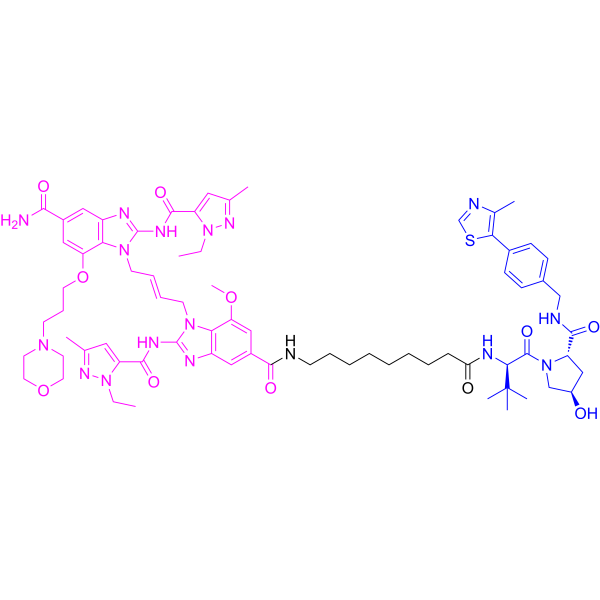
UNC9036 is a STING PROTAC degrader with a DC50 value of 227 nM. UNC9036 binds to and activates STING, recruits the VHL E3 ligase to target phosphorylated STING for ubiquitination and proteasomal degradation. UNC9036 inhibits downstream innate immune signaling events triggered by cytoplasmic DNA sensing, including IRF3 phosphorylation. UNC9036 reduces the antiviral response of cells infected with HSV-1. UNC9036 can be used in the research of renal cell carcinoma.
(Pink: STING ligand (HY-184545); Blue: VHL ligand (HY-138678B); Black: linker (HY-W105727)).
For research use only. We do not sell to patients.
- CAS No.: 3094059-54-5
- Formula: C73H95N17O11S
- Molecular Weight:1418.71
-
Storage:
Please store the product under the recommended conditions in the Certificate of Analysis.

To place orders, for customer services and technical support, please contact: MedChemExpress USA
Tel: 609-228-6898 E-mail: [email protected] [email protected]


-
 Biological Activity
-
Chemical Information
- Protocols
- Purity & Documentation
- References
-
 Help & FAQs


All PROTACs Isoforms
More

Biological Activity
Description
IC50 & Target
[1]|
VHL |
HSV-1 |
In Vitro
UNC9036 (5 μM; 24 h) degrades wild-type, cGAMP binding-deficient (R238A), and TBK1 phosphorylation-deficient (S366A) STING, but does not degrade trafficking-deficient (EDAA) or palmitoylation/activation-deficient (CACA) STING in Caki-1 renal cell carcinoma cells[1].
MedChemExpress (MCE) has not independently confirmed the accuracy of these methods. They are for reference only.
-
Cell Line:Caki-1 renal cell carcinoma cells
-
Concentration:0-31.6 μM (dose-response); 1 μM (time-course)
-
Incubation Time:24 h (dose-response); 0-24 h (time-course)
-
Result:Triggered dose-dependent and time-dependent degradation of STING protein.
Achieved a DC50 of 227 nM and a Dmax of 91.68% after 24 h treatment.
Induced significant STING degradation within 8 h of treatment with 1 μM.
-
Cell Line:Caki-1 renal cell carcinoma cells (transfected with Flag-tagged wild-type or mutant STING)
-
Concentration:5 μM
-
Incubation Time:24 h
-
Result:Degraded wild-type STING, R238A (2'3'-cGAMP binding-deficient) mutant, and S366A (TBK1 phosphorylation-deficient) mutant.
Failed to degrade EDAA (trafficking-deficient) mutant and CACA (palmitoylation/activation-deficient) mutant.
-
Cell Line:Caki-1 renal cell carcinoma cells
-
Concentration:0.316 μM UNC9036; 5 μg/mL ISD90, 5 μg/mL 2'3'-cGAMP, 5 μg/mL poly(I:C)
-
Incubation Time:6 h pre-incubation; 0-6 h stimulation
-
Result:Reduced STING protein abundance.
Suppressed ISD90-induced and 2'3'-cGAMP-induced phosphorylated IRF3 signals.
Minimally affected poly(I:C)-induced phosphorylated IRF3 signals.


Chemical Information
-
CAS No. 3094059-54-5
-
Molecular Weight 1418.71
-
Formula C73H95N17O11S
-
SMILES
O=C(NCCCCCCCCC(N[C@@H](C(N1[C@H](C(NCC2=CC=C(C=C2)C3=C(C)N=CS3)=O)C[C@H](C1)O)=O)C(C)(C)C)=O)C4=CC5=C(C(OC)=C4)N(C/C=C/CN6C(NC(C7=CC(C)=NN7CC)=O)=NC8=C6C(OCCCN9CCOCC9)=CC(C(N)=O)=C8)C(NC(C%10=CC(C)=NN%10CC)=O)=N5
-
Shipping
Room temperature in continental US; may vary elsewhere.
-
Storage
Please store the product under the recommended conditions in the Certificate of Analysis.


Purity & Documentation
References

Calculators
Concentration (start) × Volume (start) = Concentration (final) × Volume (final)


UNC9036 Related Classifications
HY-158048 Related Classifications
 Powered by Bioz