



SEW2871
Based on 11 publication(s) in Google Scholar
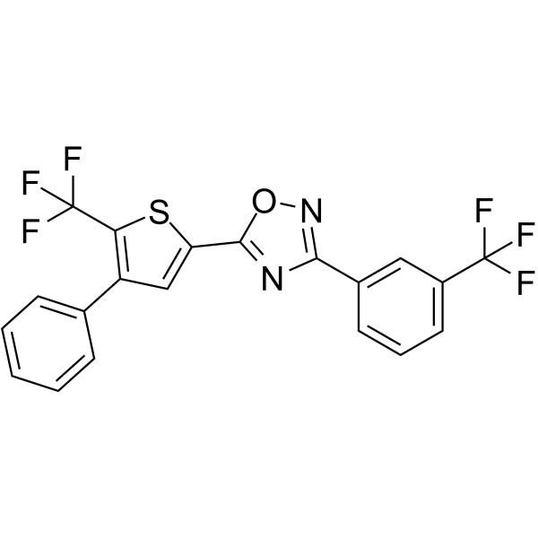
SEW2871 is an orally active, potent, highly selective S1P1 (sphingosine-1-phosphate type 1 receptor) agonist, with an EC50 of 13.8 nM. SEW2871 activates ERK, Akt, and Rac signaling pathways and induces S1P1 internalization and recycling. SEW2871 reduces lymphocyte numbers in blood. SEW2871 can be used for the research of diabetes, Alzheimer’s disease, liver fibrosis, and inflammatory responses.
For research use only. We do not sell to patients.
- Purity : 99.88%
- CAS No.: 256414-75-2
- Formula: C20H10F6N2OS
- Molecular Weight:440.36
-
Storage:Powder -20°C, 3 years ; In solvent -80°C, 6 months , -20°C, 1 month

To place orders, for customer services and technical support, please contact: MedChemExpress USA
Tel: 609-228-6898 E-mail: [email protected] [email protected]


-
 Biological Activity
-
Chemical Information
-
Solvent & Solubility
- Protocols
- Purity & Documentation
- References
-
 Help & FAQs


-
HY-101395
W146S1PR1 Antagonist -
HY-10569
PonesimodS1P1 Agonist -
HY-108496
Sphingosine-1-phosphateS1PR Agonist -
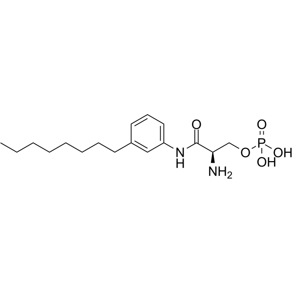
HY-108490
VPC 23019S1P1/3 Antagonist -
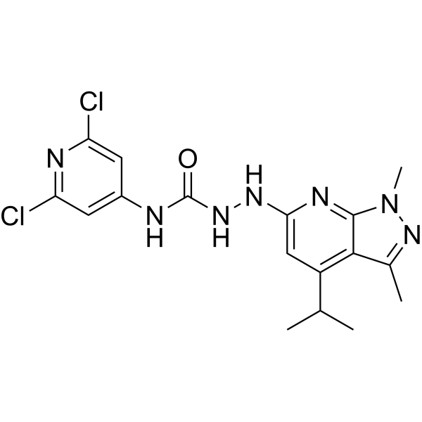
HY-100675
JTE-013S1P2 Antagonist -
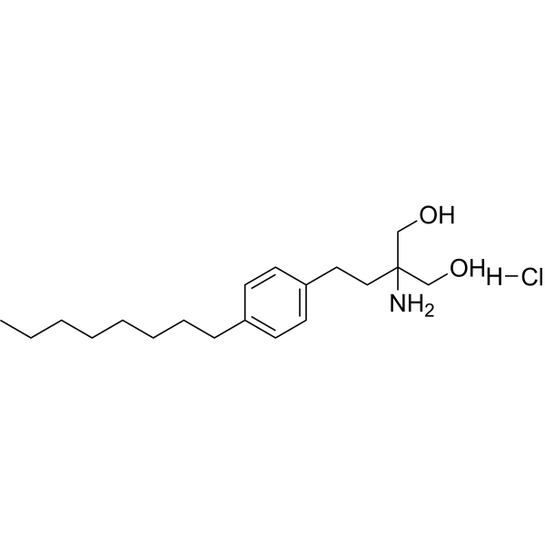
HY-12005
Fingolimod hydrochlorideS1P Receptors Modulator -
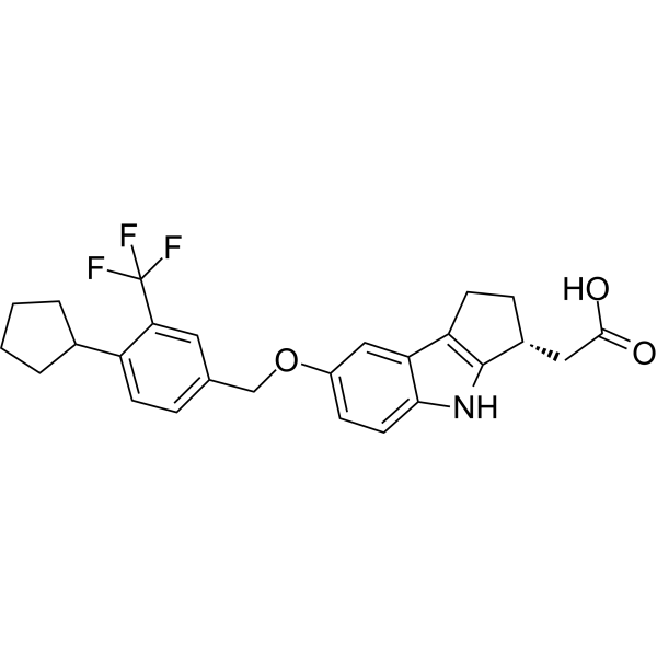
HY-12789
EtrasimodS1P1 Receptor Antagonist -
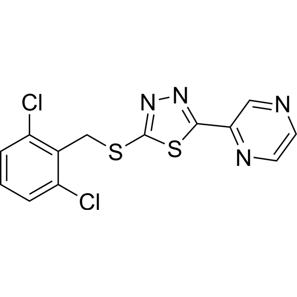
HY-18723
Yoda 1Piezo1 Agonist -
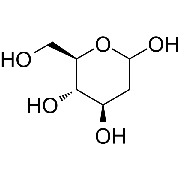
HY-13966
2-Deoxy-D-glucoseGlycolysis Inhibitor -
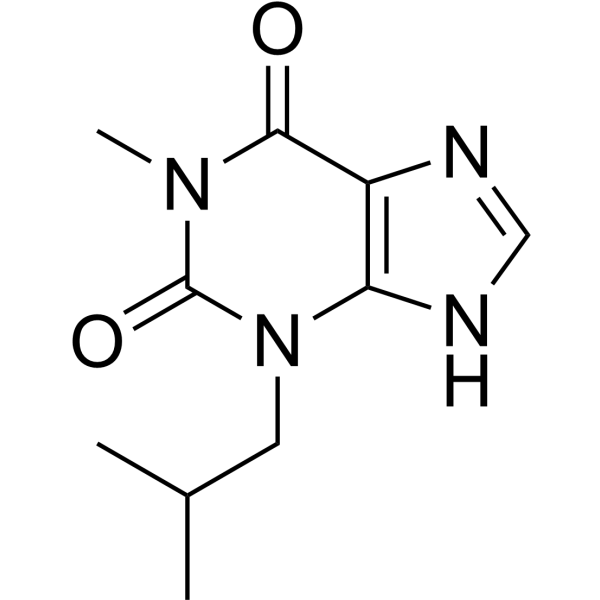
HY-12318
IBMXPDE Inhibitor -
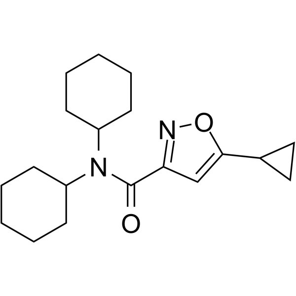
HY-101419
CYM-5541S1P3 Receptor Agonist -

HY-Y0320
Dimethyl sulfoxide, meets analytical specification of Ch.P.Aprotic Solvent
Publications Citing Use of MedChemExpress (MCE) SEW2871
More- Nat Commun. 2024 Sep 6;15(1):7791. [Abstract]
- Adv Sci (Weinh). 2026 Apr;13(24):e19500. [Abstract]
- J Renin Angiotensin Aldosterone Syst. 2025 Oct 16.
- Neurosci Bull. 2025 Jun;41(6):951-969. [Abstract]
- Am J Physiol Cell Physiol. 2023 Nov 1;325(5):C1354-C1368. [Abstract]
- iScience. 2024 Oct 30;27(12):111290. [Abstract]
- Naunyn Schmiedebergs Arch Pharmacol. 2026 May 29. [Abstract]
- Reprod Biomed Online. 2023 Nov;47(5):103304. [Abstract]
- bioRxiv. 2026 Jun 29.
- bioRxiv. 2024 Jul 26:2024.07.26.605282. [Abstract]
- Research Square Preprint. 2023 Dec 1.
 Customer Validation & Images

-
In Vivo Imaging

-
In Vivo Efficacy Study

-
In Vivo Efficacy Study

-
IF

-
WB


Biological Activity
Description
IC50 & Target
|
S1PR1 13.8 nM (EC50) |
Cellular Effect
|
Cell Line

|
Type | Value | Description | References |
|---|---|---|---|---|
| CHO | IC50 |
>10000 nM
Compound: 10
|
Displacement of [33P]sphingosine 1 phosphate from human S1P3 receptor expressed in CHO cells
Displacement of [33P]sphingosine 1 phosphate from human S1P3 receptor expressed in CHO cells
|
[PMID: 15615513] |
| CHO | IC50 |
>10000 nM
Compound: 10
|
Displacement of [33P]sphingosine 1 phosphate from human S1P4 receptor expressed in CHO cells
Displacement of [33P]sphingosine 1 phosphate from human S1P4 receptor expressed in CHO cells
|
[PMID: 15615513] |
| CHO | IC50 |
37 nM
Compound: 10
|
Displacement of [33P]sphingosine 1 phosphate from human S1P1 receptor expressed in CHO cells
Displacement of [33P]sphingosine 1 phosphate from human S1P1 receptor expressed in CHO cells
|
[PMID: 15615513] |
| CHO | IC50 |
4600 nM
Compound: 10
|
Displacement of [33P]sphingosine 1 phosphate from human S1P5 receptor expressed in CHO cells
Displacement of [33P]sphingosine 1 phosphate from human S1P5 receptor expressed in CHO cells
|
[PMID: 15615513] |
| CHO | IC50 |
>10000 nM
Compound: 10
|
Displacement of [33P]sphingosine 1 phosphate from human S1P2 receptor expressed in CHO cells
Displacement of [33P]sphingosine 1 phosphate from human S1P2 receptor expressed in CHO cells
|
[PMID: 15615513] |
| CHO | IC50 |
>10 nM
Compound: SEW-2871
|
Displacement of [33P]S1P from human S1P3R expressed in CHO cell membranes
Displacement of [33P]S1P from human S1P3R expressed in CHO cell membranes
|
[PMID: 24900286] |
| CHO | IC50 |
37 nM
Compound: SEW-2871
|
Displacement of [33P]S1P from human S1P1R expressed in CHO cell membranes
Displacement of [33P]S1P from human S1P1R expressed in CHO cell membranes
|
[PMID: 24900286] |
| CHO-K1 | EC50 |
>25 μM
Compound: 3, SEW2871
|
Agonist activity at human S1P3 receptor expressed in CHO-K1 cells co-expressing Gq/i5 G-protein assessed as calcium mobilization by FLIPR assay
Agonist activity at human S1P3 receptor expressed in CHO-K1 cells co-expressing Gq/i5 G-protein assessed as calcium mobilization by FLIPR assay
|
[PMID: 22104144] |
| U2OS | EC50 |
0.27 μM
Compound: 3, SEW2871
|
Agonist activity at human S1P1 receptor expressed in human U2OS cells co-expressing eGFP assessed as receptor internalization into cytoplasm using Hoechst dye staining
Agonist activity at human S1P1 receptor expressed in human U2OS cells co-expressing eGFP assessed as receptor internalization into cytoplasm using Hoechst dye staining
|
[PMID: 22104144] |
In Vitro
SEW2871 exerts a powerful migratory effect by increasing smooth muscle α-actin, procollagen αI and αIII, and total hydroxyproline contents in LX-2 cells (a human hepatic stellate cell line)[2].
MedChemExpress (MCE) has not independently confirmed the accuracy of these methods. They are for reference only.
In Vivo
? SEW2871 (0.5 mg/kg, IP, daily for 2 weeks) inhibited β amyloid (Aβ1-42)-induced spatial memory impairment and hippocampal neuronal loss in a rat model of Alzheimer’s disease[2].
? SEW2871 (0-0.3 mg/kg, IV) attenuates LPS-induced acute inflammatory lung injury, produces dose-dependent alveolar and vascular barrier protection in C57Bl/6 mice[2].
? SEW2871 is effective in protecting kidneys against ischaemia-reperfusion injury by reducing CD4+ T cell infiltration in mice[3].
MedChemExpress (MCE) has not independently confirmed the accuracy of these methods. They are for reference only.
-
Animal Model:IL-10–/– (interleukin (IL)-10 gene-deficient) mice, a murine model of Crohn's disease (CD)[3]
-
Dosage:20 mg/kg
-
Administration:Gavage, once daily for 2 weeks
-
Result:Ameliorated established colitis in IL-10–/– mice, associated with a reduction of serum amyloid A concentration, a decreased colon MPO concentration, a depletion of the peripheral CD4+CD45+ T cells and a reduction of the homing of T cells into colon LP. Suppressed typical cytokines of T helper type 1 (Th1) and Th17 cells and p-STAT-3 expression, and significantly decreased TNF-α, IFN-γ, IL-1β and IL-17A mRNA levels.


Chemical Information
-
CAS No. 256414-75-2
-
Appearance Solid
-
Molecular Weight 440.36
-
Formula C20H10F6N2OS
-
Color White to light yellow
-
SMILES
FC(C1=CC(C2=NOC(C3=CC(C4=CC=CC=C4)=C(C(F)(F)F)S3)=N2)=CC=C1)(F)F
-
Shipping
Room temperature in continental US; may vary elsewhere.
-
Storage
Powder -20°C 3 years In solvent -80°C 6 months -20°C 1 month


Publications (11)
-
Journal Impact Factor
-
Most Recent
-
Nat Commun
Astrocyte-derived clusterin disrupts glial physiology to obstruct remyelination in mouse models of demyelinating diseases. [Abstract]2024 Sep 6;15(1):7791. PMID: 39242637 -
Adv Sci (Weinh)
Diabetes Mellitus Facilitates Gallstone Formation Through CXCR2-NETs-Mediated Liver-Bile Barrier Damage. [Abstract]2026 Apr;13(24):e19500. PMID: 41698066
SEW2871 purchased from MedChemExpress. Usage Cited in: Adv Sci (Weinh). 2026 Apr;13(24):e19500. [Abstract]
Representative images of gallbladders from mice fed a lithogenic diet (LD), lithogenic diet with SEW2871 (0.5 mg/kg, daily, 4 weeks) intraperitoneal injection (LD+SEW2871), diabetic lithogenic diet (DMLD) and diabetic lithogenic diet with SEW2871 intraperitoneal injection.
-
-
Neurosci Bull
Alpha-synuclein Fibrils Inhibit Activation of the BDNF/ERK Signaling Loop in the mPFC to Induce Parkinson's Disease-like Alterations with Depression. [Abstract]2025 Jun;41(6):951-969. PMID: 39609371
SEW2871 purchased from MedChemExpress. Usage Cited in: Neurosci Bull. 2025 Jun;41(6):951-969. [Abstract]
In the forced swimming test, the rats in the SEW2871 group swam for a longer duration and spent less time immobilized. Rats were given SEW2871 (0.5 mg/kg) or the isovolumetric solvent daily by intraperitoneal injection for 14 days after mode completion. Meanwhile, SEW2871 (1 μmol/L) or isovolumetric solvent was added to the primary PFC neurons and incubated for 48 h, after which tests were carried out.
SEW2871 purchased from MedChemExpress. Usage Cited in: Neurosci Bull. 2025 Jun;41(6):951-969. [Abstract]
In the new object recognition test, the rats in the SEW2871 group spent more time exploring the new object and had a higher recognition index than those in the PFF group.
SEW2871 purchased from MedChemExpress. Usage Cited in: Neurosci Bull. 2025 Jun;41(6):951-969. [Abstract]
We applied immunofluorescence staining to the mPFC region and found that treatment with SEW2871 partially restored the expression levels of BDNF.
SEW2871 purchased from MedChemExpress. Usage Cited in: Neurosci Bull. 2025 Jun;41(6):951-969. [Abstract]
SEW2871 upregulates the levels of phosphorylation of ERK and its downstream proteins. The Lewy body-like inclusions showed the opposite alterations.

SEW2871 purchased from MedChemExpress. Usage Cited in: Neurosci Bull. 2025 Jun;41(6):951-969. [Abstract]
SEW2871 somewhat mitigates the decrease in DA content.
-
Am J Physiol Cell Physiol
Glycolytic enzyme PFKFB3 regulates sphingosinae 1-phosphate receptor 1 in proangiogenic glomerular endothelial cells under diabetic condition. [Abstract]2023 Nov 1;325(5):C1354-C1368. PMID: 37781737 -
iScience
Broad and diverse roles of sphingosine-1-phosphate/sphingosine-1-phosphate receptors in the prostate. [Abstract]2024 Oct 30;27(12):111290. PMID: 39618500 -
Naunyn Schmiedebergs Arch Pharmacol
Label-free cell phenotypic profiling of sphingosine-1-phosphate receptor 1 and discovery of its agonist from natural products. [Abstract]2026 May 29. PMID: 42209736 -
Reprod Biomed Online
Blocking sphingosine 1-phosphate receptor 1 with modulators reduces immune cells infiltration and alleviates endometriosis in mice. [Abstract]2023 Nov;47(5):103304. PMID: 37757611 -
-
bioRxiv
2024 Jul 26:2024.07.26.605282. PMID: 39091869 -


Solvent & Solubility
In Vitro:
DMF : 100 mg/mL (227.09 mM; Need ultrasonic)
DMSO : 25 mg/mL (56.77 mM; Need ultrasonic; Hygroscopic DMSO has a significant impact on the solubility of product, please use newly opened DMSO)
Ethanol : 20 mg/mL (45.42 mM; Need ultrasonic)
Please refer to the solubility information to select the appropriate solvent. Once prepared, please aliquot and store the solution to prevent product inactivation from repeated freeze-thaw cycles.
Storage method and period of stock solution: -80°C, 6 months; -20°C, 1 month. When stored at -80°C, please use it within 6 months. When stored at -20°C, please use it within 1 month.
Please refer to the solubility information to select the appropriate solvent. Once prepared, please aliquot and store the solution to prevent product inactivation from repeated freeze-thaw cycles.
Storage method and period of stock solution: -80°C, 6 months; -20°C, 1 month. When stored at -80°C, please use it within 6 months. When stored at -20°C, please use it within 1 month.
Concentration (start) × Volume (start) = Concentration (final) × Volume (final)
In Vivo:
Select the appropriate dissolution method based on your experimental animal and administration route.
- For the following dissolution methods, please ensure to first prepare a clear stock solution using an In Vitro approach and then sequentially add co-solvents:
- To ensure reliable experimental results, the clarified stock solution can be appropriately stored based on storage conditions. As for the working solution for In Vivo experiments, it is recommended to prepare freshly and use it on the same day.
- The percentages shown for the solvents indicate their volumetric ratio in the final prepared solution. If precipitation or phase separation occurs during preparation, heat and/or sonication can be used to aid dissolution.
Add each solvent one by one: 10% DMSO 40% PEG300 5% Tween-80 45% Saline
Solubility: ≥ 2.08 mg/mL (4.72 mM); Clear solution
This protocol yields a clear solution of ≥ 2.08 mg/mL (saturation unknown).
Taking 1 mL working solution as an example, add 100 μL DMSO stock solution (20.8 mg/mL) to 400 μL PEG300, and mix evenly; then add 50 μL Tween-80 and mix evenly; then add 450 μL Saline to adjust the volume to 1 mL.
Preparation of Saline: Dissolve 0.9 g sodium chloride in ddH₂O and dilute to 100 mL to obtain a clear Saline solution.
Add each solvent one by one: 10% EtOH 90% Corn Oil
Solubility: ≥ 2 mg/mL (4.54 mM); Clear solution
This protocol yields a clear solution of ≥ 2 mg/mL (saturation unknown). If the continuous dosing period exceeds half a month, please choose this protocol carefully.
Taking 1 mL working solution as an example, add 100 μL EtOH stock solution (20.0 mg/mL) to 900 μL Corn oil, and mix evenly.
In Vivo Dissolution Calculator
Please enter the basic information of animal experiments:
-
-
-
-
Recommended: Prepare an additional quantity of animals to account for potential losses during experiments.
Please enter your animal formula composition:
-
%DMSO +
Recommended: Keep the proportion of DMSO in working solution below 2% if your animal is weak.
-
%+
-
+%Tween-80 + +
-
%Saline +
The co-solvents required include: DMSO, . All of co-solvents are available by MedChemExpress (MCE). , Tween 80. All of co-solvents are available by MedChemExpress (MCE).
Working solution concentration: 0.22 mg/mL
Method for preparing stock solution: mg drug dissolved in μL DMSO. Stock solution concentration: mg/mL.
1. Take μL DMSO stock solution;
2. Add μL .
μL , mix evenly;
3. Then add μL Tween 80, mix evenly;
4. Then add μL
Please ensure that the stock solution in the first step is dissolved to a clear state, and add co-solvents in sequence. You can use ultrasonic heating (ultrasonic cleaner, recommended frequency 20-40 kHz), vortexing, etc. to assist dissolution.


Purity & Documentation
-
Data Sheet (280 KB)
-
SDS (479 KB)
- English - EN (479 KB)
- Français - FR (479 KB)
- Deutsch - DE (479 KB)
- Norwegian - NO (479 KB)
- Español - ES (479 KB)
- Swedish - SV (479 KB)
- Italian - IT (479 KB)
- Korean - KR (479 KB)
- Portuguese - PT (479 KB)
-
Handling Instructions (2659 KB)
References
[1]. Park SJ, et al. Sphingosine 1-Phosphate Receptor Modulators and Drug Discovery. Biomol Ther (Seoul). 2017 Jan 1;25(1):80-90. [Content Brief]
[2]. Jo E, et al. S1P1-selective in vivo-active agonists from high-throughput screening: off-the-shelf chemical probes of receptor interactions, signaling, and fate. Chem Biol. 2005 Jun;12(6):703-15. [Content Brief]
[3]. Dong J, et al. Oral treatment with SEW2871, a sphingosine-1-phosphate type 1 receptor agonist, ameliorates experimental colitis in interleukin-10 gene deficient mice. Clin Exp Immunol. 2014 Jul;177(1):94-101. [Content Brief]


Complete Stock Solution Preparation Table
Please refer to the solubility information to select the appropriate solvent. Once prepared, please aliquot and store the solution to prevent product inactivation from repeated freeze-thaw cycles.
Storage method and period of stock solution: -80°C, 6 months; -20°C, 1 month. When stored at -80°C, please use it within 6 months. When stored at -20°C, please use it within 1 month.
| Optional Solvent | Concentration Solvent Mass | 1 mg | 5 mg | 10 mg | 25 mg |
|---|---|---|---|---|---|
| Ethanol / DMSO / DMF | 1 mM | 2.2709 mL | 11.3543 mL | 22.7087 mL | 56.7717 mL |
| 5 mM | 0.4542 mL | 2.2709 mL | 4.5417 mL | 11.3543 mL | |
| 10 mM | 0.2271 mL | 1.1354 mL | 2.2709 mL | 5.6772 mL | |
| 15 mM | 0.1514 mL | 0.7570 mL | 1.5139 mL | 3.7848 mL | |
| 20 mM | 0.1135 mL | 0.5677 mL | 1.1354 mL | 2.8386 mL | |
| 25 mM | 0.0908 mL | 0.4542 mL | 0.9083 mL | 2.2709 mL | |
| 30 mM | 0.0757 mL | 0.3785 mL | 0.7570 mL | 1.8924 mL | |
| 40 mM | 0.0568 mL | 0.2839 mL | 0.5677 mL | 1.4193 mL | |
| DMSO / DMF | 50 mM | 0.0454 mL | 0.2271 mL | 0.4542 mL | 1.1354 mL |
| DMF | 60 mM | 0.0378 mL | 0.1892 mL | 0.3785 mL | 0.9462 mL |
| 80 mM | 0.0284 mL | 0.1419 mL | 0.2839 mL | 0.7096 mL | |
| 100 mM | 0.0227 mL | 0.1135 mL | 0.2271 mL | 0.5677 mL |


SEW2871 Related Classifications
HY-W008947 Related Classifications
 Powered by Bioz