VTP50469 mesylate
Based on 13 publication(s) in Google Scholar
VTP50469 mesylate is a potent, and selective Menin-MLL1 inhibitor that effectively targets MLL-rearranged and NPM1c+ leukemia. VTP50469 mesylate selectively kills cell lines with MLL rearrangements and NPM1c+ mutations. VTP50469 mesylate displaces Menin from protein complexes and inhibits MLL's chromatin occupancy at specific genes, leading to significant changes in gene expression, differentiation, and apoptosis. VTP50469 demonstrates dramatic reductions in leukemia burden in patient-derived xenograft models of MLL-r acute myeloid leukemia and MLL-r acute lymphoblastic leukemia, with some mice remaining disease-free for over a year post-treatment.
Nos produits utilisent uniquement pour la recherche. Nous ne vendons pas aux patients.
- CAS No.: 2169919-27-9
- Formule: C34H55FN6O10S3
- Masse moléculaire:823.03
-
Stockage:
Please store the product under the recommended conditions in the Certificate of Analysis.
Publications Citing Use of MedChemExpress (MCE) VTP50469 mesylate
More- Blood. 2026 Feb 24:blood.2025031486. [Abstract]
- Blood. 2024 Nov 7;144(19):2018-2032. [Abstract]
- Nat Cell Biol. 2023 Sep;25(9):1346-1358. [Abstract]
- Nat Commun. 2025 Mar 18;16(1):2641. [Abstract]
- Blood Cancer J. 2022 Jan 11;12(1):5. [Abstract]
- Leukemia. 2026 Mar 25. [Abstract]
- Leukemia. 2025 Jan;39(1):75-86. [Abstract]
- Blood Adv. 2026 Jul 14;10(13):4757-4771. [Abstract]
- Int J Oncol. 2020 Oct;57(4):1057-1071. [Abstract]
- bioRxiv. 2026 Jun 19.
- bioRxiv. 2026 Feb 17.
- bioRxiv. 2025 Nov 12:2025.11.10.687648. [Abstract]
- bioRxiv. 2023 Oct 1.
-
Cell Proliferation/Viability Assay
-
Cell Proliferation/Viability Assay
-
Cell Proliferation/Viability Assay
-
WB
-
WB
Activité biologique
Description
Chemical Information
-
CAS No. 2169919-27-9
-
Masse moléculaire 823.03
-
Formule C34H55FN6O10S3
-
SMILES
CS(=O)(O)=O.CC(C)N(C(C)C)C(C(C=C(C=C1)F)=C1OC2=CN=CN=C2N(C3)CC3(CC4)CCN4C[C@@H]5CC[C@H](CC5)NS(C)(=O)=O)=O.CS(=O)(O)=O
-
Synonyms
SNDX-50469 mesylate
-
Livraison
Room temperature in continental US; may vary elsewhere.
-
Stockage
Please store the product under the recommended conditions in the Certificate of Analysis.
Publications (13)
-
Journal Impact Factor
-
Most Recent
-
Blood
Overcoming menin inhibitor resistance in AML cells with combinations including BET proteins and a dual BRG1/BRM inhibitor. [Abstract]2026 Feb 24:blood.2025031486. PMID: 41734382 -
Blood
Epigenetic Regulation of Non-canonical Menin Targets Modulates Menin Inhibitor Response in Acute Myeloid Leukemia. [Abstract]2024 Nov 7;144(19):2018-2032. PMID: 39158067 -
Nat Cell Biol
2023 Sep;25(9):1346-1358. PMID: 37591951
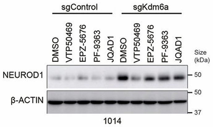
VTP50469 mesylate purchased from MedChemExpress. Usage Cited in: Nat Cell Biol. 2023 Sep;25(9):1346-1358. [Abstract]
Immunoblot analysis of 1014 Kdm6a isogenic cells treated with inhibitors that block the function of epigenetic modifiers that normally maintain gene expression including VTP50469 (500 nM), EPZ-5676 (1 μM), PF-9363 (100 nM), JQAD1 or DMSO for 6 days.
-
Nat Commun
Guanine nucleotide biosynthesis blockade impairs MLL complex formation and sensitizes leukemias to menin inhibition. [Abstract]2025 Mar 18;16(1):2641. PMID: 40102405
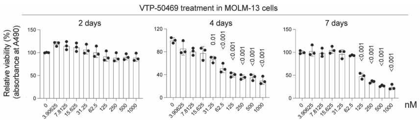
VTP50469 mesylate purchased from MedChemExpress. Usage Cited in: Nat Commun. 2025 Mar 18;16(1):2641. [Abstract]
Relative viability of MOLM-13 cells following treatment with different concentrations of VTP50469 (3.91-1000 nM) over 2-7 days.
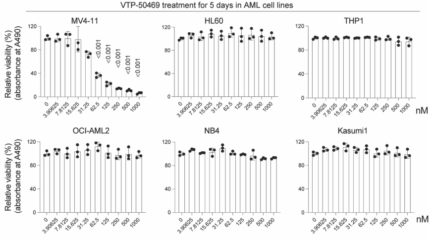
VTP50469 mesylate purchased from MedChemExpress. Usage Cited in: Nat Commun. 2025 Mar 18;16(1):2641. [Abstract]
Relative viability of human AML cell lines upon treatment with different concentrations of VTP50469 (3.91-1000 nM) over 5 days.
-
Blood Cancer J
Effective Menin inhibitor-based combinations against AML with MLL rearrangement or NPM1 mutation (NPM1c). [Abstract]2022 Jan 11;12(1):5. PMID: 35017466
VTP50469 mesylate purchased from MedChemExpress. Usage Cited in: Blood Cancer J. 2022 Jan 11;12(1):5. [Abstract]
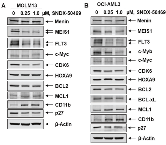
A, B MOLM13 and OCI-AML3 cells were treated with the indicated concentrations of VTP50469 (SNDX-50469; 0, 0.25, 1 μM) for 48 h. Following this, total cell lysates were prepared and immunoblot analyses were conducted. The expression levels of β-Actin in the lysates served as the loading control.
-
Leukemia
A Perturb-seq map of a differentiation hub reveals synergistic vulnerabilities in KMT2A-rearranged acute myeloid leukemia. [Abstract]2026 Mar 25. PMID: 41882099 -
Leukemia
NPM1-fusion proteins promote myeloid leukemogenesis through XPO1-dependent HOX activation. [Abstract]2025 Jan;39(1):75-86. PMID: 39443736
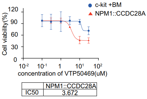
VTP50469 mesylate purchased from MedChemExpress. Usage Cited in: Leukemia. 2025 Jan;39(1):75-86. [Abstract]
NPM1::CCDC28A cells and mouse cKit+ bone marrow cells were incubated with selinexor or VTP50469 (0, 0.1, 0.3, 1, 3, 10, and 30 μM) at the indicated concentration for 72 h. Cell viability was assessed using the Cell Counting Kit-8.
-
Blood Adv
Combining menin and MEK inhibition to target poor prognosis KMT2A-rearranged RAS pathway-mutant acute myeloid leukemia. [Abstract]2026 Jul 14;10(13):4757-4771. PMID: 42085603 -
Int J Oncol
Menin‑MLL inhibitors induce ferroptosis and enhance the anti‑proliferative activity of auranofin in several types of cancer cells. [Abstract]2020 Oct;57(4):1057-1071. PMID: 32945449 -
-
-
bioRxiv
2025 Nov 12:2025.11.10.687648. PMID: 41292793 -
Protocole
-
RT-PCR
Reverse transcription technology uses RNA as a template to synthesize DNA. RT-PCR is simple, specific and sensitive, and can be used to detect gene expression levels and expression differences in cells; detect RNA virus content; clone cDNA sequences of specific genes.
-
RNA extraction experimental
By lysing cells, releasing RNA, and removing impurities such as proteins and DNA, high-purity RNA products are finally obtained. The commonly used traditional method is the guanidine isothiocyanate/phenol/chloroform method (Trizol), which is suitable for a variety of animal materials including animal tissues, microorganisms, cultured cells, etc., and most plant materials.
-
Annexin V plus membrane-impermeant dye apoptosis staining
Annexin V-based apoptosis assays rely on the detection of phosphatidylserine (PS) externalization from the inner leaflet of the plasma membrane to the outer leaflet, an early biochemical hallmark of apoptosis. Fluorescently labeled Annexin V binds PS in a calcium-dependent manner, enabling identification of early apoptotic cells by flow cytometry or fluorescence microscopy. When combined with a membrane-impermeant DNA-binding dye (e. g. , propidium iodide), this approach allows discrimination between viable (Annexin V−/dye−), early apoptotic (Annexin V+/dye−), and late apoptotic or necrotic (Annexin V+/dye+) cell populations by assessing membrane integrity and PS exposure.
-
Patient-Derived Orthotopic Xenograft (PDOX)
Patient-derived orthotopic xenograft (PDOX) modeling implants fresh patient tumor tissue or patient-derived tumor cells into the anatomically corresponding organ or tissue site of immunodeficient mice, usually by surgical orthotopic implantation, to preserve patient tumor histology, local microenvironmental context, invasion, metastatic behavior, and treatment-response features better than subcutaneous implantation. PDOX readouts include tumor engraftment, orthotopic tumor growth, local invasion, metastasis, recurrence after resection, histologic similarity to the donor tumor, biomarker retention, molecular concordance, survival, and response or resistance to therapy. PDOX models are used for preclinical drug testing and individualized therapy evaluation, but engraftment success varies by tumor type and specimen quality.
-
Real Time qPCR (Q-PCR)
Real-time quantitative PCR (qPCR) quantifies an amplifiable nucleic-acid target by monitoring fluorescence during PCR cycling rather than measuring product only after amplification. The increase in fluorescence tracks accumulation of PCR product, and the quantification cycle (Cq; historically also Ct/CP) is related to the initial amount of target: samples containing more starting target generally reach the defined fluorescence threshold in fewer cycles.
-
Apoptosis
Apoptosis, also called programmed cell death, is generally characterized by distinct morphological characteristics.
-
TUNEL staining for apoptotic DNA fragmentation
TUNEL staining detects DNA strand breaks by using terminal deoxynucleotidyl transferase to add labeled nucleotides to exposed 3′-OH DNA termini, generating either microscopic staining in fixed cells or tissue sections, or fluorescence/cytometric signal in cell suspensions. TUNEL positivity reflects DNA fragmentation but should not be interpreted alone as definitive apoptosis, because TUNEL can also label necrotic, autolytic, mechanically damaged, or DNA-repair-associated DNA breaks.
-
Patient-Derived Xenograft (PDX)
Patient-derived xenograft (PDX) models are generated by engrafting primary human tumor tissue directly into immunodeficient mice, allowing in vivo propagation of patient tumor biology without initial in vitro adaptation. These models are used to preserve key histopathological and molecular characteristics of the original tumor and enable assessment of tumor growth dynamics and therapeutic response in a living organism. The biological readout is tumor engraftment and subsequent growth in the murine host, which reflects the ability of human tumor cells to survive, vascularize, and expand in an immunocompromised microenvironment.
-
Apoptosis Solutions
Apoptosis is a regulated, generally non-lytic cell-death pathway that removes unwanted, damaged, infected, or abnormal cells through coordinated morphological changes, caspase activation, DNA fragmentation, and membrane remodeling. The intrinsic apoptosis pathway is controlled mainly by mitochondrial outer membrane permeabilization, BCL-2 family proteins, cytochrome c release, apoptosome formation, caspase-9 activation, and downstream executioner caspase-3/7 activation. The extrinsic apoptosis pathway is initiated by death receptors such as Fas, TNFR, and TRAIL receptors, which recruit adaptor proteins and activate caspase-8 before engaging executioner caspases or mitochondrial amplification through BID cleavage. Apoptosis is linked to many phenotypes, including cancer cell killing, tissue homeostasis, immune regulation, neurodegeneration, infection response, and treatment-induced cytotoxicity; unresolved questions include how apoptosis interacts with necroptosis, pyroptosis, ferroptos
-
Splenic/Portal-Vein Liver Metastasis Xenograft
Splenic and portal-vein liver metastasis xenograft models deliver tumor cells into the portal circulation so that cells reach the liver first and form hepatic metastatic lesions; splenic injection uses the spleen as an access route to the portal system, while direct portal-vein injection introduces cells into the portal vein without requiring splenectomy. The assay detects liver colonization, intrahepatic tumor growth, tumor distribution, treatment response, survival, and liver-metastasis microenvironment changes; readouts include bioluminescence or fluorescence imaging, gross liver nodule counts, liver weight or tumor burden, histology, and survival.
-
Subcutaneous Cell-Line-Derived Xenograft
Subcutaneous cell-line-derived xenograft (CDX) models are established by implanting cultured human cancer cell lines into immunodeficient mice, where the injected cells form localized tumors that can be monitored in vivo as a measure of tumorigenic potential, growth kinetics, and treatment response. These models are widely used in oncology research because they allow reproducible tumor formation and enable comparative assessment of tumor growth between different cell lines or genetic manipulations in a controlled in vivo microenvironment. Subcutaneous implantation of cancer cells in immunodeficient mice is a standard approach for evaluating tumor growth behavior and therapeutic response across multiple cancer types, including prostate, esophageal, pancreatic, and colon cancer models.
-
Orthotopic Cell-Line Xenograft
Orthotopic cell-line xenograft models involve implantation of human cancer cell lines into the anatomically corresponding organ of immunodeficient mice to reproduce tumor growth within a native microenvironment, enabling more clinically relevant tumor behavior compared with subcutaneous models. These models are widely used because orthotopic placement better recapitulates tumor progression, including invasion and metastatic spread, which are often underrepresented in heterotopic implantation systems. Compared with conventional xenografts, orthotopic implantation is described as more technically complex but provides improved simulation of tumor-microenvironment interactions and metastatic behavior, making it particularly valuable for translational oncology research. Surgical orthotopic implantation approaches have been emphasized as enabling faithful reproduction of clinical cancer features, including metastasis and disease progression patterns that align with the tumor’s organ of origi
-
Intraperitoneal/Peritoneal Dissemination Xenograft
Intraperitoneal (IP) or peritoneal dissemination xenograft models are based on the introduction of human cancer cells into the peritoneal cavity of immunodeficient mice, where they attach to peritoneal surfaces, form multicellular aggregates or spheroids, and progressively generate disseminated tumor nodules that mimic advanced peritoneal metastatic disease. These models are widely used to study ovarian cancer progression, tumor-microenvironment interactions, and intraperitoneal therapeutic responses, often incorporating bioluminescence or fluorescence imaging to longitudinally monitor tumor burden in vivo. The biological principle relies on the capacity of tumor cells such as SKOV3 or related ovarian carcinoma lines to survive in suspension, aggregate within ascites-like fluid, adhere to mesothelial surfaces, and invade peritoneal organs, thereby recapitulating human peritoneal carcinomatosis patterns observed in advanced disease.
Pureté et documentation
Références
Calculators
Concentration (start) × Volume (start) = Concentration (final) × Volume (final)