



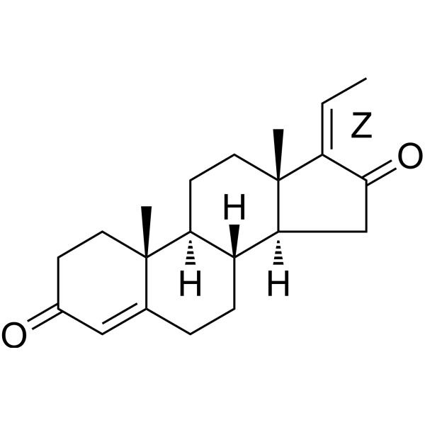
(Z)-Guggulsterone
Based on 8 publication(s) in Google Scholar
(Z)-Guggulsterone, a constituent of Indian Ayurvedic medicinal plant Commiphora mukul, inhibits the growth of human prostate cancer cells by causing apoptosis. (Z)-Guggulsterone inhibits angiogenesis by suppressing the VEGF–VEGF-R2–Akt signaling axis. (Z)-Guggulsterone is also a potent FXR antagonist. (Z)-Guggulsterone reduces ACE2 expression and SARS-CoV-2 infection.
For research use only. We do not sell to patients.
- Purity : 99.41%
- CAS No.: 39025-23-5
- Formula: C21H28O2
- Molecular Weight:312.45
-
Storage:Powder -20°C, 3 years , 4°C, 2 years ; In solvent -80°C, 6 months , -20°C, 1 month

To place orders, for customer services and technical support, please contact: MedChemExpress USA
Tel: 609-228-6898 E-mail: [email protected] [email protected]


-
 Biological Activity
-
Chemical Information
-
Solvent & Solubility
- Protocols
- Purity & Documentation
- References
-
 Help & FAQs


-
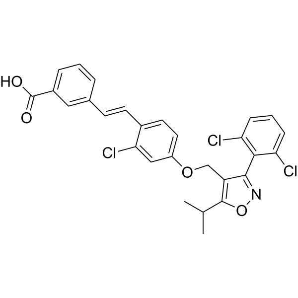
HY-50108
GW 4064FXR Agonist -
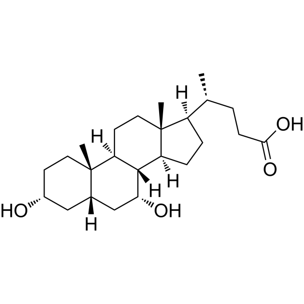
HY-76847
Chenodeoxycholic AcidEndogenous Metabolite -
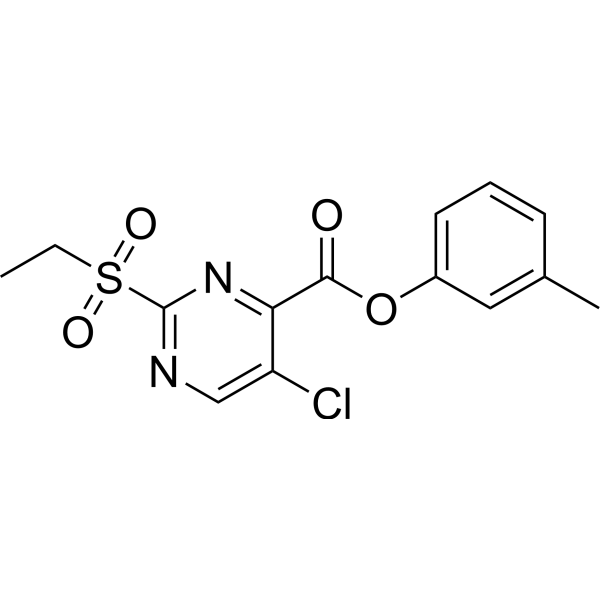
HY-111534
SBI-115TGR5 (GPCR19) Antagonist -
HY-12222
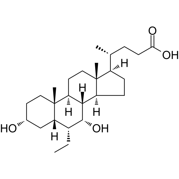
Obeticholic acidFXR Agonist -
HY-110267
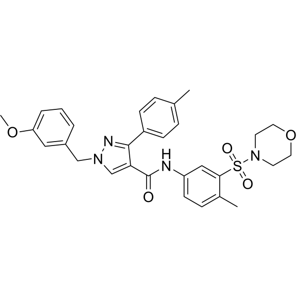
DY268FXR Antagonist -

HY-Y0320
Dimethyl sulfoxide, meets analytical specification of Ch.P.Aprotic Solvent -
HY-B0318
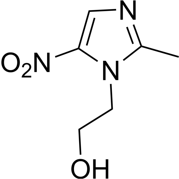
MetronidazoleAntibiotic -
HY-B0522
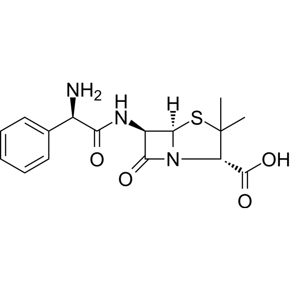
AmpicillinBacterial Inhibitor -
HY-B0671
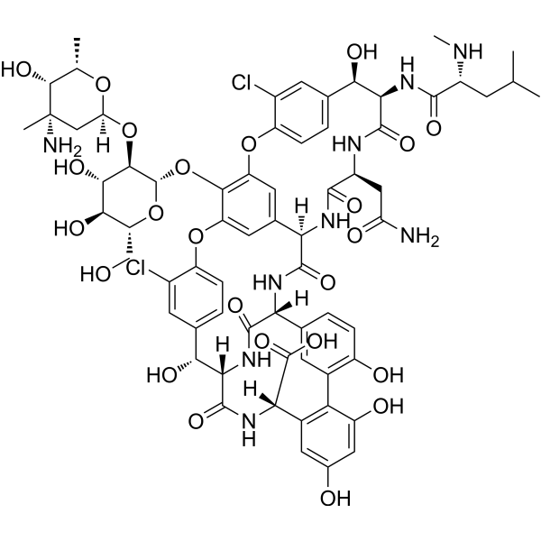
VancomycinAntibiotic -
HY-134541
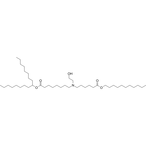
SM-102Ionizable Amino Lipid -
HY-B0470
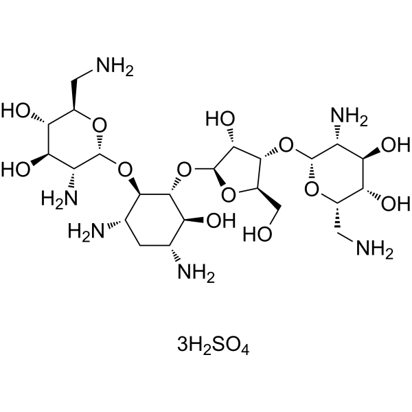
Neomycin sulfatePhospholipase C Inhibitor, Angiogenesis Modulator -
HY-N7781
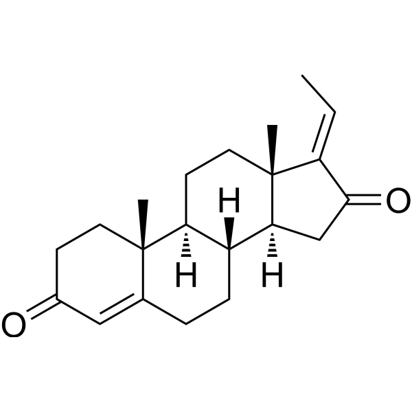
(-)-(E)-GuggulsteroneFXR Antagonist
Publications Citing Use of MedChemExpress (MCE) (Z)-Guggulsterone
More- Bone Res. 2025 Jan 30;13(1):20. [Abstract]
- Research (Wash D C). 2022 Nov 2:2022:9784081. [Abstract]
- Cell Death Dis. 2024 Nov 14;15(11):826. [Abstract]
- Pharmacol Res. 2024 Aug:206:107275. [Abstract]
- Int J Biol Macromol. 2025 May;307(Pt 4):142276. [Abstract]
- Chin Med. 2026 Jun 8;21(1):165. [Abstract]
- Eur J Pharmacol. 2026 Jul 10:1029:178995. [Abstract]
- Preprints. 2020, 2020090120.
 Customer Validation & Images

-
Cell Migration/Invasion Assay

-
WB

-
IF

-
Cell Proliferation/Viability Assay

-
Histological Imaging/Staining

All VEGFR Isoforms
More

Biological Activity
Description
IC50 & Target
[1]|
VEGF-R2 |
Cellular Effect
|
Cell Line

|
Type | Value | Description | References |
|---|---|---|---|---|
| HEK293 | IC50 |
12 μM
Compound: Gs
|
Inhibition of FXR in HEK293 cells in presence of CDCA by GAL4 transactivation activity
Inhibition of FXR in HEK293 cells in presence of CDCA by GAL4 transactivation activity
|
[PMID: 17292610] |
In Vitro
(Z)-Guggulsterone (10, 20 μM; 24 or 48 hours) causes a decrease in the level of VEGF-R2 protein in HUVEC[1].
(Z)-Guggulsterone (10 μM; 24 h) reduces ACE2 and SHP levels in primary airway and intestinal organoids, and reduces SARS-CoV-2 infection in multiple cell types via FXR-mediated ACE2 regulation[2].
MedChemExpress (MCE) has not independently confirmed the accuracy of these methods. They are for reference only. Further protocols information, click here.
-
Cell Line:Vascular endothelial growth factor (VEGF)
-
Concentration:10, 20 μM
-
Incubation Time:24 or 48 hours
-
Result:Caused a decrease in the level of VEGF-R2 protein in HUVEC.
In Vivo
MedChemExpress (MCE) has not independently confirmed the accuracy of these methods. They are for reference only.
-
Animal Model:Male nude mice (5–6 weeks old) s.c. implanted with DU145 cell-containing Matrigel plugs
-
Dosage:1 mg
-
Administration:Oral; 5 times/week
-
Result:Resulted in a statistically significantly decrease in tumor volume and wet tumor weight.


Chemical Information
-
CAS No. 39025-23-5
-
Appearance Solid
-
Molecular Weight 312.45
-
Formula C21H28O2
-
Color White to off-white
-
SMILES
C[C@@]1(/C2=C/C)[C@](CC2=O)([H])[C@@]3([H])[C@]([C@@]4(C(CC3)=CC(CC4)=O)C)([H])CC1.[Z]
-
Structure Classification
-
Initial Source
-
Shipping
Room temperature in continental US; may vary elsewhere.
-
Storage
Powder -20°C 3 years 4°C 2 years In solvent -80°C 6 months -20°C 1 month


Publications (8)
-
Journal Impact Factor
-
Most Recent
-
Bone Res
Nuclear farnesoid X receptor protects against bone loss by driving osteoblast differentiation through stabilizing RUNX2. [Abstract]2025 Jan 30;13(1):20. PMID: 39885145 -
Research (Wash D C)
Activation of Pancreatic Acinar FXR Protects against Pancreatitis via Osgin1-Mediated Restoration of Efficient Autophagy. [Abstract]2022 Nov 2:2022:9784081. PMID: 36405253 -
Cell Death Dis
FXR deficiency induced ferroptosis via modulation of the CBP-dependent p53 acetylation to suppress breast cancer growth and metastasis. [Abstract]2024 Nov 14;15(11):826. PMID: 39543094
(Z)-Guggulsterone purchased from MedChemExpress. Usage Cited in: Cell Death Dis. 2024 Nov 14;15(11):826. [Abstract]
(Z)-Guggulsterone (Z-GS) (20, 40 μM) treatment significantly reduced the invasion abilities of breast cancer cells pretreated with TGF-β1.
(Z)-Guggulsterone purchased from MedChemExpress. Usage Cited in: Cell Death Dis. 2024 Nov 14;15(11):826. [Abstract]
The proteins levels of E-Cadherin, N-Cadherin, and vimentin in MDA-MB-231 cells were measured by western blotting assays treated with (Z)-Guggulsterone (Z-GS) (20, 40 μM).
(Z)-Guggulsterone purchased from MedChemExpress. Usage Cited in: Cell Death Dis. 2024 Nov 14;15(11):826. [Abstract]
The relative levels of lipid ROS were detected by the C11-BODIPY probe in MDA-MB-231 cells treated with (Z)-Guggulsterone (Z-GS) (20, 40 μM).
(Z)-Guggulsterone purchased from MedChemExpress. Usage Cited in: Cell Death Dis. 2024 Nov 14;15(11):826. [Abstract]
The viability of MDA-MB-231 cells cultured with (Z)-Guggulsterone (Z-GS) (20, 40 μM) in the presence or absence of various cell death inhibitors was detected by CCK8.
(Z)-Guggulsterone purchased from MedChemExpress. Usage Cited in: Cell Death Dis. 2024 Nov 14;15(11):826. [Abstract]
Tumor metastasis was determined by H&E staining assay treated with (Z)-Guggulsterone (Z-GS) (30 mg/kg, i.p.).
-
Pharmacol Res
Lactobacillus rhamnosus GG ameliorates triptolide-induced liver injury through modulation of the bile acid-FXR axis. [Abstract]2024 Aug:206:107275. PMID: 38908615 -
Int J Biol Macromol
Salidroside attenuates NASH through regulating bile acid-FXR/TGR5 signaling pathway via targeting gut microbiota. [Abstract]2025 May;307(Pt 4):142276. PMID: 40118401 -
Chin Med
Calculus Bovis ameliorates primary sclerosing cholangitis via a dual-pronged mechanism restoring bile acid and lipid homeostasis in the gut-liver axis. [Abstract]2026 Jun 8;21(1):165. PMID: 42260527 -
Eur J Pharmacol
Isoastragaloside II modulates PPAR-α/FXR signaling and bile acid metabolism to ameliorate cholestatic liver diseases (CLD). [Abstract]2026 Jul 10:1029:178995. PMID: 42167668 -


Solvent & Solubility
In Vitro:
DMSO : 10 mg/mL (32.01 mM; ultrasonic and warming and heat to 60°C; Hygroscopic DMSO has a significant impact on the solubility of product, please use newly opened DMSO)
Please refer to the solubility information to select the appropriate solvent. Once prepared, please aliquot and store the solution to prevent product inactivation from repeated freeze-thaw cycles.
Storage method and period of stock solution: -80°C, 6 months; -20°C, 1 month. When stored at -80°C, please use it within 6 months. When stored at -20°C, please use it within 1 month.
Please refer to the solubility information to select the appropriate solvent. Once prepared, please aliquot and store the solution to prevent product inactivation from repeated freeze-thaw cycles.
Storage method and period of stock solution: -80°C, 6 months; -20°C, 1 month. When stored at -80°C, please use it within 6 months. When stored at -20°C, please use it within 1 month.
Concentration (start) × Volume (start) = Concentration (final) × Volume (final)
In Vivo:
Select the appropriate dissolution method based on your experimental animal and administration route.
- For the following dissolution methods, please ensure to first prepare a clear stock solution using an In Vitro approach and then sequentially add co-solvents:
- To ensure reliable experimental results, the clarified stock solution can be appropriately stored based on storage conditions. As for the working solution for In Vivo experiments, it is recommended to prepare freshly and use it on the same day.
- The percentages shown for the solvents indicate their volumetric ratio in the final prepared solution. If precipitation or phase separation occurs during preparation, heat and/or sonication can be used to aid dissolution.
Add each solvent one by one: 10% DMSO 90% (20% SBE-β-CD in Saline)
Solubility: ≥ 1 mg/mL (3.20 mM); Clear solution
This protocol yields a clear solution of ≥ 1 mg/mL (saturation unknown).
Taking 1 mL working solution as an example, add 100 μL DMSO stock solution (10.0 mg/mL) to 900 μL 20% SBE-β-CD in Saline, and mix evenly.
Preparation of 20% SBE-β-CD in Saline (4°C, storage for one week): 2 g SBE-β-CD powder is dissolved in 10 mL Saline, completely dissolve until clear.
For the following dissolution methods, please prepare the working solution directly:
It is recommended to prepare fresh solutions and use them promptly within a short period of time.
The percentages shown for the solvents indicate their volumetric ratio in the final prepared solution. If precipitation or phase separation occurs during preparation, heat and/or sonication can be used to aid dissolution.
Add each solvent one by one: 50% PEG300 50% Saline
Solubility: 10 mg/mL (32.01 mM); Suspended solution; Need ultrasonic
In Vivo Dissolution Calculator
Please enter the basic information of animal experiments:
-
-
-
-
Recommended: Prepare an additional quantity of animals to account for potential losses during experiments.
Please enter your animal formula composition:
-
%DMSO +
Recommended: Keep the proportion of DMSO in working solution below 2% if your animal is weak.
-
%+
-
+%Tween-80 + +
-
%Saline +
The co-solvents required include: DMSO, . All of co-solvents are available by MedChemExpress (MCE). , Tween 80. All of co-solvents are available by MedChemExpress (MCE).
Working solution concentration: 0.22 mg/mL
Method for preparing stock solution: mg drug dissolved in μL DMSO. Stock solution concentration: mg/mL.
1. Take μL DMSO stock solution;
2. Add μL .
μL , mix evenly;
3. Then add μL Tween 80, mix evenly;
4. Then add μL
Please ensure that the stock solution in the first step is dissolved to a clear state, and add co-solvents in sequence. You can use ultrasonic heating (ultrasonic cleaner, recommended frequency 20-40 kHz), vortexing, etc. to assist dissolution.

Protocols
-
RNA extraction experimental
By lysing cells, releasing RNA, and removing impurities such as proteins and DNA, high-purity RNA products are finally obtained. The commonly used traditional method is the guanidine isothiocyanate/phenol/chloroform method (Trizol), which is suitable for a variety of animal materials including animal tissues, microorganisms, cultured cells, etc., and most plant materials.
-
Research Protocol for Infectious Diseases
Infectious-disease experiments test how pathogens interact with host barriers, innate immune receptors, inflammatory signaling, pathogen replication, and tissue injury; pattern-recognition receptors such as TLRs, RIG-I-like receptors, NOD-like receptors, and inflammasomes detect microbial molecules and activate NF-κB, interferon, and cytokine responses. The central hypothesis is that infection severity reflects the balance between pathogen burden and host response: protective inflammation restricts pathogen growth, whereas excessive or mislocalized inflammation contributes to tissue damage and disease phenotype. Unresolved questions include which host pathways are protective versus pathogenic, why some infection models fail to translate to human disease, and which combined readouts best predict clinically relevant infection outcomes.
-
Apoptosis
Apoptosis, also called programmed cell death, is generally characterized by distinct morphological characteristics.
-
TUNEL staining for apoptotic DNA fragmentation
TUNEL staining detects DNA strand breaks by using terminal deoxynucleotidyl transferase to add labeled nucleotides to exposed 3′-OH DNA termini, generating either microscopic staining in fixed cells or tissue sections, or fluorescence/cytometric signal in cell suspensions. TUNEL positivity reflects DNA fragmentation but should not be interpreted alone as definitive apoptosis, because TUNEL can also label necrotic, autolytic, mechanically damaged, or DNA-repair-associated DNA breaks.
-
Human pluripotent stem cell endothelial-cell differentiation
Human pluripotent stem cell endothelial differentiation is based on stepwise developmental patterning: early activation of WNT/GSK3β inhibition promotes mesodermal or vascular progenitor entry, followed by endothelial specification using VEGF-related signaling, BMP4, FGF2, Notch modulation, or cAMP depending on the published protocol. Endothelial differentiation is read out by acquisition of CD31, CD34, VE-cadherin/CD144, KDR/VEGFR2, vWF, Tie2, NOS3, acetylated LDL uptake, tube/network formation, barrier function, and in vivo vessel-forming capacity where tested.
-
Vascular/Branching Fractal Analysis
Vascular/branching fractal analysis quantifies the geometric complexity of vessel trees or vascular networks from segmented 2D images, commonly by converting vessels into binary and/or skeletonized maps and estimating fractal dimension using box-counting or related approaches. Fractal dimension is interpreted as an image-derived readout of vascular branching complexity, space filling, or density, and has been applied to retinal photographs, fluorescein angiography, OCT angiography, capillary perfusion maps, and in vitro Matrigel angiogenesis networks. The assay readout is generated from vessel-positive pixels after image preprocessing, vessel segmentation, binarization, and optional skeletonization; reported outputs include fractal dimension, vessel density, branchpoint density, endpoint density, vessel length density, tortuosity, and generation-based branching metrics when VESGEN-style analysis is used. The biological interpretation is limited to quantitative vascular patterning and s
-
Annexin V plus membrane-impermeant dye apoptosis staining
Annexin V-based apoptosis assays rely on the detection of phosphatidylserine (PS) externalization from the inner leaflet of the plasma membrane to the outer leaflet, an early biochemical hallmark of apoptosis. Fluorescently labeled Annexin V binds PS in a calcium-dependent manner, enabling identification of early apoptotic cells by flow cytometry or fluorescence microscopy. When combined with a membrane-impermeant DNA-binding dye (e. g. , propidium iodide), this approach allows discrimination between viable (Annexin V−/dye−), early apoptotic (Annexin V+/dye−), and late apoptotic or necrotic (Annexin V+/dye+) cell populations by assessing membrane integrity and PS exposure.
-
Endothelial Tube Formation Assay
Endothelial tube formation assay evaluates the ability of endothelial cells to attach, migrate, align, and organize into capillary-like networks when cultured on gelled basement membrane extract or Matrigel; the readout is the morphology and quantity of tube-like networks, which reflects an in vitro endothelial morphogenesis step related to angiogenesis. Basement membrane extract/Matrigel provides laminin-rich extracellular matrix cues that support endothelial differentiation into capillary-like structures, but it can contain biologically active growth factors, so growth-factor-reduced matrix is preferred when testing defined angiogenic stimulators or inhibitors.
-
Apoptosis Solutions
Apoptosis is a regulated, generally non-lytic cell-death pathway that removes unwanted, damaged, infected, or abnormal cells through coordinated morphological changes, caspase activation, DNA fragmentation, and membrane remodeling. The intrinsic apoptosis pathway is controlled mainly by mitochondrial outer membrane permeabilization, BCL-2 family proteins, cytochrome c release, apoptosome formation, caspase-9 activation, and downstream executioner caspase-3/7 activation. The extrinsic apoptosis pathway is initiated by death receptors such as Fas, TNFR, and TRAIL receptors, which recruit adaptor proteins and activate caspase-8 before engaging executioner caspases or mitochondrial amplification through BID cleavage. Apoptosis is linked to many phenotypes, including cancer cell killing, tissue homeostasis, immune regulation, neurodegeneration, infection response, and treatment-induced cytotoxicity; unresolved questions include how apoptosis interacts with necroptosis, pyroptosis, ferroptos
-
Endothelial Cell Migration/Angiogenic Sprouting Assay
Endothelial cell migration and angiogenic sprouting assays are in vitro (and partially ex vivo-adapted) functional models that quantify the ability of endothelial cells to undergo coordinated migration, extracellular matrix invasion, and multicellular organization into capillary-like sprouts in response to pro-angiogenic stimuli such as VEGF, bFGF, or conditioned microenvironments. These assays are used to model early angiogenic events including tip-cell formation, directional migration, and lumen-like sprout extension, which collectively reflect angiogenic activation and vascular morphogenesis processes observed in vivo.


Purity & Documentation
-
Data Sheet (277 KB)
-
SDS (393 KB)
- English - EN (393 KB)
- Français - FR (393 KB)
- Deutsch - DE (393 KB)
- Norwegian - NO (393 KB)
- Español - ES (393 KB)
- Swedish - SV (393 KB)
- Italian - IT (393 KB)
- Korean - KR (393 KB)
- Portuguese - PT (393 KB)
-
Handling Instructions (2659 KB)
References
[1]. Xiao D, et al. z-Guggulsterone, a constituent of Ayurvedic medicinal plant Commiphora mukul, inhibits angiogenesis in vitro and in vivo. Mol Cancer Ther. 2008 Jan;7(1):171-80. [Content Brief]
[2]. Brevini T, et al. FXR inhibition may protect from SARS-CoV-2 infection by reducing ACE2. Nature. 2022 Dec 5. [Content Brief]


Complete Stock Solution Preparation Table
Please refer to the solubility information to select the appropriate solvent. Once prepared, please aliquot and store the solution to prevent product inactivation from repeated freeze-thaw cycles.
Storage method and period of stock solution: -80°C, 6 months; -20°C, 1 month. When stored at -80°C, please use it within 6 months. When stored at -20°C, please use it within 1 month.
| Optional Solvent | Concentration Solvent Mass | 1 mg | 5 mg | 10 mg | 25 mg |
|---|---|---|---|---|---|
| DMSO | 1 mM | 3.2005 mL | 16.0026 mL | 32.0051 mL | 80.0128 mL |
| 5 mM | 0.6401 mL | 3.2005 mL | 6.4010 mL | 16.0026 mL | |
| 10 mM | 0.3201 mL | 1.6003 mL | 3.2005 mL | 8.0013 mL | |
| 15 mM | 0.2134 mL | 1.0668 mL | 2.1337 mL | 5.3342 mL | |
| 20 mM | 0.1600 mL | 0.8001 mL | 1.6003 mL | 4.0006 mL | |
| 25 mM | 0.1280 mL | 0.6401 mL | 1.2802 mL | 3.2005 mL | |
| 30 mM | 0.1067 mL | 0.5334 mL | 1.0668 mL | 2.6671 mL |


(Z)-Guggulsterone Related Classifications
HY-110066 Related Classifications
 Powered by Bioz