



2-APQC
Based on 2 publication(s) in Google Scholar
2-APQC is an orally active and selective agonist of Sirtuin-3 (SIRT3) (Kd=2.756 μM), antagonizes Isoproterenol/ISO (HY-B0468)-induced cytotoxicity. 2-APQC activates the SIRT3-PYCR1 axis to enhance mitochondrial proline metabolism and inhibit the ROS-p38MAPK pathway by inhibiting signaling pathways such as mTOR-p70S6K, JNK, and TGF-β/Smad3. 2-APQC also activates the AMPK-Parkin axis to alleviate myocardial hypertrophy and fibrosis and protect cardiac function. 2-APQC can be used in the study of heart failure.
For research use only. We do not sell to patients.
- Purity : 99.70%
- CAS No.: 500271-63-6
- Formula: C23H24FN5O
- Molecular Weight:405.47
-
Storage:Powder -20°C, 3 years ; In solvent -80°C, 6 months , -20°C, 1 month


To place orders, for customer services and technical support, please contact: MedChemExpress USA
Tel: 609-228-6898 E-mail: [email protected] [email protected]


-
 Biological Activity
-
Chemical Information
-
Solvent & Solubility
- Protocols
- Purity & Documentation
- References
-
 Help & FAQs


-

HY-160818
MC3138SIRT5 Inhibitor -
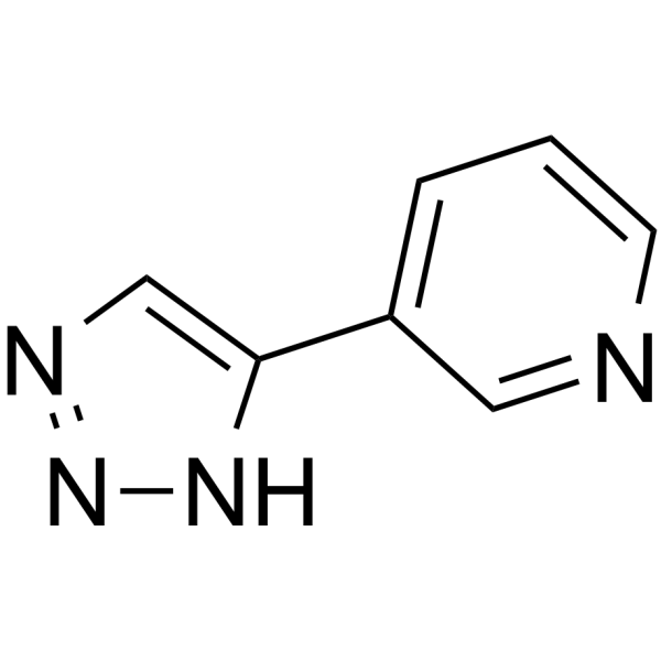
HY-108331
3-TYPSIRT3 Inhibitor -
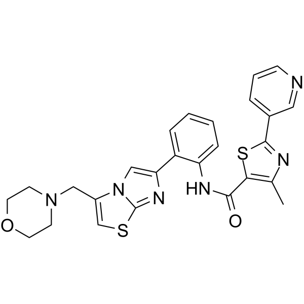
HY-15262
SRT 2104SIRT1 Activator -
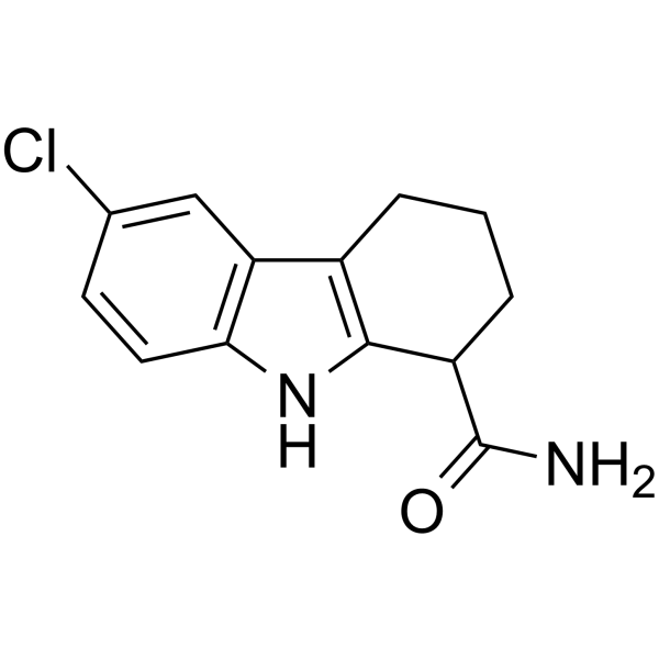
HY-15452
SelisistatSirT1/Sir2 Inhibitor -
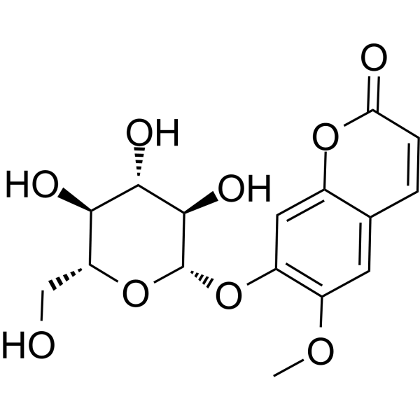
HY-N0341
ScopolinSIRT1 Activator -

HY-119376
MDL-800SIRT6 Activator -

HY-13259
MG-132Proteasome Inhibitor -
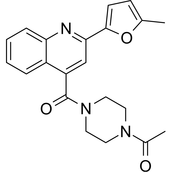
HY-117006
E1231SIRT1 Activator -
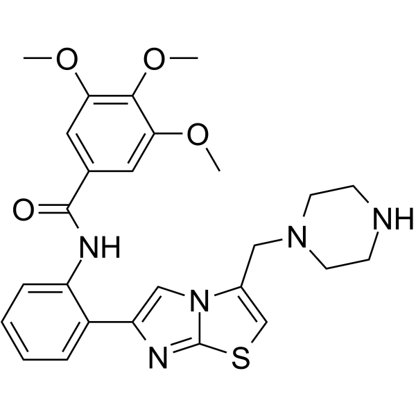
HY-124037
SRT 1460Sirtuin Activator -
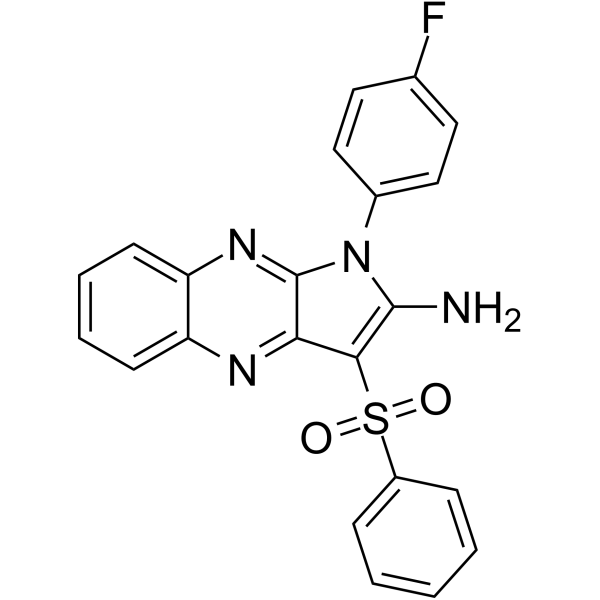
HY-104073
CAY10602SIRT1 Activator -
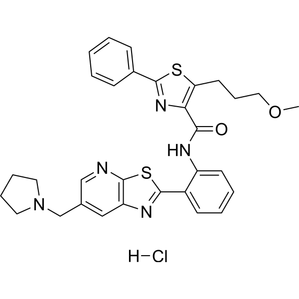
HY-19758A
Sirtuin modulator 1Sirtuin Modulator -
HY-N0003
HonokiolAutophagy Inducer
Publications Citing Use of MedChemExpress (MCE) 2-APQC
More Customer Validation & Images

-
WB
-
Cell Migration/Invasion Assay
-
Cell Imaging/Staining


Biological Activity
Description
IC50 & Target
|
SIRT3 2.756 (Kd) |
In Vitro
2-APQC (1, 10, 100 μM; 24 h) activates SIRT3 deacetylation activity in H9c2 cells in a concentration-dependent manner[1].
2-APQC (40 μM; 24 h) has no significant cytotoxicity to H9c2 cells[1].
2-APQC (1-100 μM; 24 h pretreatment + ISO treatment for 48 h) improves Isoproterenol/ISO (HY-B0468)-induced decrease in H9c2 cell viability, attenuates ISO-induced cardiomyocyte hypertrophy at a dose of 10 μM, and reduces the expression of α-SMA and collagen I[1].
2-APQC (10 μM; 24 h) reduces AcK68-MnSOD2, AcK122-MnSOD2 and acetylated lysine levels in H9c2 cells, without significantly change SIRT3 protein expression[1].
MedChemExpress (MCE) has not independently confirmed the accuracy of these methods. They are for reference only. Further protocols information, click here.
-
Cell Line:H9c2 cells
-
Concentration:10 μM
-
Incubation Time:24 h
-
Result:Decreased the levels of acetylated lysine, AcK68-MnSOD2, and AcK122-MnSOD2, indicating enhanced SIRT3 deacetylation activity.
Didn't change the expression of SIRT3 protein level.
In Vivo
2-APQC (42 mg/kg; intraperitoneal injection; once a day; 4 weeks) can improve cardiac function and reduce myocardial injury in the ISO-induced wild-type male mouse heart failure model, while this protective effect disappears in SIRT3 knockout mice[1].
MedChemExpress (MCE) has not independently confirmed the accuracy of these methods. They are for reference only.
-
Animal Model:Male SD rats (220-250 g), ISO-induced heart failure model[1]
-
Dosage:10, 20, 30 mg/kg
-
Administration:Gavage, once daily, 4 weeks; while ISO groups were injected subcutaneously with 5 mg/kg/d for 2 weeks. After the model was successfully established, 3 mL/kg/d normal saline was fed to rats.
-
Result:Significantly improved cardiac function as evidenced by increased EF and FS, decreased LVESD and LVPWD, and reduced BNP levels.
Alleviated myocardial hypertrophy and fibrosis, as shown by decreased heart weight index, reduced collagen fiber deposition in Sirius Red staining, and smaller myocyte cross-sectional area in WGA staining.
Serum levels of LDH, AST, CK-MB, and α-HBDH were decreased, indicating reduced myocardial injury.


Chemical Information
-
CAS No. 500271-63-6
-
Appearance Solid
-
Molecular Weight 405.47
-
Formula C23H24FN5O
-
Color White to off-white
-
SMILES
CCCCCNC(C1=C(N(C2=NC3=C(N=C21)C=CC=C3)CC4=CC=C(C=C4)F)N)=O
-
Shipping
Room temperature in continental US; may vary elsewhere.
-
Storage
Powder -20°C 3 years In solvent -80°C 6 months -20°C 1 month


Publications (2)
-
Journal Impact Factor
-
Most Recent
-
Free Radic Biol Med
Thiomyristoyl promotes type 2 diabetic wound healing and inhibits scarring via the PPARγ/Sirt3/SOD2 axis. [Abstract]2026 Jun 8:254:31-45. PMID: 42264197 -
Am J Transl Res
Sirtuin 3 modulation by high phosphates: a potential mechanism in muscle aging and sarcopenia. [Abstract]2025 Jun 15;17(6):4187-4197. PMID: 40672597
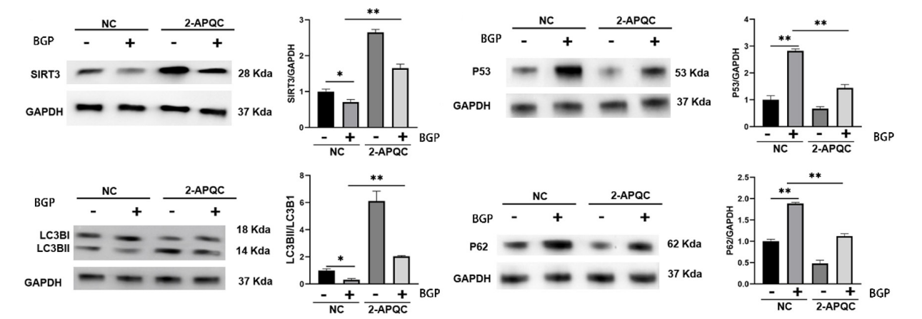
2-APQC purchased from MedChemExpress. Usage Cited in: Am J Transl Res. 2025 Jun 15;17(6):4187-4197. [Abstract]
To further elucidate the role of SIRT3 in phosphate-induced cellular senescence, C2C12 myoblasts were treated with 2-APQC, a selective SIRT3 activator. Western blot analysis revealed that BGP treatment significantly upregulated the expression of senescence markers P53 and P62, while reducing the ratio of autophagy-related proteins LC3BII/LC3BI.
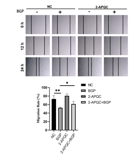
2-APQC purchased from MedChemExpress. Usage Cited in: Am J Transl Res. 2025 Jun 15;17(6):4187-4197. [Abstract]
Elevated phosphate levels compromise cellular processes essential for wound healing and tissue regeneration. However, co-treatment with the SIRT3 activator 2-APQC partially restored the migration potential of C2C12 cells, indicating that SIRT3 plays a protective role in maintaining cellular motility under phosphate stress.
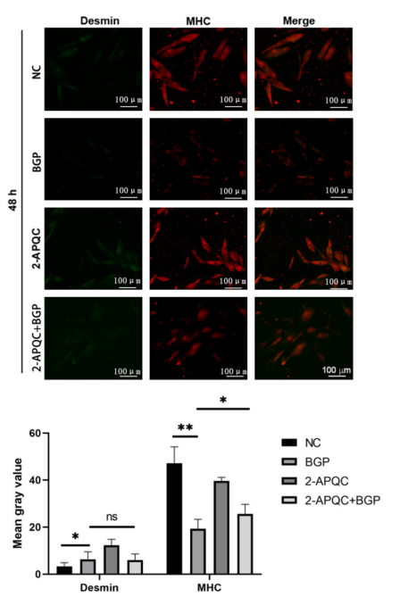
2-APQC purchased from MedChemExpress. Usage Cited in: Am J Transl Res. 2025 Jun 15;17(6):4187-4197. [Abstract]
Treatment with 2-APQC significantly increased MHC expression in phosphate-treated cells, alongside a notable co-localization of MHC with desmin, a protein that stabilizes muscle fibers during differentiation.


Solvent & Solubility
In Vitro:
DMSO : 50 mg/mL (123.31 mM; Need ultrasonic; Hygroscopic DMSO has a significant impact on the solubility of product, please use newly opened DMSO)
Please refer to the solubility information to select the appropriate solvent. Once prepared, please aliquot and store the solution to prevent product inactivation from repeated freeze-thaw cycles.
Storage method and period of stock solution: -80°C, 6 months; -20°C, 1 month. When stored at -80°C, please use it within 6 months. When stored at -20°C, please use it within 1 month.
Please refer to the solubility information to select the appropriate solvent. Once prepared, please aliquot and store the solution to prevent product inactivation from repeated freeze-thaw cycles.
Storage method and period of stock solution: -80°C, 6 months; -20°C, 1 month. When stored at -80°C, please use it within 6 months. When stored at -20°C, please use it within 1 month.
Concentration (start) × Volume (start) = Concentration (final) × Volume (final)
In Vivo:
Select the appropriate dissolution method based on your experimental animal and administration route.
- For the following dissolution methods, please ensure to first prepare a clear stock solution using an In Vitro approach and then sequentially add co-solvents:
- To ensure reliable experimental results, the clarified stock solution can be appropriately stored based on storage conditions. As for the working solution for In Vivo experiments, it is recommended to prepare freshly and use it on the same day.
- The percentages shown for the solvents indicate their volumetric ratio in the final prepared solution. If precipitation or phase separation occurs during preparation, heat and/or sonication can be used to aid dissolution.
Add each solvent one by one: 10% DMSO 40% PEG300 5% Tween-80 45% Saline
Solubility: ≥ 2.5 mg/mL (6.17 mM); Clear solution
This protocol yields a clear solution of ≥ 2.5 mg/mL (saturation unknown).
Taking 1 mL working solution as an example, add 100 μL DMSO stock solution (25.0 mg/mL) to 400 μL PEG300, and mix evenly; then add 50 μL Tween-80 and mix evenly; then add 450 μL Saline to adjust the volume to 1 mL.
Preparation of Saline: Dissolve 0.9 g sodium chloride in ddH₂O and dilute to 100 mL to obtain a clear Saline solution.
Add each solvent one by one: 10% DMSO 90% (20% SBE-β-CD in Saline)
Solubility: ≥ 2.5 mg/mL (6.17 mM); Clear solution
This protocol yields a clear solution of ≥ 2.5 mg/mL (saturation unknown).
Taking 1 mL working solution as an example, add 100 μL DMSO stock solution (25.0 mg/mL) to 900 μL 20% SBE-β-CD in Saline, and mix evenly.
Preparation of 20% SBE-β-CD in Saline (4°C, storage for one week): 2 g SBE-β-CD powder is dissolved in 10 mL Saline, completely dissolve until clear.
In Vivo Dissolution Calculator
Please enter the basic information of animal experiments:
-
-
-
-
Recommended: Prepare an additional quantity of animals to account for potential losses during experiments.
Please enter your animal formula composition:
-
%DMSO +
Recommended: Keep the proportion of DMSO in working solution below 2% if your animal is weak.
-
%+
-
+%Tween-80 + +
-
%Saline +
The co-solvents required include: DMSO, . All of co-solvents are available by MedChemExpress (MCE). , Tween 80. All of co-solvents are available by MedChemExpress (MCE).
Working solution concentration: 0.22 mg/mL
Method for preparing stock solution: mg drug dissolved in μL DMSO. Stock solution concentration: mg/mL.
1. Take μL DMSO stock solution;
2. Add μL .
μL , mix evenly;
3. Then add μL Tween 80, mix evenly;
4. Then add μL
Please ensure that the stock solution in the first step is dissolved to a clear state, and add co-solvents in sequence. You can use ultrasonic heating (ultrasonic cleaner, recommended frequency 20-40 kHz), vortexing, etc. to assist dissolution.

Protocols
-
Fibrosis/Collagen Morphometry
Fibrosis and collagen morphometry is based on the quantitative visualization of fibrillar collagen deposition in tissue sections using histochemical stains such as Sirius Red (Picrosirius Red) or Masson's trichrome, followed by image-based or polarization-enhanced analysis to estimate collagen proportional area as a surrogate of extracellular matrix accumulation during fibrotic remodeling. Sirius Red combined with polarized light microscopy enhances detection of collagen fibers due to birefringence properties, enabling more specific visualization of collagen type I and III fibrils compared to conventional bright-field histology, while whole-section or region-restricted digital morphometry reduces field-selection bias in fibrosis assessment. Alternative quantitative approaches include second harmonic generation (SHG) and two-photon excited fluorescence microscopy, which enable label-free detection of fibrillar collagen and have been validated against histological staining and biochemica
-
Collagen: Sirius Red Staining
Sirius Red or picrosirius red staining is a histochemical method for visualizing collagen-rich extracellular matrix in tissue sections, and collagen fibers are detected as red-stained structures under bright-field microscopy with enhanced birefringence under polarized light. Picrosirius red is useful for assessing total collagen organization, distribution, and fibrosis burden, but polarized color should not be interpreted as a definitive collagen type I versus type III readout because color is affected by fiber orientation, thickness, and packing.
-
Cell Cytotoxicity Assay
Cytotoxicity assays are usually based on the assessment of cell membrane damage, which can also be indirectly detected by measuring cell viability. Detection methods include MTT assay, CKK-8 assay, LDH assay and ATP assay, etc.
-
Mammalian live/dead viability and cytotoxicity staining
Live/dead viability and cytotoxicity staining assays are based on the simultaneous detection of intracellular esterase activity in metabolically active (viable) cells and membrane integrity loss in non-viable cells. In commonly used dual-staining approaches, membrane-permeant fluorogenic substrates are converted by intracellular esterases into fluorescent products in live cells, while impermeant DNA-binding dyes selectively enter cells with compromised plasma membranes and label nucleic acids in dead or dying cells, enabling discrimination between viable and non-viable populations by fluorescence microscopy or flow cytometry.
-
ROS/oxidative-stress fluorescent staining
ROS/oxidative-stress fluorescent staining uses cell-permeant fluorogenic probes that become fluorescent after oxidation inside cells or tissues; commonly used examples include DCFH-DA/DCFDA for broad cellular oxidant detection, DHE for superoxide-related signal detection, MitoSOX for mitochondrial superoxide-related signal detection, and CellROX probes for oxidative-stress-associated fluorescence readouts. The assay detects probe oxidation rather than a single ROS species unless the probe and analysis method have been chemically validated for that species. DCFH-DA enters cells, is deacetylated by intracellular esterases to DCFH, and produces fluorescent DCF after oxidation, so the readout is used as an operational measure of total cellular oxidative stress rather than a species-specific ROS measurement. DHE and MitoSOX can report superoxide-related oxidation, but red fluorescence alone can include non-specific ethidium-like oxidation products; HPLC or optimized spectral approaches are
-
Connective Tissue: Masson's Trichrome/Collagen Trichrome Staining
Masson’s Trichrome (collagen/trichrome staining) is a histological technique that differentially stains tissue compartments using sequential acidic dyes to distinguish collagen from muscle and cytoplasmic components based on dye affinity and tissue permeability differences, enabling visualization of fibrosis and connective tissue architecture in histological sections. The classical formulation typically uses Weigert's iron hematoxylin for nuclear staining, Biebrich scarlet-acid fuchsin for cytoplasm and muscle, and aniline blue (or light green variants) for collagen, producing a characteristic blue/green collagen signal contrasted against red cytoplasm and dark nuclei. The staining principle relies on selective displacement of smaller dye molecules by larger anionic dyes in collagen-rich regions under controlled acidified conditions, which enhances collagen-specific dye retention. This property makes the method widely used for fibrosis assessment in organs such as heart, liver, lung, a
-
Research Protocol for Cardiovascular Diseases
Cardiovascular disease can be modeled as maladaptive cardiac remodeling, where ischemic injury or pressure overload activates inflammatory signaling, fibroblast activation, extracellular-matrix deposition, cardiomyocyte hypertrophy, vascular remodeling, and progressive ventricular dysfunction. The TGF-β/SMAD axis is a central profibrotic pathway after myocardial injury and pressure overload, while innate immune and cytokine pathways regulate leukocyte recruitment, scar formation, and adverse remodeling. Key unresolved questions include which inflammatory signals are reparative versus harmful, when fibrosis is protective versus maladaptive, and whether pathway inhibition improves function without weakening necessary infarct healing or compensatory remodeling.
-
Mitophagy Solutions
Mitophagy is the selective autophagic degradation of mitochondria and functions as a mitochondrial quality-control pathway that removes damaged, depolarized, excess, or developmentally programmed mitochondria. The pathway links mitochondrial damage recognition, autophagosome recruitment, lysosomal delivery, and mitochondrial turnover to phenotypes such as mitochondrial homeostasis, oxidative-stress control, metabolic remodeling, differentiation, and neurodegeneration-related mitochondrial fidelity. The best-characterized damage-induced pathway is the PINK1-Parkin axis. Parkin is recruited selectively to impaired mitochondria and promotes their autophagic elimination, while mitochondrial depolarization stabilizes PINK1 on damaged mitochondria, recruits Parkin, and activates Parkin-dependent mitophagy. PINK1 also phosphorylates ubiquitin to activate Parkin E3 ubiquitin ligase activity, and PINK1-driven ubiquitin phosphorylation creates a feed-forward signal for recruiting autophagy machi
-
Cell Viability Determination by MTT Colorimetric Assay
The following protocol uses the MTT colorimetric assay as a classic literature-established method for assessing cell viability/metabolic activity in cultured mammalian cells. MTT[3-(4,5-dimethylthiazol-2-yl)-2,5-diphenyltetrazolium bromide] is reduced by metabolically active cells to a colored formazan product; the amount of formazan is quantified spectrophotometrically and provides an indirect measure of metabolically active viable cells. Importantly, MTT reduction reflects cellular oxidoreductase/metabolic activity rather than an absolute direct count of living cells, so changes in cellular metabolism can alter the signal independently of cell number.


Purity & Documentation
-
Data Sheet (283 KB)
-
SDS (252 KB)
- English - EN (252 KB)
- Français - FR (252 KB)
- Deutsch - DE (252 KB)
- Norwegian - NO (252 KB)
- Español - ES (252 KB)
- Swedish - SV (252 KB)
- Italian - IT (252 KB)
- Korean - KR (252 KB)
- Portuguese - PT (252 KB)
-
Handling Instructions (2659 KB)
References


Complete Stock Solution Preparation Table
Please refer to the solubility information to select the appropriate solvent. Once prepared, please aliquot and store the solution to prevent product inactivation from repeated freeze-thaw cycles.
Storage method and period of stock solution: -80°C, 6 months; -20°C, 1 month. When stored at -80°C, please use it within 6 months. When stored at -20°C, please use it within 1 month.
| Optional Solvent | Concentration Solvent Mass | 1 mg | 5 mg | 10 mg | 25 mg |
|---|---|---|---|---|---|
| DMSO | 1 mM | 2.4663 mL | 12.3314 mL | 24.6627 mL | 61.6568 mL |
| 5 mM | 0.4933 mL | 2.4663 mL | 4.9325 mL | 12.3314 mL | |
| 10 mM | 0.2466 mL | 1.2331 mL | 2.4663 mL | 6.1657 mL | |
| 15 mM | 0.1644 mL | 0.8221 mL | 1.6442 mL | 4.1105 mL | |
| 20 mM | 0.1233 mL | 0.6166 mL | 1.2331 mL | 3.0828 mL | |
| 25 mM | 0.0987 mL | 0.4933 mL | 0.9865 mL | 2.4663 mL | |
| 30 mM | 0.0822 mL | 0.4110 mL | 0.8221 mL | 2.0552 mL | |
| 40 mM | 0.0617 mL | 0.3083 mL | 0.6166 mL | 1.5414 mL | |
| 50 mM | 0.0493 mL | 0.2466 mL | 0.4933 mL | 1.2331 mL | |
| 60 mM | 0.0411 mL | 0.2055 mL | 0.4110 mL | 1.0276 mL | |
| 80 mM | 0.0308 mL | 0.1541 mL | 0.3083 mL | 0.7707 mL | |
| 100 mM | 0.0247 mL | 0.1233 mL | 0.2466 mL | 0.6166 mL |


2-APQC Related Classifications
HY-158426 Related Classifications
 Powered by Bioz