



Astragaloside II
Based on 2 publication(s) in Google Scholar
Astragaloside II is an orally active Cycloartane-type triterpene glycoside. Astragaloside II can be extracted from Astragalus membranaceus. Astragaloside II inhibits Autophagy, decreases pro-inflammatory cytokines (IL-6, IL-1β), HIF-α, p-p65, p-IκB and increases SOD. Astragaloside II regulates immunity and reduces inflammatory responses. Astragaloside II can be used in the research of diseases such as liver cancer, osteoporosis, immunosuppressive diseases, and ulcerative colitis.
For research use only. We do not sell to patients.
- Purity : 99.66%
- CAS No.: 84676-89-1
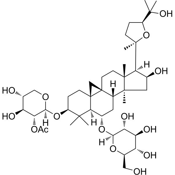
- Formula: C43H70O15
- Molecular Weight:827.01
-
Storage:Powder -20°C, 3 years , 4°C, 2 years ; In solvent -80°C, 2 years , -20°C, 1 year

To place orders, for customer services and technical support, please contact: MedChemExpress USA
Tel: 609-228-6898 E-mail: [email protected] [email protected]


-
 Biological Activity
-
Chemical Information
-
Solvent & Solubility
- Protocols
- Purity & Documentation
- References
-
 Help & FAQs


-
HY-N0431
Astragaloside IVMMP Inhibitor -
HY-N0432
Astragaloside IWnt/β-catenin Activator -
HY-N0434
Astragaloside IIINatural Product -
HY-18085
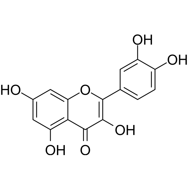
QuercetinFlavonoid Antioxidant -
HY-N0039
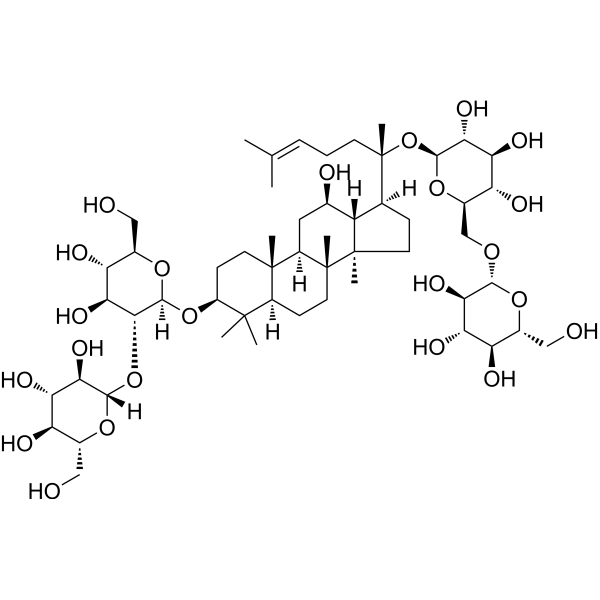
Ginsenoside Rb1Autophagy Inducer -
HY-N0519
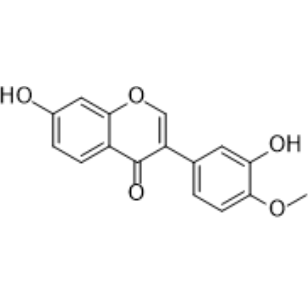
CalycosinPhytoestrogen -
HY-N0520
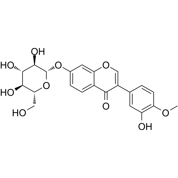
Calycosin-7-O-β-D-glucosideBioactive Isoflavone -

HY-N0433R
Astragaloside II (Standard)Natural Product -
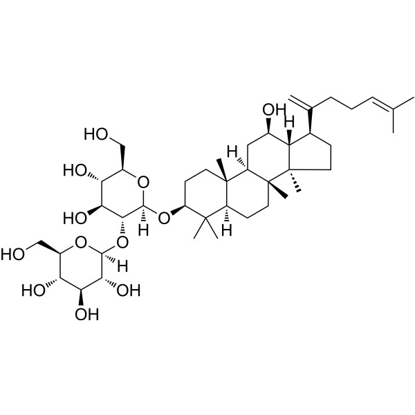
HY-N2515
Ginsenoside Rk1Apoptosis Inducer -
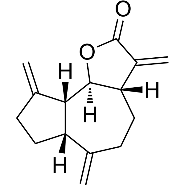
HY-N0591
Dehydrocostus LactoneSesquiterpene -
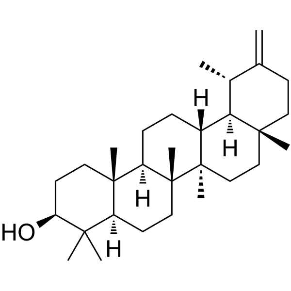
HY-N1178
TaraxasterolAnti-Inflammatory Agent; LXRα Activator -
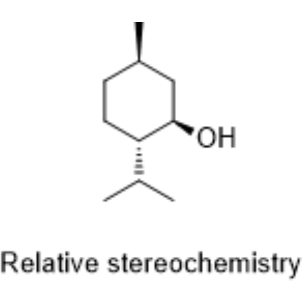
HY-Y1683
DL-MentholUGT Inhibitor
Publications Citing Use of MedChemExpress (MCE) Astragaloside II
More Customer Validation & Images

-
Cell Proliferation/Viability Assay


Biological Activity
Description
In Vitro
Astragaloside II (0.1 nM-10 μM; 48-72 h) significantly promotes the cell viability of rat primary osteoblasts in a concentration-dependent manner[1].
Astragaloside II (10-30 nM; 48-96 h) significantly enhances the proliferation of primary splenocytes induced by ConA, alloantigen or anti-CD3[3].
Astragaloside II (80 μM; 48 h) sensitizes Bel-7402/FU cells to 5-fluorouracil (HY-90006)-induced cell death via suppression of autophagy[4].
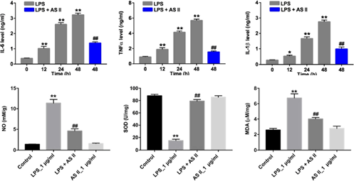
Astragaloside II (1 μM; 48 h) inhibits the levels of HIF-α, p-p65 and p-IκB in Lipopolysaccharide-stimulated CCD-18Co cells, exerting an anti-inflammatory effect[5].
MedChemExpress (MCE) has not independently confirmed the accuracy of these methods. They are for reference only. Further protocols information, click here.
-
Cell Line:Bel-7402/FU
-
Concentration:40 μM, 80 μM, 160 μM, 320 μM, 80 μM
-
Incubation Time:48 h
-
Result:Decreased the expression of autophagy-related proteins LC3-II and Beclin-1.
-
Cell Line:CCD-18Co
-
Concentration:0.1 μM, 0.33 μM, 1 μM, 3 μM
-
Incubation Time:48 h
-
Result:Significantly decreased the viability of CCD-18Co cells at 3 μM.
Had very limited cytotoxicity at 1 μM.
In Vivo
MedChemExpress (MCE) has not independently confirmed the accuracy of these methods. They are for reference only.
-
Animal Model:Male BABL/c mice (20-22 g) with DSS-induced ulcerative colitis[5]
-
Dosage:30 mg/kg (dissolved in 2% Tween 20 in PBS), 50 mg/kg (dissolved in 2% Tween 20 in PBS)
-
Administration:Oral gavage, once per day, for 10 days
-
Result:Significantly reduced the DAI score.
Markedly prevented the body weight loss, increased the colon length, decreased the levels of pro-inflammatory cytokines IL-6, TNF-α, IL-1β, NO, MPO and MDA, and increased the level of SOD in colon tissues.
Decreased the expression of inflammatory proteins HIF-α, p-p65 and p-IκB.


Chemical Information
-
CAS No. 84676-89-1
-
Appearance Solid
-
Molecular Weight 827.01
-
Formula C43H70O15
-
Color White to off-white
-
SMILES
O[C@H]1[C@H](O)[C@@H](OC(C)=O)[C@]([H])(O[C@@H]2C(C)(C)[C@@]([C@@H](O[C@]3([H])O[C@H](CO)[C@@H](O)[C@H](O)[C@H]3O)C[C@]4([H])[C@@]56CC[C@@]7(C)[C@@]4(C)C[C@H](O)[C@@]7([C@]8(C)O[C@H](C(O)(C)C)CC8)[H])([H])[C@]5(C6)CC2)OC1
-
Synonyms
Astrasieversianin VIII
-
Structure Classification
-
Shipping
Room temperature in continental US; may vary elsewhere.
-
Storage
Powder -20°C 3 years 4°C 2 years In solvent -80°C 2 years -20°C 1 year


Publications (2)
-
Journal Impact Factor
-
Most Recent
-
J Mol Histol
Astragaloside II suppresses colorectal cancer progression by upregulating LGALS4 expression. [Abstract]2026 Jun 29;57(4):199. PMID: 42319491 -
Am J Transl Res
Astragaloside II alleviates the symptoms of experimental ulcerative colitis in vitro and in vivo. [Abstract]2019 Nov 15;11(11):7074-7083. PMID: 31814910
Astragaloside II purchased from MedChemExpress. Usage Cited in: Am J Transl Res. 2019 Nov 15;11(11):7074-7083. [Abstract]
AS II (Astragaloside II) attenuates oxidative stress damage in LPS-stimulated CCD-18Co cells via decreasing the production of inflammatory factors. CCD-18Co cells are treated with LPS (1 μg/mL) for 0, 12, 24 and 48 h. In addition, CCD-18Co cells are treated with 1 μM AS II and 1 μg/mL LPS for 48 h. The level of IL-6 in the culture media is measured with ELISA. The level of TNF-α in the culture media is measured with ELISA. The level of IL-β in the culture media is measured with ELISA. CCD-18Co c


Solvent & Solubility
In Vitro:
DMSO : 100 mg/mL (120.92 mM; Need ultrasonic; Hygroscopic DMSO has a significant impact on the solubility of product, please use newly opened DMSO)
Please refer to the solubility information to select the appropriate solvent. Once prepared, please aliquot and store the solution to prevent product inactivation from repeated freeze-thaw cycles.
Storage method and period of stock solution: -80°C, 2 years; -20°C, 1 year. When stored at -80°C, please use it within 2 years. When stored at -20°C, please use it within 1 year.
Please refer to the solubility information to select the appropriate solvent. Once prepared, please aliquot and store the solution to prevent product inactivation from repeated freeze-thaw cycles.
Storage method and period of stock solution: -80°C, 2 years; -20°C, 1 year. When stored at -80°C, please use it within 2 years. When stored at -20°C, please use it within 1 year.
Concentration (start) × Volume (start) = Concentration (final) × Volume (final)
In Vivo:
Select the appropriate dissolution method based on your experimental animal and administration route.
- For the following dissolution methods, please ensure to first prepare a clear stock solution using an In Vitro approach and then sequentially add co-solvents:
- To ensure reliable experimental results, the clarified stock solution can be appropriately stored based on storage conditions. As for the working solution for In Vivo experiments, it is recommended to prepare freshly and use it on the same day.
- The percentages shown for the solvents indicate their volumetric ratio in the final prepared solution. If precipitation or phase separation occurs during preparation, heat and/or sonication can be used to aid dissolution.
Add each solvent one by one: 10% DMSO 40% PEG300 5% Tween-80 45% Saline
Solubility: ≥ 2.5 mg/mL (3.02 mM); Clear solution
This protocol yields a clear solution of ≥ 2.5 mg/mL (saturation unknown).
Taking 1 mL working solution as an example, add 100 μL DMSO stock solution (25.0 mg/mL) to 400 μL PEG300, and mix evenly; then add 50 μL Tween-80 and mix evenly; then add 450 μL Saline to adjust the volume to 1 mL.
Preparation of Saline: Dissolve 0.9 g sodium chloride in ddH₂O and dilute to 100 mL to obtain a clear Saline solution.
In Vivo Dissolution Calculator
Please enter the basic information of animal experiments:
-
-
-
-
Recommended: Prepare an additional quantity of animals to account for potential losses during experiments.
Please enter your animal formula composition:
-
%DMSO +
Recommended: Keep the proportion of DMSO in working solution below 2% if your animal is weak.
-
%+
-
+%Tween-80 + +
-
%Saline +
The co-solvents required include: DMSO, . All of co-solvents are available by MedChemExpress (MCE). , Tween 80. All of co-solvents are available by MedChemExpress (MCE).
Working solution concentration: 0.22 mg/mL
Method for preparing stock solution: mg drug dissolved in μL DMSO. Stock solution concentration: mg/mL.
1. Take μL DMSO stock solution;
2. Add μL .
μL , mix evenly;
3. Then add μL Tween 80, mix evenly;
4. Then add μL
Please ensure that the stock solution in the first step is dissolved to a clear state, and add co-solvents in sequence. You can use ultrasonic heating (ultrasonic cleaner, recommended frequency 20-40 kHz), vortexing, etc. to assist dissolution.

Protocols
-
LPS-Induced Endotoxemia/Systemic Inflammation
Lipopolysaccharide (LPS)-induced endotoxemia is a widely used in vivo model of acute systemic inflammation in which LPS, a Gram-negative bacterial endotoxin, activates innate immune signaling primarily through TLR4, leading to rapid and transient induction of pro-inflammatory cytokines such as TNF-α, IL-6, and IL-1β in circulation and tissues. This cytokine surge is commonly used as a measurable readout of systemic inflammatory activation and immune dysregulation, and is typically assessed within hours after intraperitoneal LPS administration in mouse models of endotoxemia. The model captures key features of systemic inflammatory response syndrome, including cytokine release, immune cell activation, and downstream tissue responses, and has been used to evaluate anti-inflammatory interventions such as cytokine modulation, lipid mediators, and immune cell-targeting therapies.
-
DSS-Induced Colitis
Dextran sulfate sodium (DSS)-induced colitis is generated by administering DSS in mouse drinking water, producing epithelial injury, barrier disruption, weight loss, diarrhea, fecal blood, colon shortening, histologic mucosal damage, and inflammatory mediator changes; the model is mainly used to study acute or chronic intestinal inflammation resembling selected features of ulcerative colitis. DSS injury is interpreted through clinical and tissue readouts rather than a single molecular endpoint: daily body weight, stool consistency, and bleeding are combined into a disease activity index, while colon length, histology, cytokines, myeloperoxidase activity, intestinal permeability, and tight-junction markers provide complementary measures of inflammation and barrier damage.
-
TNBS-Induced Colitis
TNBS-induced colitis is produced by intrarectal delivery of 2,4,6-trinitrobenzene sulfonic acid in ethanol, where ethanol disrupts the mucosal barrier and TNBS haptenates colonic proteins, generating immune-mediated colonic inflammation with weight loss, diarrhea, ulceration, transmural injury, inflammatory-cell infiltration, and cytokine responses. The model is used as an experimental intestinal inflammation model with Crohn’s disease–like features, especially when Th1-type responses, IL-12–dependent inflammation, chronic relapsing inflammation, or fibrosis-related endpoints are studied.
-
Autophagy
Autophagy is a process in which eukaryotic cells use lysosomes to degrade their own cytoplasmic proteins and damaged organelles under the regulation of autophagy related gene (Atg). Microtubule-associated proteins light chain 3 (LC3) is recognized as autophagy marker, which transfers from cytoplasmic LC3 (LC3-I) to membrane type (LC3-II). LC3-II/I ratio could be detected by Western Blot and fluorescence microscopy.
-
Lysosome and acidic-vesicle live-cell staining
Lysosome and acidic-vesicle live-cell staining detects acidic intracellular compartments by using membrane-permeant acidotropic probes that accumulate in low-pH vesicles, including lysosomes, late endosomes, autolysosomes, and acidic phagosomes. LysoTracker staining is commonly used as an intensity-based readout of acidic lysosomal compartment abundance or enlargement, while acridine orange produces green fluorescence in less concentrated compartments and red fluorescence after concentration-dependent accumulation in acidic vesicular organelles. Loss or reduction of acridine-orange red signal can be used as a readout of lysosomal membrane permeabilization or reduced acidic-vesicle integrity. This protocol is designed for live cultured cells and can be adapted for fluorescence microscopy, high-content imaging, plate-reader readout, or flow cytometry when the selected literature supports the readout. Because these dyes report acidotropic accumulation rather than lysosome identity alone,
-
Research Protocol for Inflammation-related Diseases
The NLRP3 inflammasome is a cytosolic innate immune signaling platform that integrates priming signals and danger-signal activation to promote caspase-1 activation, maturation of IL-1β and IL-18, and gasdermin D-mediated pyroptotic cell death. The core experimental logic is to determine whether inflammatory disease phenotypes are driven by increased NLRP3 expression, ASC-containing inflammasome assembly, caspase-1 cleavage, GSDMD cleavage, and extracellular release of IL-1β/IL-18 rather than by nonspecific cell injury alone. The pathway is strongly linked to inflammation-related disease phenotypes because monosodium urate crystals activate NALP3/NLRP3 inflammasome signaling in gout-like crystal inflammation, cholesterol crystals activate NLRP3 inflammasomes in atherogenesis models, and DSS-induced intestinal inflammation has been reported to involve NLRP3 inflammasome activity. However, experimental colitis studies also show context-dependent protective effects of NLRP3 inflammasome co
-
Liver Cancer Modeling
Liver cancer can be classified into primary liver cancer and secondary liver cancer. Secondary liver cancer is the metastatic liver cancer. Primary liver cancer includes hepatocellular carcinoma (HCC), intrahepatic cholangiocarcinoma (ICC) and fibrolamellar HCC, of which HCC is the most common form, accounting for approximately 90% of primary liver cancers[1]. HCC mouse models include chemical agent-induced models, transplanted tumor models, and genetic engineered models.
-
Macroautophagy Solutions
Macroautophagy is a conserved lysosome-dependent degradation pathway in which cytoplasmic material is sequestered into double-membrane autophagosomes and delivered to lysosomes for degradation and recycling. The pathway supports cellular homeostasis during nutrient limitation, organelle stress, protein-aggregate accumulation, infection, differentiation, and tissue remodeling by coupling cargo sequestration, autophagosome maturation, lysosomal fusion, and degradation of cargo-derived macromolecules. The core molecular sequence includes initiation by nutrient- and stress-regulated autophagy machinery, autophagosome nucleation, LC3/ATG8-family conjugation to autophagosomal membranes, cargo selection through receptors such as SQSTM1/p62, autophagosome-lysosome fusion, and lysosomal degradation. LC3 was identified as a mammalian homolog of yeast Atg8 that localizes to autophagosomal membranes after processing, and p62/SQSTM1 was shown to connect ubiquitinated cargo with autophagic degradati
-
Pyroptosis Solutions
Pyroptosis is a lytic inflammatory cell-death pathway executed by gasdermin pores, most classically through inflammasome-mediated activation of caspase-1, cleavage of gasdermin D, membrane pore formation, LDH release, and secretion of IL-1β and IL-18. The canonical pathway is commonly modeled by priming cells with an inflammatory signal such as LPS to induce pro-IL-1β and inflammasome components, followed by an activation signal such as ATP or nigericin to activate NLRP3, ASC speck formation, caspase-1 cleavage, GSDMD cleavage, cytokine release, and pyroptotic membrane rupture. The non-canonical pathway is triggered when cytosolic LPS activates mouse caspase-11 or human caspase-4/5, leading to GSDMD cleavage and pyroptosis, and this can secondarily activate NLRP3-dependent IL-1β release. Pyroptosis is linked to inflammatory injury, infection, cancer, liver disease, ocular disease, placental inflammation, and other disease phenotypes, but unresolved questions include which gasdermin fam


Purity & Documentation
-
Data Sheet (303 KB)
-
SDS (252 KB)
- English - EN (252 KB)
- Français - FR (252 KB)
- Deutsch - DE (252 KB)
- Norwegian - NO (252 KB)
- Español - ES (252 KB)
- Swedish - SV (252 KB)
- Italian - IT (252 KB)
- Korean - KR (252 KB)
- Portuguese - PT (252 KB)
-
Handling Instructions (2659 KB)
References
[1]. Kong XH, et al. Astragaloside II induces osteogenic activities of osteoblasts through the bone morphogenetic protein-2/MAPK and Smad1/5/8 pathways. Int J Mol Med. 2012 Jun;29(6):1090-8. [Content Brief]
[2]. Huang C, et al. Reversal of P-glycoprotein-mediated multidrug resistance of human hepatic cancer cells by Astragaloside II. J Pharm Pharmacol. 2012 Dec;64(12):1741-50. [Content Brief]
[3]. Chun-ping Wan, et al. Astragaloside II triggers T cell activation through regulation of CD45 protein tyrosine phosphatase activity. Acta Pharmacol Sin. 2013 Apr;34(4):522-30. [Content Brief]
[4]. Wang M, et al. Astragaloside II sensitizes human hepatocellular carcinoma cells to 5-fluorouracil via suppression of autophagy. J Pharm Pharmacol. 2017 Jun;69(6):743-752. [Content Brief]
[5]. Qiao C, et al. Astragaloside II alleviates the symptoms of experimental ulcerative colitis in vitro and in vivo. Am J Transl Res. 2019 Nov 15;11(11):7074-7083. [Content Brief]


Complete Stock Solution Preparation Table
Please refer to the solubility information to select the appropriate solvent. Once prepared, please aliquot and store the solution to prevent product inactivation from repeated freeze-thaw cycles.
Storage method and period of stock solution: -80°C, 2 years; -20°C, 1 year. When stored at -80°C, please use it within 2 years. When stored at -20°C, please use it within 1 year.
| Optional Solvent | Concentration Solvent Mass | 1 mg | 5 mg | 10 mg | 25 mg |
|---|---|---|---|---|---|
| DMSO | 1 mM | 1.2092 mL | 6.0459 mL | 12.0918 mL | 30.2294 mL |
| 5 mM | 0.2418 mL | 1.2092 mL | 2.4184 mL | 6.0459 mL | |
| 10 mM | 0.1209 mL | 0.6046 mL | 1.2092 mL | 3.0229 mL | |
| 15 mM | 0.0806 mL | 0.4031 mL | 0.8061 mL | 2.0153 mL | |
| 20 mM | 0.0605 mL | 0.3023 mL | 0.6046 mL | 1.5115 mL | |
| 25 mM | 0.0484 mL | 0.2418 mL | 0.4837 mL | 1.2092 mL | |
| 30 mM | 0.0403 mL | 0.2015 mL | 0.4031 mL | 1.0076 mL | |
| 40 mM | 0.0302 mL | 0.1511 mL | 0.3023 mL | 0.7557 mL | |
| 50 mM | 0.0242 mL | 0.1209 mL | 0.2418 mL | 0.6046 mL | |
| 60 mM | 0.0202 mL | 0.1008 mL | 0.2015 mL | 0.5038 mL | |
| 80 mM | 0.0151 mL | 0.0756 mL | 0.1511 mL | 0.3779 mL | |
| 100 mM | 0.0121 mL | 0.0605 mL | 0.1209 mL | 0.3023 mL |


Astragaloside II Related Classifications
HY-N0433 Related Classifications
 Powered by Bioz