



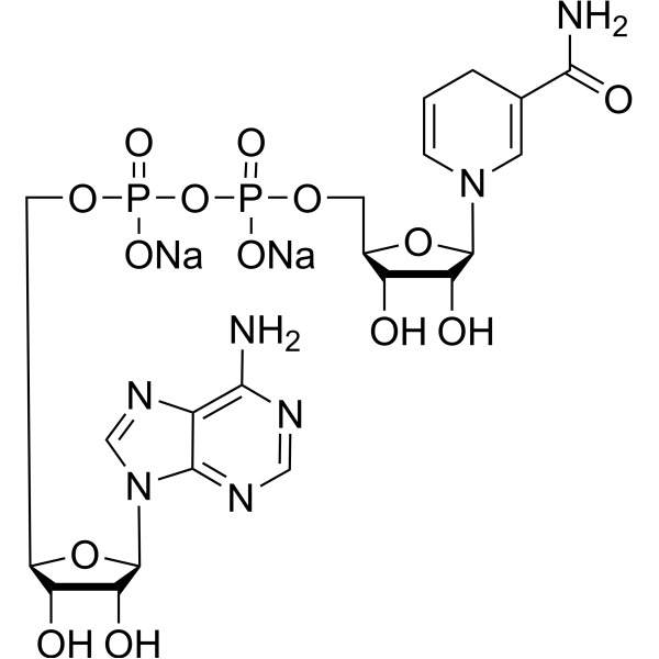
NADH disodium salt
Based on 11 publication(s) in Google Scholar
NADH disodium salt (Disodium NADH) is an orally effective reduced coenzyme. NADH disodium salt plays a role in regenerating electron donors during cellular energy metabolism, including glycolysis, β-oxidation, and the tricarboxylic acid (TCA) cycle. NADH disodium salt regulates calcium homeostasis by promoting the opening of IP3-gated Ca2+ channels and inhibiting Ryanodine receptor, and affects mitochondrial function through direct interaction with VDAC. NADH disodium salt is used in cytoprotective studies related to cerebral ischemic injury, neurodegenerative diseases, aging, and signal transduction.
For research use only. We do not sell to patients.
- Purity : 99.98%
- CAS No.: 606-68-8
- Formula: C21H27N7Na2O14P2
- Molecular Weight:709.40
-
Storage:
4°C, sealed storage, away from moisture
* In solvent : -80°C, 6 months; -20°C, 1 month (sealed storage, away from moisture)

To place orders, for customer services and technical support, please contact: MedChemExpress USA
Tel: 609-228-6898 E-mail: [email protected] [email protected]


-
 Biological Activity
-
Chemical Information
-
Solvent & Solubility
- Protocols
- Purity & Documentation
- References
-
 Help & FAQs


-
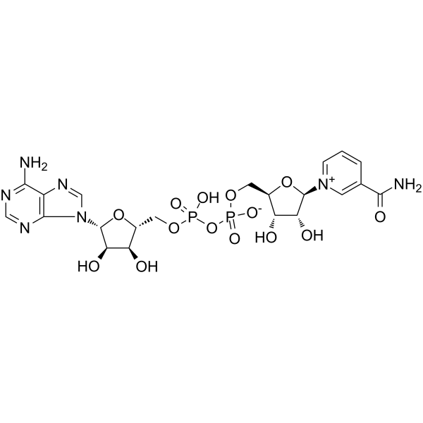
HY-B0445
NAD+Endogenous Metabolite -
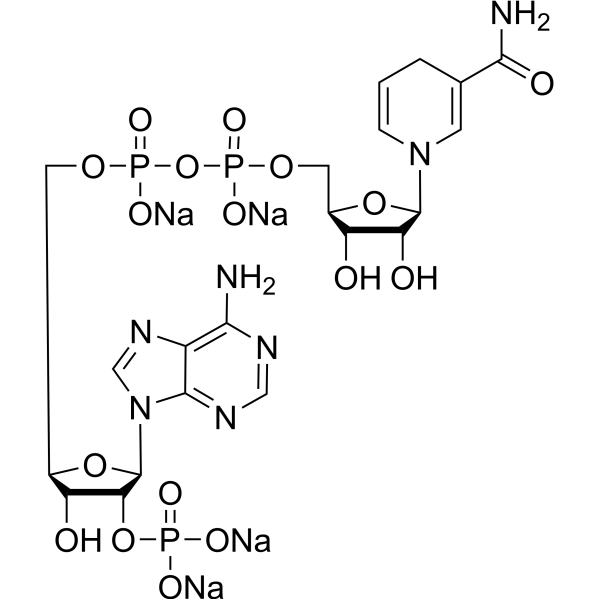
HY-F0003
NADPH tetrasodium saltCofactor -
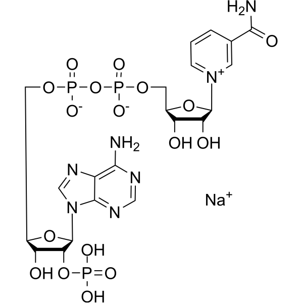
HY-F0002
NADP sodium saltRedox Coenzyme -

HY-113324
NADPHReducing Agent -
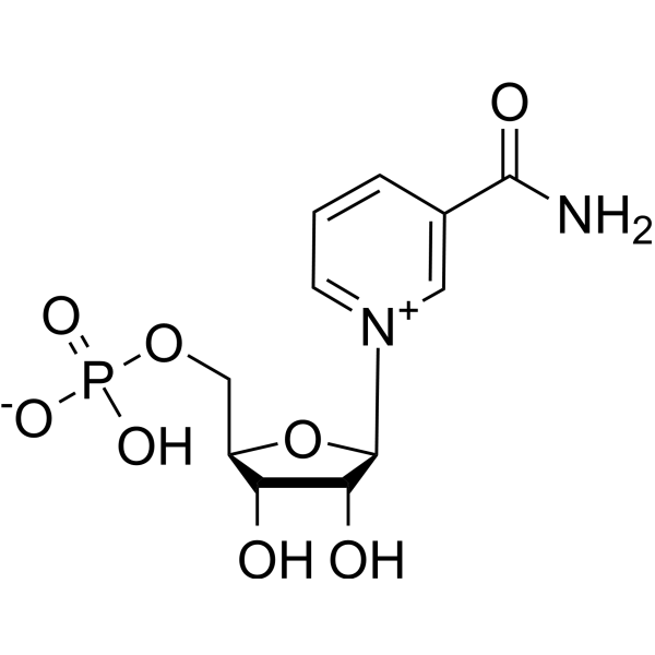
HY-F0004
FosribnicotinamideNAD+ Intermediate -
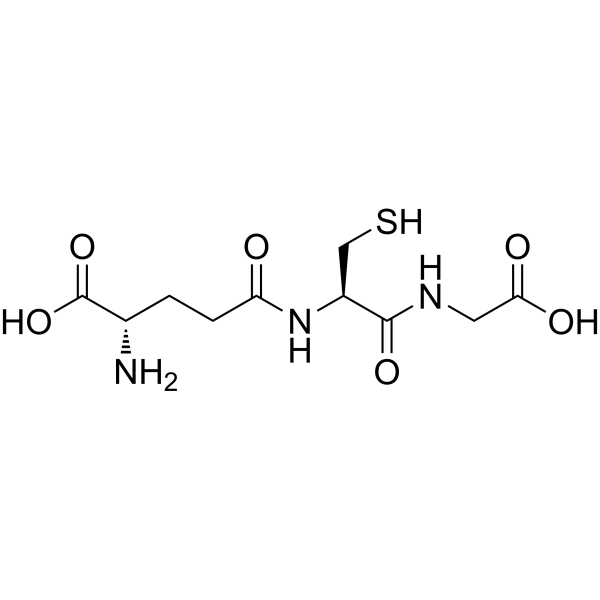
HY-D0187
L-Glutathione reducedAntioxidant -
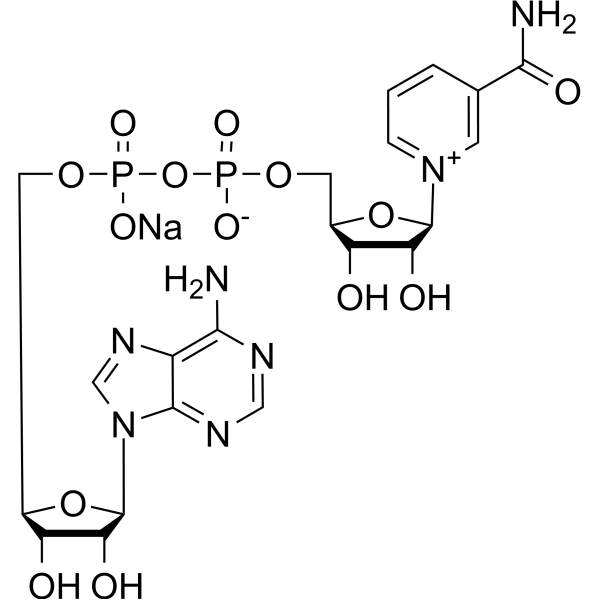
HY-B0445A
NAD sodiumCofactor, Macrophage M2 Polarization Inducer -
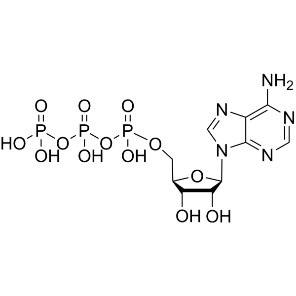
HY-B2176
ATPEndogenous Metabolite -

HY-F0001R
NADH disodium salt (Standard)Reference Standard -
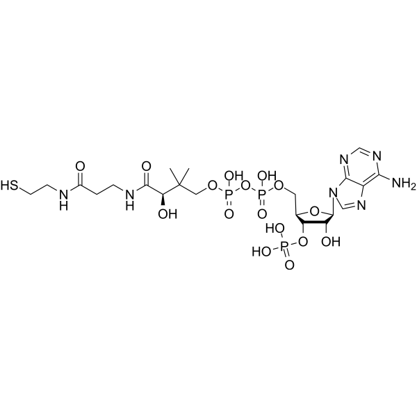
HY-128851
Coenzyme ACofactor -

HY-13259
MG-132Proteasome Inhibitor -

HY-10219
RapamycinmTOR Inhibitor
Publications Citing Use of MedChemExpress (MCE) NADH disodium salt
More- Cell Mol Immunol. 2024 Jun;21(6):561-574. [Abstract]
- Food Chem. 2023 Oct 15:423:136274. [Abstract]
- Biomater Res. 2026 Apr 29.
- Free Radic Biol Med. 2025 Jan:226:56-69. [Abstract]
- J Ginseng Res. 2025 Sep;49(5):541-552. [Abstract]
- Mol Plant Pathol. 2025 Dec;26(12):e70196. [Abstract]
- Microorganisms. 2026 May 9;14(5):1070. [Abstract]
- ACS Chem Biol. 2026 Jul 2.
- Biochemistry. 2023 Dec 5;62(23):3396-3410. [Abstract]
- Chemrxiv. 2025 Aug 28.
- Research Square Preprint. 2023 Sep 15.
All Endogenous Metabolite Isoforms
MoreAll Calcium Channel Isoforms
More

Biological Activity
Description
IC50 & Target
|
Human Endogenous Metabolite |
In Vitro
NADH disodium salt (5 mM; 1-20 min) decreases Na+ binding capacity in purified V-ATPase, with Kd values of 20 µM and 15 µM in the presence and absence of ATP (HY-B2176), respectively[3].
β-Nicotinamide adenine dinucleotide reduced (10 µM - 10 mM) dipotassium inhibits PARP-1 (HY-P73337)-mediated cell death and increases intracellular NAD+ levels in mouse astrocytes and can be transported into cells via P2X7 receptor-mediated uptake[1].
MedChemExpress (MCE) has not independently confirmed the accuracy of these methods. They are for reference only. Further protocols information, click here.
In Vivo
MedChemExpress (MCE) has not independently confirmed the accuracy of these methods. They are for reference only.
-
Animal Model:Rat model of 2-hour transient focal ischemia[1]
-
Dosage:10, 20 mg/kg
-
Administration:intranasal administration; single dose; given 2 hours after ischemic onset
-
Result:Significantly reduced the formation of brain infarction.
Improved neurological deficits by more than 80%.


Chemical Information
-
CAS No. 606-68-8
-
Appearance Solid
-
Molecular Weight 709.40
-
Formula C21H27N7Na2O14P2
-
Color Off-white to yellow
-
SMILES
O[C@H]1[C@@H](O)[C@H](N2C=C(C(N)=O)CC=C2)O[C@@H]1COP(OP(OC[C@@H]3[C@@H](O)[C@@H](O)[C@H](N(C4=NC=N5)C=NC4=C5N)O3)(O[Na])=O)(O[Na])=O
-
Synonyms
Disodium NADH
-
Structure Classification
-
Initial Source
-
Shipping
Room temperature in continental US; may vary elsewhere.
-
Storage
4°C, sealed storage, away from moisture
* In solvent : -80°C, 6 months; -20°C, 1 month (sealed storage, away from moisture)


Publications (11)
-
Journal Impact Factor
-
Most Recent
-
Cell Mol Immunol
2024 Jun;21(6):561-574. PMID: 38570588 -
Food Chem
Improvement of catalytic activity of sorbose dehydrogenase for deoxynivalenol degradation by rational design. [Abstract]2023 Oct 15:423:136274. PMID: 37159968 -
-
Free Radic Biol Med
TSPO exacerbates sepsis-induced cardiac dysfunction by inhibiting p62-Mediated autophagic flux via the ROS-RIP1/RIP3-exosome axis. [Abstract]2025 Jan:226:56-69. PMID: 39542185 -
J Ginseng Res
Total ginsenosides and ginsenoside Rb2 delay hepatocyte senescence by regulating NAD+ metabolism and promoting IDO2/QPRT expression. [Abstract]2025 Sep;49(5):541-552. PMID: 40843012 -
Mol Plant Pathol
Negative Immune Regulator CAD7 Functions as a Small-Molecule Aldehyde Reductase and Increases Histamine Accumulation in Arabidopsis. [Abstract]2025 Dec;26(12):e70196. PMID: 41456913 -
Microorganisms
Characterization of RmlABCD Enzymes from Marine Bacteria and Efficient Synthesis of dTDP-L-Rhamnose. [Abstract]2026 May 9;14(5):1070. PMID: 42197457 -
-
Biochemistry
Full-Length NAD+-I Riboswitches Bind a Single Cofactor but Cannot Discriminate against Adenosine Triphosphate. [Abstract]2023 Dec 5;62(23):3396-3410. PMID: 37947391 -
-


Solvent & Solubility
In Vitro:
H2O : 100 mg/mL (140.96 mM; Need ultrasonic)
DMSO : 100 mg/mL (140.96 mM; Need ultrasonic; Hygroscopic DMSO has a significant impact on the solubility of product, please use newly opened DMSO)
Please refer to the solubility information to select the appropriate solvent. Once prepared, please aliquot and store the solution to prevent product inactivation from repeated freeze-thaw cycles.
Storage method and period of stock solution: -80°C, 6 months; -20°C, 1 month (sealed storage, away from moisture). When stored at -80°C, please use it within 6 months. When stored at -20°C, please use it within 1 month.
* Note: If you choose water as the stock solution, please dilute it to the working solution, then filter and sterilize it with a 0.22 μm filter before use.
Please refer to the solubility information to select the appropriate solvent. Once prepared, please aliquot and store the solution to prevent product inactivation from repeated freeze-thaw cycles.
Storage method and period of stock solution: -80°C, 6 months; -20°C, 1 month (sealed storage, away from moisture). When stored at -80°C, please use it within 6 months. When stored at -20°C, please use it within 1 month.
* Note: If you choose water as the stock solution, please dilute it to the working solution, then filter and sterilize it with a 0.22 μm filter before use.
Concentration (start) × Volume (start) = Concentration (final) × Volume (final)
In Vivo:
Select the appropriate dissolution method based on your experimental animal and administration route.
- For the following dissolution methods, please ensure to first prepare a clear stock solution using an In Vitro approach and then sequentially add co-solvents:
- To ensure reliable experimental results, the clarified stock solution can be appropriately stored based on storage conditions. As for the working solution for In Vivo experiments, it is recommended to prepare freshly and use it on the same day.
- The percentages shown for the solvents indicate their volumetric ratio in the final prepared solution. If precipitation or phase separation occurs during preparation, heat and/or sonication can be used to aid dissolution.
Add each solvent one by one: 10% DMSO 40% PEG300 5% Tween-80 45% Saline
Solubility: 2.5 mg/mL (3.52 mM); Clear solution; Need ultrasonic
This protocol yields a clear solution of 2.5 mg/mL.
Taking 1 mL working solution as an example, add 100 μL DMSO stock solution (25.0 mg/mL) to 400 μL PEG300, and mix evenly; then add 50 μL Tween-80 and mix evenly; then add 450 μL Saline to adjust the volume to 1 mL.
Preparation of Saline: Dissolve 0.9 g sodium chloride in ddH₂O and dilute to 100 mL to obtain a clear Saline solution.
Add each solvent one by one: 10% DMSO 90% (20% SBE-β-CD in Saline)
Solubility: 2.5 mg/mL (3.52 mM); Clear solution; Need ultrasonic
This protocol yields a clear solution of 2.5 mg/mL.
Taking 1 mL working solution as an example, add 100 μL DMSO stock solution (25.0 mg/mL) to 900 μL 20% SBE-β-CD in Saline, and mix evenly.
Preparation of 20% SBE-β-CD in Saline (4°C, storage for one week): 2 g SBE-β-CD powder is dissolved in 10 mL Saline, completely dissolve until clear.
In Vivo Dissolution Calculator
Please enter the basic information of animal experiments:
-
-
-
-
Recommended: Prepare an additional quantity of animals to account for potential losses during experiments.
Working solution concentration: 0.22 mg/mL
This product has good water solubility, please refer to the measured solubility data in water/PBS/Saline for details.

Protocols
-
Mitochondrial membrane-potential fluorescent assay
Mitochondrial membrane potential fluorescent assays estimate ΔΨm in living cells using lipophilic cationic dyes such as TMRM, TMRE, rhodamine 123, and JC-1, which accumulate in mitochondria according to membrane polarization; loss of signal after FCCP or CCCP treatment is interpreted as mitochondrial depolarization. TMRM/TMRE and rhodamine 123 are commonly used for semi-quantitative live-cell microscopy or flow cytometry, while JC-1 can report a shift from red aggregate fluorescence to green monomer fluorescence during depolarization; interpretation requires controls because dye concentration, quenching mode, cell type, dye efflux, and mitochondrial mass can affect fluorescence independently of ΔΨm.
-
Mitochondrial membrane-potential and mitochondrial mass staining
Mitochondrial membrane potential staining measures the electrochemical polarization across the mitochondrial inner membrane in live cells using lipophilic cationic fluorescent probes; early rhodamine-based work showed that selective mitochondrial dye accumulation is lost when the mitochondrial transmembrane potential is dissipated. JC-1 reports mitochondrial polarization by shifting from green monomer fluorescence to red J-aggregate fluorescence as dye concentration increases within energized mitochondria; therefore, the red/green fluorescence ratio is used as a relative readout of mitochondrial membrane potential. TMRE or TMRM staining provides a single-channel relative readout because these cationic rhodamine esters accumulate in polarized mitochondria, and lower fluorescence indicates reduced mitochondrial polarization when acquisition and dye-loading conditions are controlled. Mitochondrial mass staining is commonly performed with MitoTracker Green FM or related MitoTracker dyes as
-
Calcium Spark Assay
Calcium sparks are localized, transient increases in intracellular calcium concentration ([Ca2+]i) that occur in cardiac myocytes and represent elementary events underlying excitation-contraction coupling. These events are generated by the coordinated opening of clusters of ryanodine receptors (RyRs) on the sarcoplasmic reticulum membrane, leading to a brief release of Ca2+ into the cytosol. The detection and analysis of calcium sparks provide insights into the mechanisms of calcium handling and signaling in cardiac cells. Imaging techniques using fluorescent calcium indicators such as Fluo-3 are employed to visualize these subcellular calcium transients with high spatial and temporal resolution. The protocol is based on established methodologies described in primary literature for both experimental measurement and automated analysis of calcium sparks.
-
Ca2+ Staining Technique
Ca2+ staining is an experimental technique that utilizes specific fluorescent probes (such as Fluo-4 AM, Fura-2, etc.) to qualitatively or quantitatively detect dynamic changes in intracellular Ca2+ concentrations; this is achieved by monitoring the changes in fluorescent signals generated when these probes bind to free intracellular calcium ions. The underlying principle relies primarily on the presence of chelating groups within the probe's molecular structure that possess high affinity for calcium ions.


Purity & Documentation
-
Data Sheet (300 KB)
-
SDS (394 KB)
- English - EN (394 KB)
- Français - FR (394 KB)
- Deutsch - DE (394 KB)
- Norwegian - NO (394 KB)
- Español - ES (394 KB)
- Swedish - SV (394 KB)
- Italian - IT (394 KB)
- Korean - KR (394 KB)
- Portuguese - PT (394 KB)
-
Handling Instructions (2659 KB)
References


Complete Stock Solution Preparation Table
Please refer to the solubility information to select the appropriate solvent. Once prepared, please aliquot and store the solution to prevent product inactivation from repeated freeze-thaw cycles.
Storage method and period of stock solution: -80°C, 6 months; -20°C, 1 month (sealed storage, away from moisture). When stored at -80°C, please use it within 6 months. When stored at -20°C, please use it within 1 month.
| Optional Solvent | Concentration Solvent Mass | 1 mg | 5 mg | 10 mg | 25 mg |
|---|---|---|---|---|---|
| H2O / DMSO | 1 mM | 1.4096 mL | 7.0482 mL | 14.0964 mL | 35.2410 mL |
| 5 mM | 0.2819 mL | 1.4096 mL | 2.8193 mL | 7.0482 mL | |
| 10 mM | 0.1410 mL | 0.7048 mL | 1.4096 mL | 3.5241 mL | |
| 15 mM | 0.0940 mL | 0.4699 mL | 0.9398 mL | 2.3494 mL | |
| 20 mM | 0.0705 mL | 0.3524 mL | 0.7048 mL | 1.7621 mL | |
| 25 mM | 0.0564 mL | 0.2819 mL | 0.5639 mL | 1.4096 mL | |
| 30 mM | 0.0470 mL | 0.2349 mL | 0.4699 mL | 1.1747 mL | |
| 40 mM | 0.0352 mL | 0.1762 mL | 0.3524 mL | 0.8810 mL | |
| 50 mM | 0.0282 mL | 0.1410 mL | 0.2819 mL | 0.7048 mL | |
| 60 mM | 0.0235 mL | 0.1175 mL | 0.2349 mL | 0.5874 mL | |
| 80 mM | 0.0176 mL | 0.0881 mL | 0.1762 mL | 0.4405 mL | |
| 100 mM | 0.0141 mL | 0.0705 mL | 0.1410 mL | 0.3524 mL |
* Note: If you choose water as the stock solution, please dilute it to the working solution, then filter and sterilize it with a 0.22 μm filter before use.


NADH disodium salt Related Classifications
HY-F0001 Related Classifications
 Powered by Bioz