



PF-543 Citrate
Based on 28 publication(s) in Google Scholar
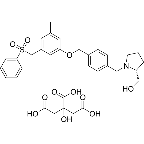
PF-543 Citrate (Sphingosine Kinase 1 Inhibitor II Citrate) is a potent, selective, reversible and sphingosine-competitive SPHK1 inhibitor with an IC50 of 2 nM and a Ki of 3.6 nM. PF-543 Citrate is >100-fold selectivity for SPHK1 over SPHK2. PF-543 Citrate is an effective potent inhibitor of sphingosine 1-phosphate (S1P) formation in whole blood with an IC50 of 26.7 nM. PF-543 Citrate induces apoptosis, necrosis, and autophagy.
For research use only. We do not sell to patients.
- Purity : 99.47%
- CAS No.: 1415562-83-2
- Formula: C33H39NO11S
- Molecular Weight:657.73
-
Storage:
4°C, sealed storage, away from moisture
* In solvent : -80°C, 6 months; -20°C, 1 month (sealed storage, away from moisture)

To place orders, for customer services and technical support, please contact: MedChemExpress USA
Tel: 609-228-6898 E-mail: [email protected] [email protected]


-
 Biological Activity
-
Chemical Information
-
Solvent & Solubility
- Protocols
- Purity & Documentation
- References
-
 Help & FAQs


-
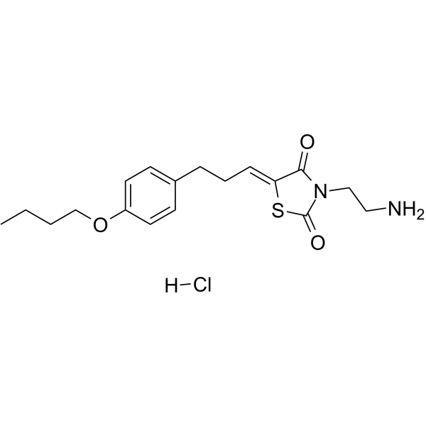
HY-15779A
K145 hydrochlorideSphK2 Inhibitor -
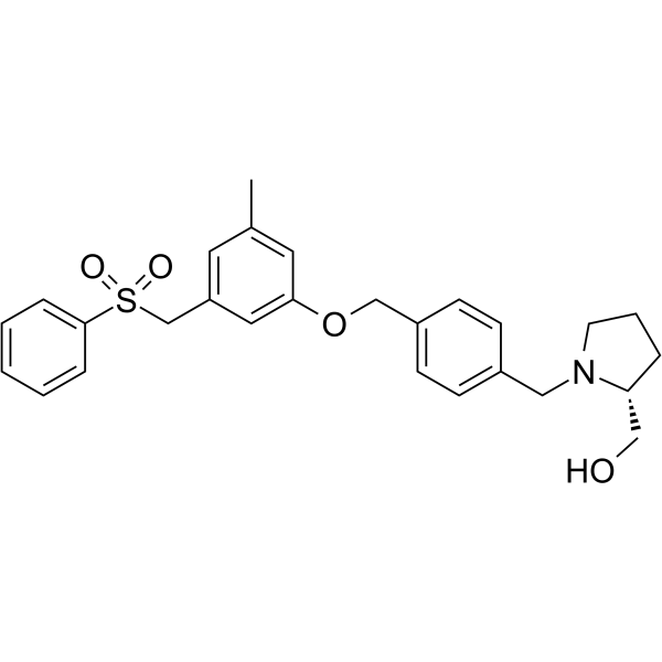
HY-15425
PF-543SPHK1 Inhibitor -
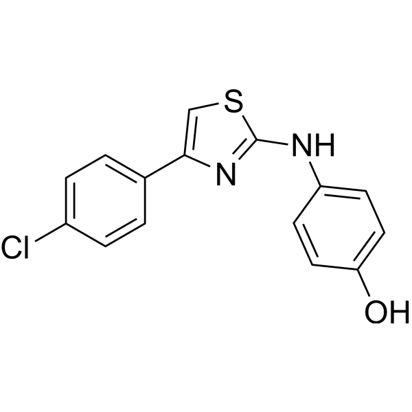
HY-13822
SKI IISphK/DES1 Inhibitor -
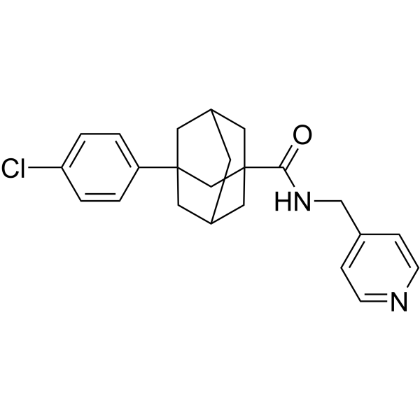
HY-16015
Opaganib -
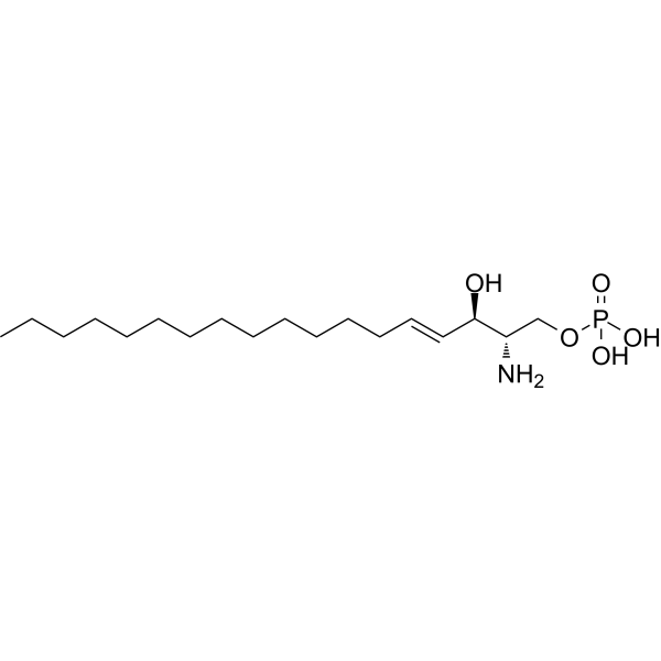
HY-108496
Sphingosine-1-phosphateS1PR Agonist -
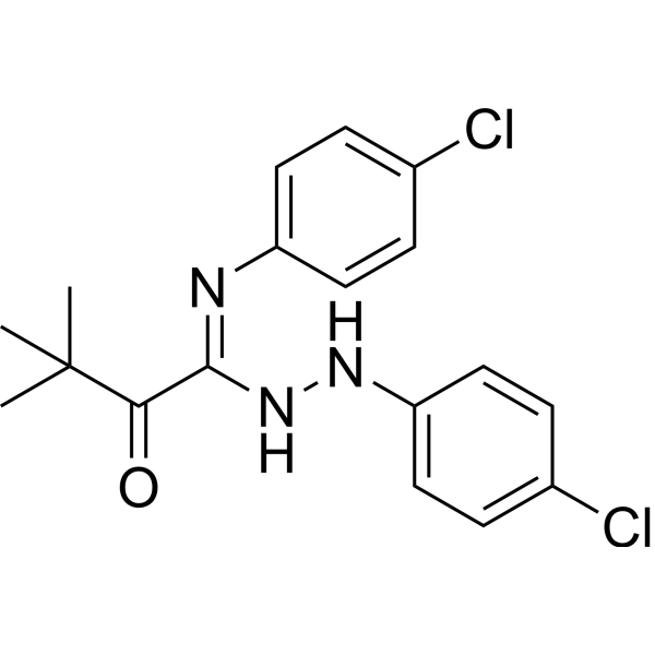
HY-19736
TY-52156LPL Receptor Antagonist -
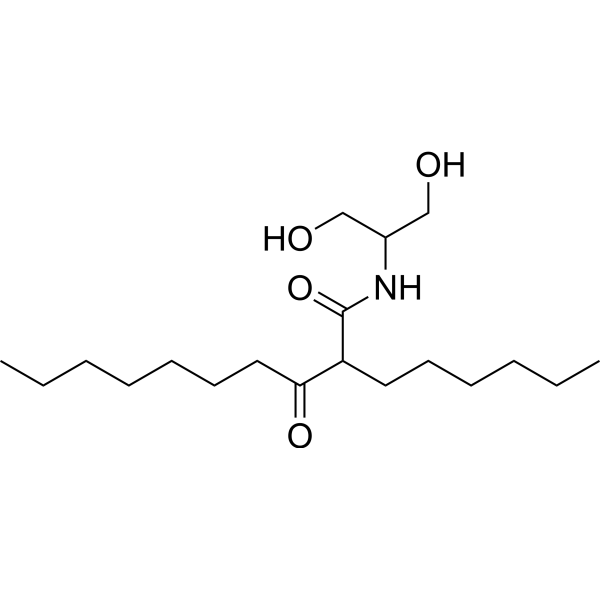
HY-124042
K6PC-5SPHK1 Activator -
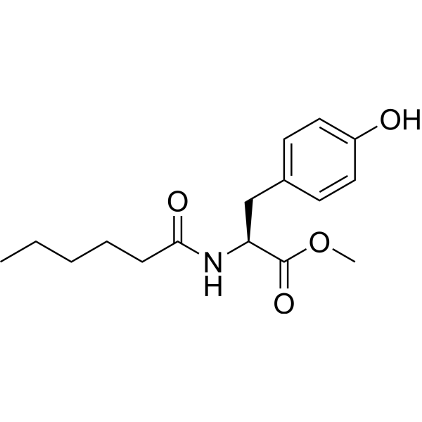
HY-101572
MHPSPHK1 Activator -
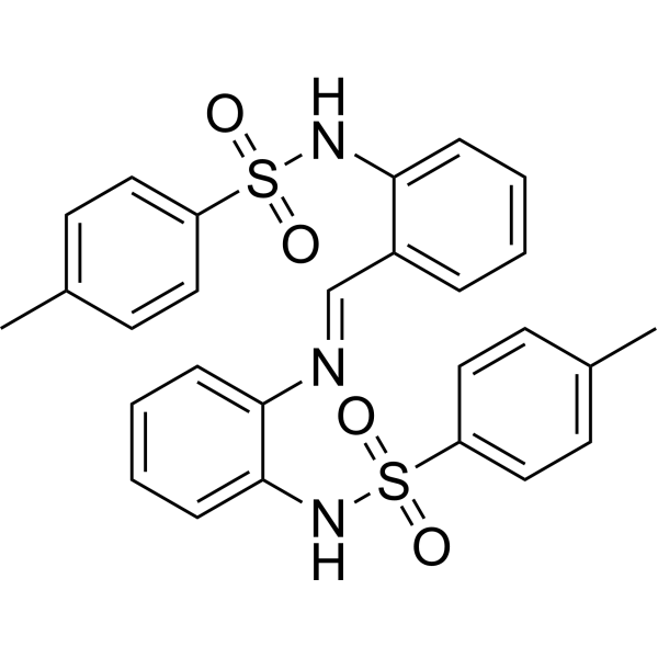
HY-19794
MP-A08SphK Inhibitor -
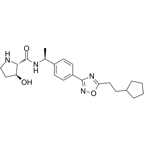
HY-101805
SK1-IN-1SPHK1 Inhibitor -
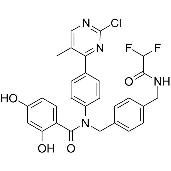
HY-12492
VER-246608 -
HY-12892
SKI-178SphK1/SphK2 Inhibitor
Publications Citing Use of MedChemExpress (MCE) PF-543 Citrate
More- Cell Metab. 2026 May 14:S1550-4131(26)00151-8. [Abstract]
- Cancer Commun (Lond). 2025 Jul 16. [Abstract]
- Mol Cell. 2020 Mar 19;77(6):1294-1306.e5. [Abstract]
- Pharmacol Res. 2026 Apr 30:108225. [Abstract]
- Cell Death Dis. 2024 Aug 1;15(8):552. [Abstract]
- Phytomedicine. 2025 Sep 19:148:157271. [Abstract]
- EBioMedicine. 2025 Dec 8;123:106058.
- Sci China Life Sci. 2026 May;69(5):1634-1649. [Abstract]
- Sci China Life Sci. 2022 Feb;65(2):341-361. [Abstract]
- Chin Med J (Engl). 2026 May 5;139(9):1375-1387. [Abstract]
- Arch Pharm Res. 2025 Aug;48(7-8):798-813. [Abstract]
- Phytother Res. 2025 Aug;39(8):3419-3431. [Abstract]
- Environ Pollut. 2025 Oct 15:383:126846. [Abstract]
- J Ethnopharmacol. 2026 Nov 15:370:121987. [Abstract]
- Pain. 2026 Apr 1;167(4):962-975. [Abstract]
- Eur J Pharmacol. 2026 May 10:1023:178866. [Abstract]
- Inflammation. 2021 Dec;44(6):2170-2179. [Abstract]
- Chem Biol Interact. 2026 Jul 1:434:112105. [Abstract]
- Sci Rep. 2020 Aug 14;10(1):13834. [Abstract]
- Cancer Sci. 2020 Jul;111(7):2259-2274. [Abstract]
- FASEB J. 2024 Jan 31;38(2):e23417. [Abstract]
- Immunotargets Ther. 2026 Jan 21;15:1-19.
- Immunotargets Ther. 2026 Jan 22:15:569823. [Abstract]
- Ren Fail. 2025 Dec;47(1):2568972. [Abstract]
- Pflugers Arch. 2025 Jun;477(6):815-826. [Abstract]
- Dig Dis Sci. 2025 Jun 5. [Abstract]
- Hum Cell. 2020 Jan;33(1):57-66. [Abstract]
- Curr Res Pharmacol Drug Discov. 2025 Jan 9:8:100212. [Abstract]
 Customer Validation & Images

-
RT-PCR
-
Bio/Physico-chemical Assay
-
In Vivo Efficacy Study
-
IF
-
Bio/Physico-chemical Assay


Biological Activity
Description
IC50 & Target
|
SphK1 |
In Vitro
PF-543 (10-1000 nM; 24?hours; PASM cells) treatment abolishes SK1 expression at nM concentrations[2].
?
PF-543 (0.1-10 μM; 24 hours; PASM cells) treatment induces caspase-3/7 activity[2].
?
PF-543 inhibits C17-S1P formation in 1483 cells with an IC50 of 1.0 nM[1].
?
SphK1 inhibition by PF-543 causes a dose-dependent depletion of the intracellular level of S1P with EC50 concentration of 8.4 nM and a concomitant elevation of the intracellular level of sphingosine in 1483 cells. The level of endogenous S1P in 1483 cells after a 1 h treatment with 200 nM PF-543 is decreased 10-fold, producing a proportional increase in the level of sphingosine[1].
MedChemExpress (MCE) has not independently confirmed the accuracy of these methods. They are for reference only. Further protocols information, click here.
-
Cell Line:Human pulmonary arterial smooth muscle (PASM) cells
-
Concentration:10 nM, 100 nM, 1000 nM
-
Incubation Time:24 hours
-
Result:Abolished SK1 expression at nM concentrations.
-
Cell Line:Human pulmonary arterial smooth muscle (PASM) cells
-
Concentration:0.1 μM, 1 μM, 10 μM
-
Incubation Time:24 hours
-
Result:Induced caspase-3/7 activity in cultured human pulmonary smooth muscle cells.
In Vivo
? Mice are initially dosed (ip) with 10 mg/kg or 30 mg/kg of PF-543 for 24 h and the T1/2 is 1.2 h in blood samples. Administration of 10 mg/kg PF-543 for 24 h to mice induces a decrease in SK1 expression in pulmonary vessels[2].
MedChemExpress (MCE) has not independently confirmed the accuracy of these methods. They are for reference only.
-
Animal Model:Female C57BL/6 J mice (7-12 week-old) with hypoxic-induced pulmonary arterial hypertension[2]
-
Dosage:1 mg/kg
-
Administration:Intraperitoneal injection; every second day; for 21 days
-
Result:Reduced right ventricular hypertrophy. The protection involves a reduction in the expression of p53 (that promotes cardiomyocyte death) and an increase in the expression of anti-oxidant nuclear factor Nrf-2.


Chemical Information
-
CAS No. 1415562-83-2
-
Appearance Solid
-
Molecular Weight 657.73
-
Formula C33H39NO11S
-
Color White to yellow
-
SMILES
OC(C(O)=O)(CC(O)=O)CC(O)=O.O=S(C1=CC=CC=C1)(CC2=CC(C)=CC(OCC3=CC=C(CN4[C@@H](CO)CCC4)C=C3)=C2)=O
-
Synonyms
Sphingosine Kinase 1 Inhibitor II Citrate
-
Shipping
Room temperature in continental US; may vary elsewhere.
-
Storage
4°C, sealed storage, away from moisture
* In solvent : -80°C, 6 months; -20°C, 1 month (sealed storage, away from moisture)


Publications (28)
-
Journal Impact Factor
-
Most Recent
-
Cell Metab
Adipokine IL-11/IL-11Ra constrains sphingolipid metabolism to limit the thermogenic capacity of beige adipocytes. [Abstract]2026 May 14:S1550-4131(26)00151-8. PMID: 42140185 -
Cancer Commun (Lond)
Targeting SPHK1 in macrophages remodels the tumor microenvironment and enhances anti-PD-1 immunotherapy efficacy in colorectal cancer liver metastasis. [Abstract]2025 Jul 16. PMID: 40665874
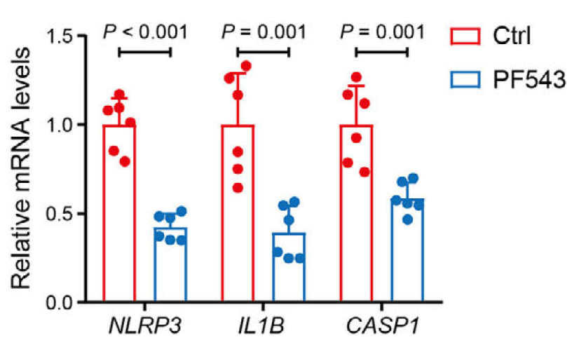
PF-543 Citrate purchased from MedChemExpress. Usage Cited in: Cancer Commun (Lond). 2025 Jul 16. [Abstract]
Relative mRNA expression of inflammasome genes in TAMs from liver tumors with or without PF-543 (5 mg/kg; i.p.) treatment were determined by qPCR.
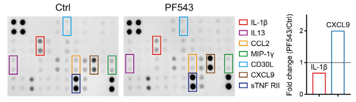
PF-543 Citrate purchased from MedChemExpress. Usage Cited in: Cancer Commun (Lond). 2025 Jul 16. [Abstract]
Representative images and relative levels of IL-1β and CXCL9 in Ctrl- and PF-543-treated liver tumors determined by a mouse inflammatory cytokine array. PF-543 (5 mg/kg; i.p.) significantly decreased IL‐1β expression in TAMs compared to control group.
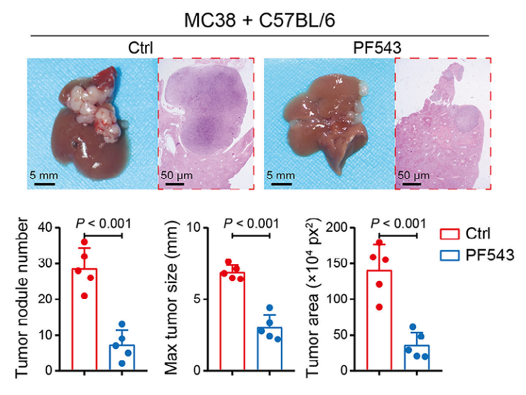
PF-543 Citrate purchased from MedChemExpress. Usage Cited in: Cancer Commun (Lond). 2025 Jul 16. [Abstract]
SPHK1 inhibitor PF-543 (5 mg/kg; i.p.; once every 3 d) reduced the number and diameter of liver metastatic tumors in mice.
PF-543 Citrate purchased from MedChemExpress. Usage Cited in: Cancer Commun (Lond). 2025 Jul 16. [Abstract]
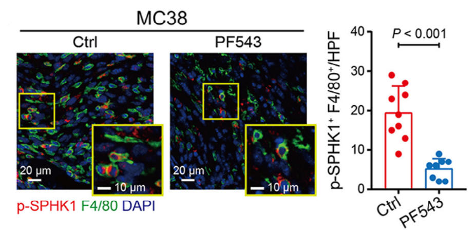
Representative IF images and quantification of co-staining of p-SPHK1 (red) and F4/80 (green) in liver tumor tissues from C57BL/6 mice with or without PF-543 (5 mg/kg; i.p.; once every 3 d). PF-543 treatment decreased number of phosphorylated SPHK1+ TAMs (activated SPHK1) in the liver tumor microenvironment.
PF-543 Citrate purchased from MedChemExpress. Usage Cited in: Cancer Commun (Lond). 2025 Jul 16. [Abstract]
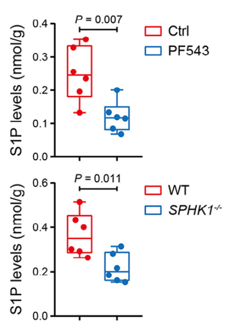
SPHK1 inhibitor PF-543 (5 mg/kg; i.p.; once every 3 d) markedly reduced the level of S1P in MC38 liver tumors.
-
Mol Cell
2020 Mar 19;77(6):1294-1306.e5. PMID: 32023483 -
Pharmacol Res
Lysosome-triggered nanotherapy packages osteoclast apoptotic bodies with pro-anabolic lipids to couple anti-resorption and bone formation. [Abstract]2026 Apr 30:108225. PMID: 42069319 -
Cell Death Dis
Triggering of endoplasmic reticulum stress via ATF4-SPHK1 signaling promotes glioblastoma invasion and chemoresistance. [Abstract]2024 Aug 1;15(8):552. PMID: 39090107 -
Phytomedicine
Alisol A 24-acetate protects against NASH-associated fibrosis via suppression of Kupffer cell-derived SPHK1/S1P axis. [Abstract]2025 Sep 19:148:157271. PMID: 40992063 -
-
Sci China Life Sci
SPHK1 deficiency promotes intestinal homeostasis by ameliorating ER stress-induced gastrointestinal injury during murine graft-versus-host disease. [Abstract]2026 May;69(5):1634-1649. PMID: 41627669 -
Sci China Life Sci
2022 Feb;65(2):341-361. PMID: 34047913 -
Chin Med J (Engl)
Sphingosine-1-phosphate induces pulmonary artery smooth muscle cell proliferation, migration and pulmonary arterial remodeling by modulating sonic hedgehog signaling effector FoxM1. [Abstract]2026 May 5;139(9):1375-1387. PMID: 41859863 -
Arch Pharm Res
2025 Aug;48(7-8):798-813. PMID: 40681917 -
Phytother Res
Geniposide Improves Glycolysis Driven Angiogenesis in Experimentary Arthritis by Inhibiting SphK1-PI3K-Akt-PFKFB3 Signal. [Abstract]2025 Aug;39(8):3419-3431. PMID: 40583637 -
Environ Pollut
Polycyclic aromatic hydrocarbon aggravates high-fat diet-induced metabolic dysfunction-associated steatotic liver disease through disturbing hepatocyte sphingolipid metabolism. [Abstract]2025 Oct 15:383:126846. PMID: 40681076 -
J Ethnopharmacol
Xianlian Jiedu Decoction induces apoptosis in colorectal cancer via modulation of sphingosine-1-phosphate-dependent JNK/p38 MAPK signaling. [Abstract]2026 Nov 15:370:121987. PMID: 42276393 -
Pain
Mast cell corticotropin-releasing factor receptor 1 contributes to pancreatic cancer pain via mitogen-activated protein kinase/sphingosine kinases type 1 signaling. [Abstract]2026 Apr 1;167(4):962-975. PMID: 41615309 -
Eur J Pharmacol
Inhibition of PKM2 lactylation by geniposide ameliorates synovial hyperplasia in experimental arthritis. [Abstract]2026 May 10:1023:178866. PMID: 41990904 -
Inflammation
Blocking SphK1/S1P/S1PR1 Signaling Pathway Alleviates Lung Injury Caused by Sepsis in Acute Ethanol Intoxication Mice. [Abstract]2021 Dec;44(6):2170-2179. PMID: 34109517 -
Chem Biol Interact
Glyoxylic acid-induced calcium oxalate crystal deposition drives nephrotoxicity via SPHK1-mediated ferroptosis: Insights from untargeted lipidomics. [Abstract]2026 Jul 1:434:112105. PMID: 42044864 -
Sci Rep
Follicle-stimulating hormone promotes the proliferation of epithelial ovarian cancer cells by activating sphingosine kinase. [Abstract]2020 Aug 14;10(1):13834. PMID: 32796926 -
Cancer Sci
Human alkaline ceramidase 2 promotes the growth, invasion, and migration of hepatocellular carcinoma cells via sphingomyelin phosphodiesterase acid-like 3B. [Abstract]2020 Jul;111(7):2259-2274. PMID: 32391585 -
FASEB J
SPHK1/S1P/S1PR pathway promotes the progression of peritoneal fibrosis by mesothelial-mesenchymal transition. [Abstract]2024 Jan 31;38(2):e23417. PMID: 38226856 -
-
Immunotargets Ther
Upregulated BLM and RECQL4 in Osteosarcoma: Association with Poor Prognosis, Immune Cell Infiltration, and Inhibitory Effects of Sphingosine Kinase 1 Inhibitor II/Pilaralisib. [Abstract]2026 Jan 22:15:569823. PMID: 41878019 -
Ren Fail
Hypoxia-inducible factor 2α overexpression in podocytes ameliorates lipid metabolism disorders in diabetic kidney disease by inhibiting S1P. [Abstract]2025 Dec;47(1):2568972. PMID: 41077842 -
Pflugers Arch
2025 Jun;477(6):815-826. PMID: 39899071 -
Dig Dis Sci
The Combination of PF-543 and TRAIL Effectively Induces Apoptotic Cell Death and Inhibits Stem Cell-Like Properties Through the SPHK1/S1PR1/STAT3 Pathway in TRAIL-Resistant Colorectal Cancer Cells. [Abstract]2025 Jun 5. PMID: 40467915 -
Hum Cell
2020 Jan;33(1):57-66. PMID: 31606874 -
Curr Res Pharmacol Drug Discov
The suppression of the SPHK1/S1P/S1PR3 signaling pathway diminishes EGFR activation and increases the sensitivity of non-small cell lung cancer to gefitinib. [Abstract]2025 Jan 9:8:100212. PMID: 39896887


Solvent & Solubility
In Vitro:
DMSO : 200 mg/mL (304.08 mM; Need ultrasonic; Hygroscopic DMSO has a significant impact on the solubility of product, please use newly opened DMSO)
H2O : 50 mg/mL (76.02 mM; Need ultrasonic)
Please refer to the solubility information to select the appropriate solvent. Once prepared, please aliquot and store the solution to prevent product inactivation from repeated freeze-thaw cycles.
Storage method and period of stock solution: -80°C, 6 months; -20°C, 1 month (sealed storage, away from moisture). When stored at -80°C, please use it within 6 months. When stored at -20°C, please use it within 1 month.
* Note: If you choose water as the stock solution, please dilute it to the working solution, then filter and sterilize it with a 0.22 μm filter before use.
Please refer to the solubility information to select the appropriate solvent. Once prepared, please aliquot and store the solution to prevent product inactivation from repeated freeze-thaw cycles.
Storage method and period of stock solution: -80°C, 6 months; -20°C, 1 month (sealed storage, away from moisture). When stored at -80°C, please use it within 6 months. When stored at -20°C, please use it within 1 month.
* Note: If you choose water as the stock solution, please dilute it to the working solution, then filter and sterilize it with a 0.22 μm filter before use.
Concentration (start) × Volume (start) = Concentration (final) × Volume (final)
In Vivo:
Select the appropriate dissolution method based on your experimental animal and administration route.
- For the following dissolution methods, please ensure to first prepare a clear stock solution using an In Vitro approach and then sequentially add co-solvents:
- To ensure reliable experimental results, the clarified stock solution can be appropriately stored based on storage conditions. As for the working solution for In Vivo experiments, it is recommended to prepare freshly and use it on the same day.
- The percentages shown for the solvents indicate their volumetric ratio in the final prepared solution. If precipitation or phase separation occurs during preparation, heat and/or sonication can be used to aid dissolution.
Add each solvent one by one: 10% DMSO 40% PEG300 5% Tween-80 45% Saline
Solubility: ≥ 2.5 mg/mL (3.80 mM); Clear solution
This protocol yields a clear solution of ≥ 2.5 mg/mL (saturation unknown).
Taking 1 mL working solution as an example, add 100 μL DMSO stock solution (25.0 mg/mL) to 400 μL PEG300, and mix evenly; then add 50 μL Tween-80 and mix evenly; then add 450 μL Saline to adjust the volume to 1 mL.
Preparation of Saline: Dissolve 0.9 g sodium chloride in ddH₂O and dilute to 100 mL to obtain a clear Saline solution.
Add each solvent one by one: 10% DMSO 90% (20% SBE-β-CD in Saline)
Solubility: ≥ 2.5 mg/mL (3.80 mM); Clear solution
This protocol yields a clear solution of ≥ 2.5 mg/mL (saturation unknown).
Taking 1 mL working solution as an example, add 100 μL DMSO stock solution (25.0 mg/mL) to 900 μL 20% SBE-β-CD in Saline, and mix evenly.
Preparation of 20% SBE-β-CD in Saline (4°C, storage for one week): 2 g SBE-β-CD powder is dissolved in 10 mL Saline, completely dissolve until clear.
For the following dissolution methods, please prepare the working solution directly:
It is recommended to prepare fresh solutions and use them promptly within a short period of time.
The percentages shown for the solvents indicate their volumetric ratio in the final prepared solution. If precipitation or phase separation occurs during preparation, heat and/or sonication can be used to aid dissolution.
Add each solvent one by one: PBS
Solubility: 100 mg/mL (152.04 mM); Clear solution; Need ultrasonic
In Vivo Dissolution Calculator
Please enter the basic information of animal experiments:
-
-
-
-
Recommended: Prepare an additional quantity of animals to account for potential losses during experiments.
Working solution concentration: 0.22 mg/mL
This product has good water solubility, please refer to the measured solubility data in water/PBS/Saline for details.

Protocols
-
Apoptosis
Apoptosis, also called programmed cell death, is generally characterized by distinct morphological characteristics.
-
TUNEL staining for apoptotic DNA fragmentation
TUNEL staining detects DNA strand breaks by using terminal deoxynucleotidyl transferase to add labeled nucleotides to exposed 3′-OH DNA termini, generating either microscopic staining in fixed cells or tissue sections, or fluorescence/cytometric signal in cell suspensions. TUNEL positivity reflects DNA fragmentation but should not be interpreted alone as definitive apoptosis, because TUNEL can also label necrotic, autolytic, mechanically damaged, or DNA-repair-associated DNA breaks.
-
Cell Cytotoxicity Assay
Cytotoxicity assays are usually based on the assessment of cell membrane damage, which can also be indirectly detected by measuring cell viability. Detection methods include MTT assay, CKK-8 assay, LDH assay and ATP assay, etc.
-
Annexin V plus membrane-impermeant dye apoptosis staining
Annexin V-based apoptosis assays rely on the detection of phosphatidylserine (PS) externalization from the inner leaflet of the plasma membrane to the outer leaflet, an early biochemical hallmark of apoptosis. Fluorescently labeled Annexin V binds PS in a calcium-dependent manner, enabling identification of early apoptotic cells by flow cytometry or fluorescence microscopy. When combined with a membrane-impermeant DNA-binding dye (e. g. , propidium iodide), this approach allows discrimination between viable (Annexin V−/dye−), early apoptotic (Annexin V+/dye−), and late apoptotic or necrotic (Annexin V+/dye+) cell populations by assessing membrane integrity and PS exposure.
-
Autophagy
Autophagy is a process in which eukaryotic cells use lysosomes to degrade their own cytoplasmic proteins and damaged organelles under the regulation of autophagy related gene (Atg). Microtubule-associated proteins light chain 3 (LC3) is recognized as autophagy marker, which transfers from cytoplasmic LC3 (LC3-I) to membrane type (LC3-II). LC3-II/I ratio could be detected by Western Blot and fluorescence microscopy.
-
Apoptosis Solutions
Apoptosis is a regulated, generally non-lytic cell-death pathway that removes unwanted, damaged, infected, or abnormal cells through coordinated morphological changes, caspase activation, DNA fragmentation, and membrane remodeling. The intrinsic apoptosis pathway is controlled mainly by mitochondrial outer membrane permeabilization, BCL-2 family proteins, cytochrome c release, apoptosome formation, caspase-9 activation, and downstream executioner caspase-3/7 activation. The extrinsic apoptosis pathway is initiated by death receptors such as Fas, TNFR, and TRAIL receptors, which recruit adaptor proteins and activate caspase-8 before engaging executioner caspases or mitochondrial amplification through BID cleavage. Apoptosis is linked to many phenotypes, including cancer cell killing, tissue homeostasis, immune regulation, neurodegeneration, infection response, and treatment-induced cytotoxicity; unresolved questions include how apoptosis interacts with necroptosis, pyroptosis, ferroptos
-
Lysosome and acidic-vesicle live-cell staining
Lysosome and acidic-vesicle live-cell staining detects acidic intracellular compartments by using membrane-permeant acidotropic probes that accumulate in low-pH vesicles, including lysosomes, late endosomes, autolysosomes, and acidic phagosomes. LysoTracker staining is commonly used as an intensity-based readout of acidic lysosomal compartment abundance or enlargement, while acridine orange produces green fluorescence in less concentrated compartments and red fluorescence after concentration-dependent accumulation in acidic vesicular organelles. Loss or reduction of acridine-orange red signal can be used as a readout of lysosomal membrane permeabilization or reduced acidic-vesicle integrity. This protocol is designed for live cultured cells and can be adapted for fluorescence microscopy, high-content imaging, plate-reader readout, or flow cytometry when the selected literature supports the readout. Because these dyes report acidotropic accumulation rather than lysosome identity alone,
-
Macroautophagy Solutions
Macroautophagy is a conserved lysosome-dependent degradation pathway in which cytoplasmic material is sequestered into double-membrane autophagosomes and delivered to lysosomes for degradation and recycling. The pathway supports cellular homeostasis during nutrient limitation, organelle stress, protein-aggregate accumulation, infection, differentiation, and tissue remodeling by coupling cargo sequestration, autophagosome maturation, lysosomal fusion, and degradation of cargo-derived macromolecules. The core molecular sequence includes initiation by nutrient- and stress-regulated autophagy machinery, autophagosome nucleation, LC3/ATG8-family conjugation to autophagosomal membranes, cargo selection through receptors such as SQSTM1/p62, autophagosome-lysosome fusion, and lysosomal degradation. LC3 was identified as a mammalian homolog of yeast Atg8 that localizes to autophagosomal membranes after processing, and p62/SQSTM1 was shown to connect ubiquitinated cargo with autophagic degradati


Purity & Documentation
-
Data Sheet (283 KB)
-
SDS (252 KB)
- English - EN (252 KB)
- Français - FR (252 KB)
- Deutsch - DE (252 KB)
- Norwegian - NO (252 KB)
- Español - ES (252 KB)
- Swedish - SV (252 KB)
- Italian - IT (252 KB)
- Korean - KR (252 KB)
- Portuguese - PT (252 KB)
-
Handling Instructions (2659 KB)
References
[1]. Schnute ME, et al. Modulation of cellular S1P levels with a novel, potent and specific inhibitor of sphingosine kinase-1. Biochem J. 2012 May 15;444(1):79-88. [Content Brief]
[2]. MacRitchie N, et al. Effect of the sphingosine kinase 1 selective inhibitor, PF-543 on arterial and cardiac remodelling in a hypoxic model of pulmonary arterial hypertension. Cell Signal. 2016 Aug;28(8):946-55. [Content Brief]
[3]. Hamada M, et al. Induction of autophagy by sphingosine kinase 1 inhibitor PF-543 in head and neck squamous cell carcinoma cells. Cell Death Discov. 2017 Aug 14;3:17047. [Content Brief]


Complete Stock Solution Preparation Table
Please refer to the solubility information to select the appropriate solvent. Once prepared, please aliquot and store the solution to prevent product inactivation from repeated freeze-thaw cycles.
Storage method and period of stock solution: -80°C, 6 months; -20°C, 1 month (sealed storage, away from moisture). When stored at -80°C, please use it within 6 months. When stored at -20°C, please use it within 1 month.
| Optional Solvent | Concentration Solvent Mass | 1 mg | 5 mg | 10 mg | 25 mg |
|---|---|---|---|---|---|
| H2O / DMSO | 1 mM | 1.5204 mL | 7.6019 mL | 15.2038 mL | 38.0095 mL |
| 5 mM | 0.3041 mL | 1.5204 mL | 3.0408 mL | 7.6019 mL | |
| 10 mM | 0.1520 mL | 0.7602 mL | 1.5204 mL | 3.8010 mL | |
| 15 mM | 0.1014 mL | 0.5068 mL | 1.0136 mL | 2.5340 mL | |
| 20 mM | 0.0760 mL | 0.3801 mL | 0.7602 mL | 1.9005 mL | |
| 25 mM | 0.0608 mL | 0.3041 mL | 0.6082 mL | 1.5204 mL | |
| 30 mM | 0.0507 mL | 0.2534 mL | 0.5068 mL | 1.2670 mL | |
| 40 mM | 0.0380 mL | 0.1900 mL | 0.3801 mL | 0.9502 mL | |
| 50 mM | 0.0304 mL | 0.1520 mL | 0.3041 mL | 0.7602 mL | |
| 60 mM | 0.0253 mL | 0.1267 mL | 0.2534 mL | 0.6335 mL | |
| DMSO | 80 mM | 0.0190 mL | 0.0950 mL | 0.1900 mL | 0.4751 mL |
| 100 mM | 0.0152 mL | 0.0760 mL | 0.1520 mL | 0.3801 mL |
* Note: If you choose water as the stock solution, please dilute it to the working solution, then filter and sterilize it with a 0.22 μm filter before use.


PF-543 Citrate Related Classifications
HY-15425A Related Classifications
 Powered by Bioz