



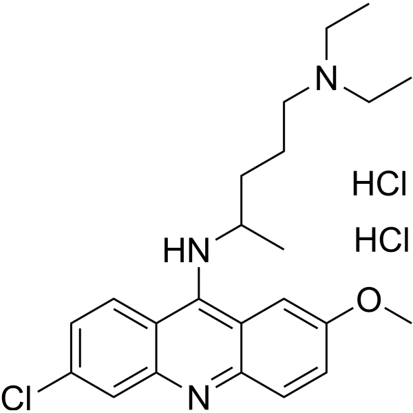
Quinacrine dihydrochloride
Based on 9 publication(s) in Google Scholar
Quinacrine (Mepacrine) dihydrochloride is an orally bioavailable antimalarial agent, which possess anticancer effect both in vitro and vivo. Quinacrine dihydrochloride suppresses NF-κB and activate p53 signaling, which results in the induction of the apoptosis.
For research use only. We do not sell to patients.
- Purity : 99.38%
- CAS No.: 69-05-6
- Formula: C23H32Cl3N3O
- Molecular Weight:472.88
-
Storage:
4°C, sealed storage, away from moisture
* In solvent : -80°C, 6 months; -20°C, 1 month (sealed storage, away from moisture)

To place orders, for customer services and technical support, please contact: MedChemExpress USA
Tel: 609-228-6898 E-mail: [email protected] [email protected]


-
 Biological Activity
-
Chemical Information
-
Solvent & Solubility
- Protocols
- Purity & Documentation
- References
-
 Help & FAQs


-
HY-13629
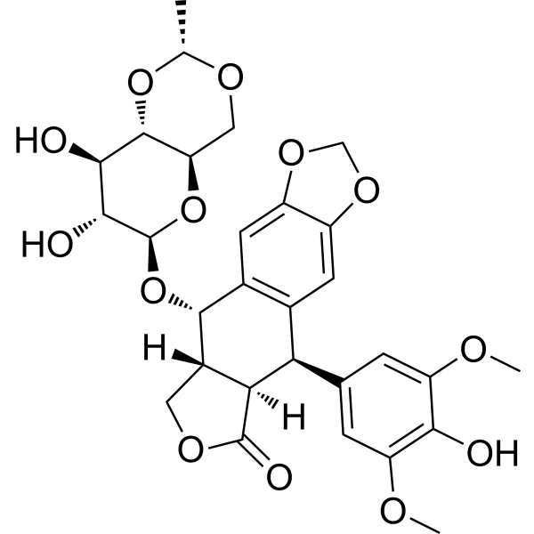
EtoposideTopoisomerase II Inhibitor -
HY-15142
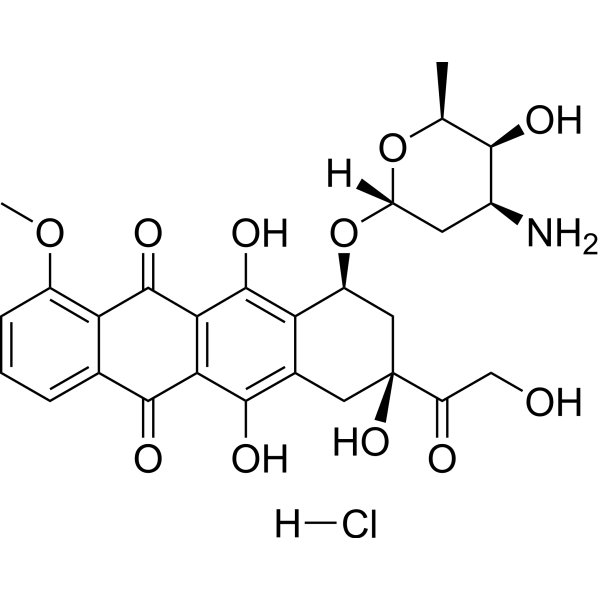
Doxorubicin hydrochlorideTopoisomerase Inhibitor -

HY-10182
LaduviglusibGSK-3 Inhibitor -

HY-18739
Phorbol 12-myristate 13-acetatePKC Activator -
HY-13502
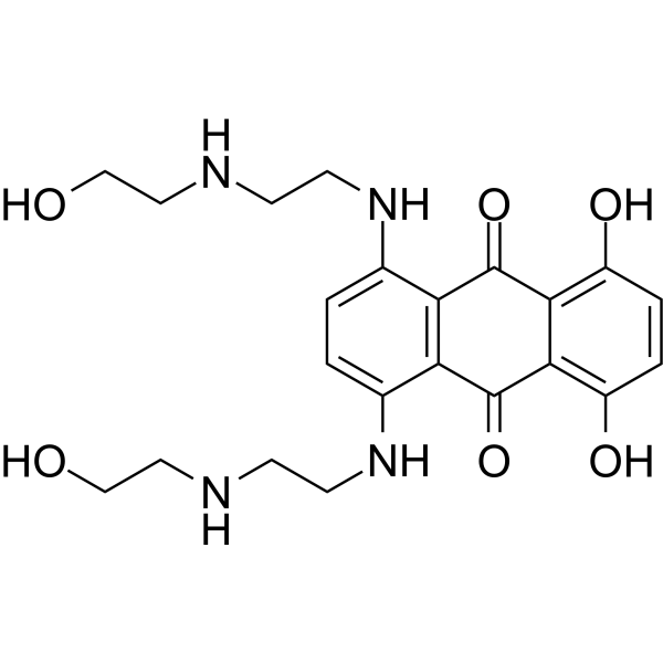
MitoxantroneTop II/PKC Inhibitor -
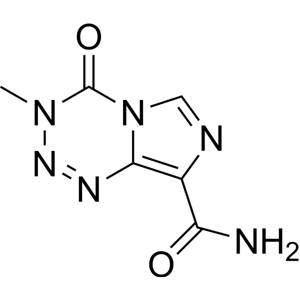
HY-17364
TemozolomideDNA Alkylator -
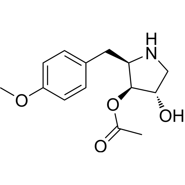
HY-18982
AnisomycinProtein Synthesis Inhibitor -
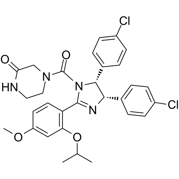
HY-10029
Nutlin-3aMDM2 Antagonist -
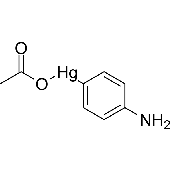
HY-148905
p-Aminophenylmercuric acetateMatrix Metalloproteinases Activator -
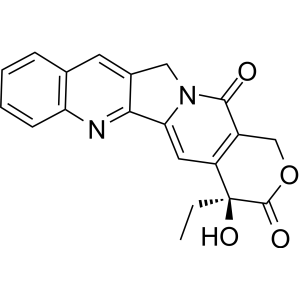
HY-16560
CamptothecinTopo I inhibitor -
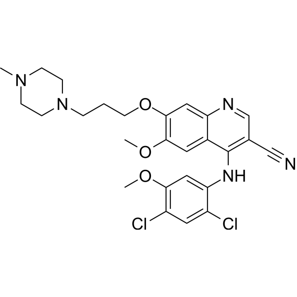
HY-10158
BosutinibSrc/Abl Inhibitor -
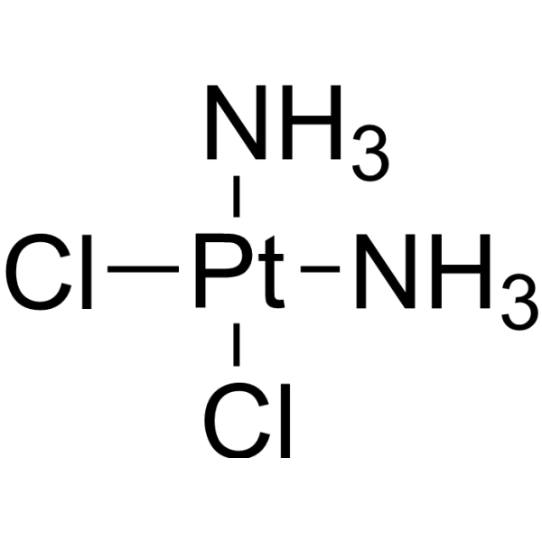
HY-17394
CisplatinDNA Alkylator
Publications Citing Use of MedChemExpress (MCE) Quinacrine dihydrochloride
More- J Biomed Sci. 2025 Feb 5;32(1):15. [Abstract]
- J Neuroinflammation. 2025 Oct 27;22(1):243. [Abstract]
- Phytomedicine. 2025 Sep:145:156981. [Abstract]
- J Pharm Anal. 2025 Dec 15.
- Part Fibre Toxicol. 2024 Mar 7;21(1):13. [Abstract]
- Brain Behav Immun. 2026 Jan 18:134:106296. [Abstract]
- Pharmaceutics. 2022 Jan 12;14(1):176. [Abstract]
- bioRxiv. 2025 Nov 11.
- University of Saskatchewan. 2020 Jun 22.
 Customer Validation & Images

-
Cell Proliferation/Viability Assay

-
Cell Proliferation/Viability Assay

-
Bio/Physico-chemical Assay

All Parasite Isoforms
More

Biological Activity
Description
IC50 & Target
|
Plasmodium |
In Vitro
Quinacrine (5-20 μM; 24 hours) inhibits the growth of SGC-7901 cells[1].
Quinacrine (7.5 and 15 μM; 24 hours) induces apoptosis in SGC-7901 cells, which is associated with mitochondria-dependent signal pathway and involves p53 upregulation and caspase-3 activation pathway[1].
Quinacrine (15 μM; 24 hours) treatment significantly increased the levels of proapoptotic proteins, including cytochrome c, Bax, and p53, and decreased the levels of antiapoptotic protein Bcl-2, thus shifting the ratio of Bax/Bcl-2 in favor of apoptosis [1].
MedChemExpress (MCE) has not independently confirmed the accuracy of these methods. They are for reference only. Further protocols information, click here.
-
Cell Line:SGC-7901 cells
-
Concentration:0, 5, 10, 15, and 20 μM
-
Incubation Time:24 hours
-
Result:Cell viability was inhibited in a dose-dependent manner, and the mean IC50 value is 16.18 μM.
-
Cell Line:SGC-7901 cells
-
Concentration:7.5 and 15 μM
-
Incubation Time:24 hours
-
Result:The percentage of apoptotic cells, including the early phase and late phase apoptosis, increased to 26.30%, compared with control group of 3.37%.
-
Cell Line:SGC-7901 cells
-
Concentration:15 μM
-
Incubation Time:24 hours
-
Result:The relative quantity of cytochrome c protein was upregulated, increased from 0.10 to 0.24.
The relative quantity of p53 protein was dramatically increased, from 0.06 to 0.19.
The Bax/Bcl-2 ratio was dramatically elevated from 1.21 to 2.59.
In Vivo
MedChemExpress (MCE) has not independently confirmed the accuracy of these methods. They are for reference only.
-
Animal Model:Female SCID mice with acute myeloid leukemia (AML)-PS model[2]
-
Dosage:100 mg/kg
-
Administration:Administered by oral gavage (po); three times a week for two consecutive weeks
-
Result:In the first AML mouse in vivo study, evaluation of circulating leukemic cells detected in blood samples (in percent of white blood cells (WBC)) at day 30/31 showed 72% human tumor cells in the control mice, whereas in mice treated with Quinacrine, this was only 2.2%.
The MST of control mice was 34 days whereas it was 46 days in Quinacrine-treated mice.


Chemical Information
-
CAS No. 69-05-6
-
Appearance Solid
-
Molecular Weight 472.88
-
Formula C23H32Cl3N3O
-
Color Light yellow to yellow
-
SMILES
CC(NC1=C(C=C(OC)C=C2)C2=NC3=CC(Cl)=CC=C31)CCCN(CC)CC.Cl.Cl
-
Synonyms
Mepacrine dihydrochloride; SN-390 dihydrochloride
-
Shipping
Room temperature in continental US; may vary elsewhere.
-
Storage
4°C, sealed storage, away from moisture
* In solvent : -80°C, 6 months; -20°C, 1 month (sealed storage, away from moisture)


Publications (9)
-
Journal Impact Factor
-
Most Recent
-
J Biomed Sci
Targeting the G-quadruplex as a novel strategy for developing antibiotics against hypervirulent drug-resistant Staphylococcus aureus. [Abstract]2025 Feb 5;32(1):15. PMID: 39905515
Quinacrine dihydrochloride purchased from MedChemExpress. Usage Cited in: J Biomed Sci. 2025 Feb 5;32(1):15. [Abstract]
The tabulated datasheet from IC50 data showing the percent inhibition of SAUSA300 cells at 10 µM of G4-ligands (Quinacrine dihydrochloride, etc.) and their IC50 values.
-
J Neuroinflammation
2025 Oct 27;22(1):243. PMID: 41146204 -
Phytomedicine
Quinacrine induces autophagy via the Dlg5/AKT pathway to inhibit osteosarcoma cell proliferation and suppresses migration and invasion through the Dlg5/Girdin pathway. [Abstract]2025 Sep:145:156981. PMID: 40541124 -
-
Part Fibre Toxicol
Exposure to high dose of polystyrene nanoplastics causes trophoblast cell apoptosis and induces miscarriage. [Abstract]2024 Mar 7;21(1):13. PMID: 38454452
Quinacrine dihydrochloride purchased from MedChemExpress. Usage Cited in: Part Fibre Toxicol. 2024 Mar 7;21(1):13. [Abstract]
Cell viability of trophoblast cells treated with 10 μM Raptinal, 15 μM Quinacrine dihydrochloride, or 500 μg/mL PS-NPs, and co-treated with 10 μM Z-VAD-FMK for 48 h. The results showed that treatment with Raptinal, Quinacrine dihydrochloride, or PS-NPs reduced trophoblast cell viability, and co-treatment with Z-VAD-FMK rescued trophoblast cell viability.
-
Brain Behav Immun
Inhibition of the NFATc2/FKBP5 axis alleviates microglial neuroinflammation by regulating arachidonic acid metabolism in Parkinson's disease. [Abstract]2026 Jan 18:134:106296. PMID: 41554345 -
Pharmaceutics
Drug Repurposing for the Identification of Compounds with Anti-SARS-CoV-2 Capability via Multiple Targets. [Abstract]2022 Jan 12;14(1):176. PMID: 35057070
Quinacrine dihydrochloride purchased from MedChemExpress. Usage Cited in: Pharmaceutics. 2022 Jan 12;14(1):176. [Abstract]
Quinacrine (Mepacrine) dihydrochloride inhibited the enzymatic activities of TMPRSS2 and PLpro, with IC50 values of 29.9 μM and 4.4 μM, respectively.
-
-


Solvent & Solubility
In Vitro:
H2O : 50 mg/mL (105.74 mM; ultrasonic and warming and heat to 60°C)
Please refer to the solubility information to select the appropriate solvent. Once prepared, please aliquot and store the solution to prevent product inactivation from repeated freeze-thaw cycles.
Storage method and period of stock solution: -80°C, 6 months; -20°C, 1 month (sealed storage, away from moisture). When stored at -80°C, please use it within 6 months. When stored at -20°C, please use it within 1 month.
* Note: If you choose water as the stock solution, please dilute it to the working solution, then filter and sterilize it with a 0.22 μm filter before use.
Please refer to the solubility information to select the appropriate solvent. Once prepared, please aliquot and store the solution to prevent product inactivation from repeated freeze-thaw cycles.
Storage method and period of stock solution: -80°C, 6 months; -20°C, 1 month (sealed storage, away from moisture). When stored at -80°C, please use it within 6 months. When stored at -20°C, please use it within 1 month.
* Note: If you choose water as the stock solution, please dilute it to the working solution, then filter and sterilize it with a 0.22 μm filter before use.
Concentration (start) × Volume (start) = Concentration (final) × Volume (final)

Protocols
-
Apoptosis
Apoptosis, also called programmed cell death, is generally characterized by distinct morphological characteristics.
-
TUNEL staining for apoptotic DNA fragmentation
TUNEL staining detects DNA strand breaks by using terminal deoxynucleotidyl transferase to add labeled nucleotides to exposed 3′-OH DNA termini, generating either microscopic staining in fixed cells or tissue sections, or fluorescence/cytometric signal in cell suspensions. TUNEL positivity reflects DNA fragmentation but should not be interpreted alone as definitive apoptosis, because TUNEL can also label necrotic, autolytic, mechanically damaged, or DNA-repair-associated DNA breaks.
-
Annexin V plus membrane-impermeant dye apoptosis staining
Annexin V-based apoptosis assays rely on the detection of phosphatidylserine (PS) externalization from the inner leaflet of the plasma membrane to the outer leaflet, an early biochemical hallmark of apoptosis. Fluorescently labeled Annexin V binds PS in a calcium-dependent manner, enabling identification of early apoptotic cells by flow cytometry or fluorescence microscopy. When combined with a membrane-impermeant DNA-binding dye (e. g. , propidium iodide), this approach allows discrimination between viable (Annexin V−/dye−), early apoptotic (Annexin V+/dye−), and late apoptotic or necrotic (Annexin V+/dye+) cell populations by assessing membrane integrity and PS exposure.
-
Apoptosis Solutions
Apoptosis is a regulated, generally non-lytic cell-death pathway that removes unwanted, damaged, infected, or abnormal cells through coordinated morphological changes, caspase activation, DNA fragmentation, and membrane remodeling. The intrinsic apoptosis pathway is controlled mainly by mitochondrial outer membrane permeabilization, BCL-2 family proteins, cytochrome c release, apoptosome formation, caspase-9 activation, and downstream executioner caspase-3/7 activation. The extrinsic apoptosis pathway is initiated by death receptors such as Fas, TNFR, and TRAIL receptors, which recruit adaptor proteins and activate caspase-8 before engaging executioner caspases or mitochondrial amplification through BID cleavage. Apoptosis is linked to many phenotypes, including cancer cell killing, tissue homeostasis, immune regulation, neurodegeneration, infection response, and treatment-induced cytotoxicity; unresolved questions include how apoptosis interacts with necroptosis, pyroptosis, ferroptos
-
Research Protocol for Inflammation-related Diseases
The NLRP3 inflammasome is a cytosolic innate immune signaling platform that integrates priming signals and danger-signal activation to promote caspase-1 activation, maturation of IL-1β and IL-18, and gasdermin D-mediated pyroptotic cell death. The core experimental logic is to determine whether inflammatory disease phenotypes are driven by increased NLRP3 expression, ASC-containing inflammasome assembly, caspase-1 cleavage, GSDMD cleavage, and extracellular release of IL-1β/IL-18 rather than by nonspecific cell injury alone. The pathway is strongly linked to inflammation-related disease phenotypes because monosodium urate crystals activate NALP3/NLRP3 inflammasome signaling in gout-like crystal inflammation, cholesterol crystals activate NLRP3 inflammasomes in atherogenesis models, and DSS-induced intestinal inflammation has been reported to involve NLRP3 inflammasome activity. However, experimental colitis studies also show context-dependent protective effects of NLRP3 inflammasome co


Purity & Documentation
-
Data Sheet (283 KB)
-
SDS (393 KB)
- English - EN (393 KB)
- Français - FR (393 KB)
- Deutsch - DE (393 KB)
- Norwegian - NO (393 KB)
- Español - ES (393 KB)
- Swedish - SV (393 KB)
- Italian - IT (393 KB)
- Korean - KR (393 KB)
- Portuguese - PT (393 KB)
-
Handling Instructions (2659 KB)
References
[1]. Xiaoyang Wu, et al. Quinacrine Inhibits Cell Growth and Induces Apoptosis in Human Gastric Cancer Cell Line SGC-7901. Curr Ther Res Clin Exp. 2012 Feb;73(1-2):52-64. [Content Brief]
[2]. Anna Eriksson, et al. Towards repositioning of quinacrine for treatment of acute myeloid leukemia - Promising synergies and in vivo effects. Leuk Res. 2017 Dec;63:41-46. [Content Brief]


Complete Stock Solution Preparation Table
Please refer to the solubility information to select the appropriate solvent. Once prepared, please aliquot and store the solution to prevent product inactivation from repeated freeze-thaw cycles.
Storage method and period of stock solution: -80°C, 6 months; -20°C, 1 month (sealed storage, away from moisture). When stored at -80°C, please use it within 6 months. When stored at -20°C, please use it within 1 month.
| Optional Solvent | Concentration Solvent Mass | 1 mg | 5 mg | 10 mg | 25 mg |
|---|---|---|---|---|---|
| H2O | 1 mM | 2.1147 mL | 10.5735 mL | 21.1470 mL | 52.8675 mL |
| 5 mM | 0.4229 mL | 2.1147 mL | 4.2294 mL | 10.5735 mL | |
| 10 mM | 0.2115 mL | 1.0574 mL | 2.1147 mL | 5.2868 mL | |
| 15 mM | 0.1410 mL | 0.7049 mL | 1.4098 mL | 3.5245 mL | |
| 20 mM | 0.1057 mL | 0.5287 mL | 1.0574 mL | 2.6434 mL | |
| 25 mM | 0.0846 mL | 0.4229 mL | 0.8459 mL | 2.1147 mL | |
| 30 mM | 0.0705 mL | 0.3525 mL | 0.7049 mL | 1.7623 mL | |
| 40 mM | 0.0529 mL | 0.2643 mL | 0.5287 mL | 1.3217 mL | |
| 50 mM | 0.0423 mL | 0.2115 mL | 0.4229 mL | 1.0574 mL | |
| 60 mM | 0.0352 mL | 0.1762 mL | 0.3525 mL | 0.8811 mL | |
| 80 mM | 0.0264 mL | 0.1322 mL | 0.2643 mL | 0.6608 mL | |
| 100 mM | 0.0211 mL | 0.1057 mL | 0.2115 mL | 0.5287 mL |
* Note: If you choose water as the stock solution, please dilute it to the working solution, then filter and sterilize it with a 0.22 μm filter before use.


Quinacrine dihydrochloride Related Classifications
HY-13735A Related Classifications
 Powered by Bioz