



Acetyl coenzyme A trilithium

Based on 1 Customer Validation
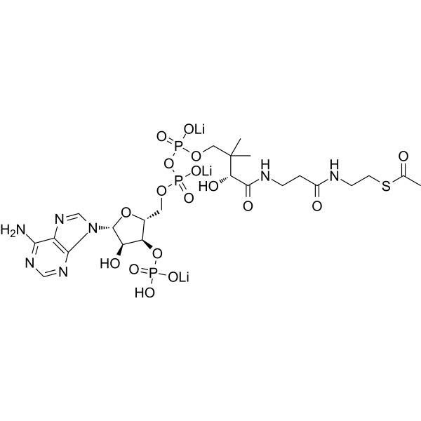
Acetyl-coenzyme A (Acetyl-CoA) trilithium is a membrane-impermeant central metabolic intermediate, participates in the TCA cycle and oxidative phosphorylation metabolism. Acetyl-coenzyme A trilithium regulates various cellular mechanisms by providing (sole donor) acetyl groups to target amino acid residues for post-translational acetylation reactions of proteins. Acetyl Coenzyme A trilithium is also a key precursor of lipid synthesis.
For research use only. We do not sell to patients.
- Purity : 97%
- CAS No.: 75520-41-1
- Formula: C23H35Li3N7O17P3S
- Molecular Weight:827.37
-
Storage:
Please store the product under the recommended conditions in the Certificate of Analysis.

To place orders, for customer services and technical support, please contact: MedChemExpress USA
Tel: 609-228-6898 E-mail: [email protected] [email protected]


-
 Biological Activity
-
Chemical Information
- Protocols
- Purity & Documentation
- References
-
 Help & FAQs


-
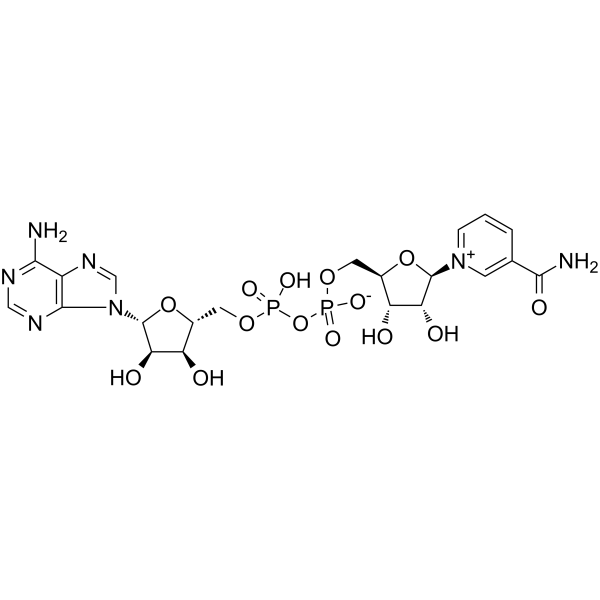
HY-B0445
NAD+Endogenous Metabolite -
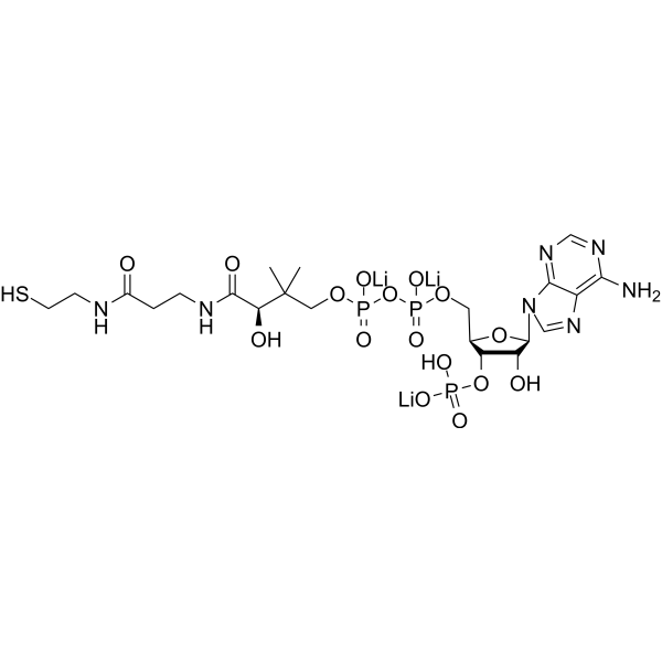
HY-128851A
Coenzyme A trilithiumCofactor -
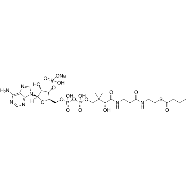
HY-138177
Butyryl-Coenzyme A sodiumButyric Acid Derivative -
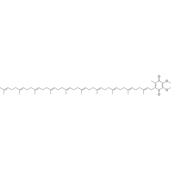
HY-N0111
Coenzyme Q10Antioxidant -
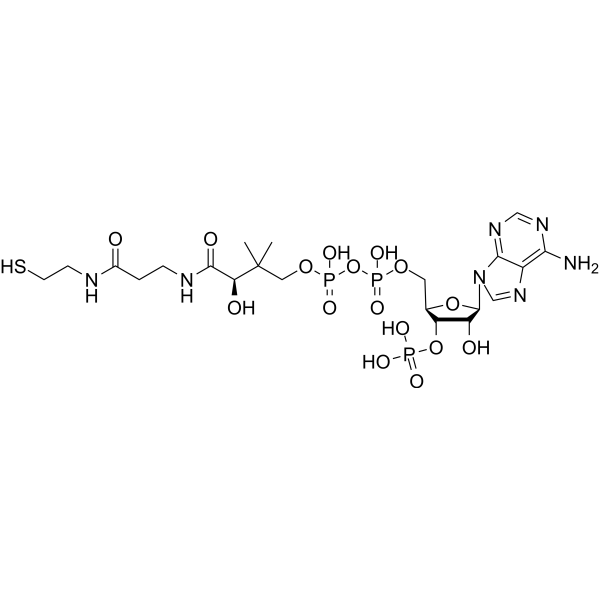
HY-128851
Coenzyme ACofactor -
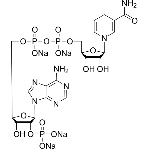
HY-F0003
NADPH tetrasodium saltCofactor -
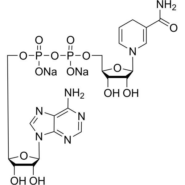
HY-F0001
NADH disodium saltReduced Coenzyme -

HY-N0111R
Coenzyme Q10 (Standard)Antioxidant -
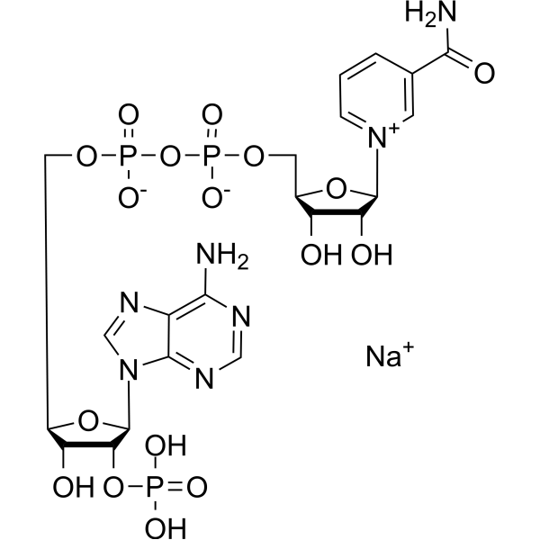
HY-F0002
NADP sodium saltRedox Coenzyme -

HY-F0001R
NADH disodium salt (Standard)Reference Standard -
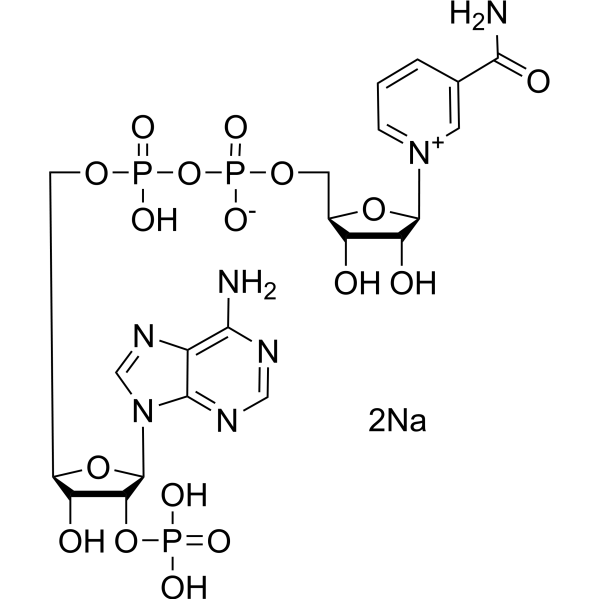
HY-F0002A
NADP disodium saltRedox Coenzyme -

HY-F0003R
NADPH tetrasodium salt (Standard)Standard of NADPH tetrasodium salt
All Endogenous Metabolite Isoforms
More

Biological Activity
Description
In Vitro
Acetyl coenzyme A trilithium increases cytoplasmic protein acetylation in starved U2OS cells while reducing starvation-induced autophagic fluxes. (U2OS cells stably expressing GFP-LC3 and are microinjected with Acetyl coenzyme A; incubated in nutrient-free conditions in the presence of 100 nM BafA1 and fixed after 3 h)[2].
MedChemExpress (MCE) has not independently confirmed the accuracy of these methods. They are for reference only. Further protocols information, click here.
In Vivo
MedChemExpress (MCE) has not independently confirmed the accuracy of these methods. They are for reference only.


Chemical Information
-
CAS No. 75520-41-1
-
Appearance Solid
-
Molecular Weight 827.37
-
Formula C23H35Li3N7O17P3S
-
Color White to off-white
-
SMILES
CC(SCCNC(CCNC([C@H](O)C(C)(C)COP(OP(OC[C@H]1O[C@@H](N(C=N2)C3=C2C(N)=NC=N3)[C@H](O)[C@@H]1OP(O)(O[Li])=O)(O[Li])=O)(O[Li])=O)=O)=O)=O
-
Synonyms
Acetyl-CoA trilithium
-
Structure Classification
-
Initial Source
-
Shipping
Room temperature in continental US; may vary elsewhere.
-
Storage
Please store the product under the recommended conditions in the Certificate of Analysis.

Protocols
-
Kinase activity and phosphorylation assays
Kinase activity assays measure the ability of kinases to transfer phosphate groups from ATP to specific substrates, while phosphorylation assays detect the presence and levels of phosphorylated proteins. Common methods include radiolabeled ATP incorporation (e. g. ,), ADP release detection via bioluminescence (e. g. ,[3]), enzyme-linked immunosorbent assays (ELISA) for phospho-specific epitopes (e. g. ,[6]), and microtiter-based formats for high-throughput screening (e. g. ,[8]). The ADP-Glo assay quantifies kinase activity by measuring ADP produced during phosphorylation using a luciferase-based system. Radiometric assays involve autoradiography or scintillation counting after incorporation of 32P-labeled ATP into substrate proteins. ELISA-based approaches rely on phospho-specific antibodies to detect activated kinases in cell lysates or purified samples.
-
Western Blot
Western blotting (WB) is a commonly used experimental method in molecular biology, biochemistry, and immunogenetics for identifying and quantifying target proteins. It combines gel electrophoresis with immunoassay, enabling researchers to analyze protein expression, post-translational modifications, and molecular weight.
-
Protocol for Kinase activity and phosphorylation assays
Kinase activity assays measure transfer of phosphate from ATP to a protein or peptide substrate, generating phosphorylated substrate, ADP, or incorporated radiolabeled phosphate as the readout; phosphorylation assays measure site-specific phosphorylation in cells or tissues as a proxy for kinase-pathway activation, inhibition, or substrate regulation. Phosphorylation can be detected by phospho-specific Western blot, immunoprecipitation kinase assay, phospho-immunofluorescence, phospho-flow cytometry, luminescent ADP detection, radiolabeled ATP incorporation, or reporter-based pathway assays, and these readouts can be applied to cancer cells, primary neurons, mouse tumors, organoids, inflammatory macrophages, ferroptosis studies, and mitophagy studies when the kinase target is biologically relevant.


Purity & Documentation
-
Data Sheet (276 KB)
-
SDS (394 KB)
- English - EN (394 KB)
- Français - FR (394 KB)
- Deutsch - DE (394 KB)
- Norwegian - NO (394 KB)
- Español - ES (394 KB)
- Swedish - SV (394 KB)
- Italian - IT (394 KB)
- Korean - KR (394 KB)
- Portuguese - PT (394 KB)
-
Handling Instructions (2659 KB)
References
[1]. Choudhary C, et al. The growing landscape of lysine acetylation links metabolism and cell signalling. Nat Rev Mol Cell Biol. 2014 Aug;15(8):536-50. [Content Brief]
[2]. Mariño G, et al. Regulation of autophagy by cytosolic acetyl-coenzyme A. Mol Cell. 2014 Mar 6;53(5):710-25. [Content Brief]
[3]. Zhu H, et al. Cardiac autophagy is a maladaptive response to hemodynamic stress. J Clin Invest. 2007 Jul;117(7):1782-93. [Content Brief]
[4]. Pietrocola F, et al. Acetyl coenzyme A: a central metabolite and second messenger. Cell Metab. 2015 Jun 2;21(6):805-21. [Content Brief]

Calculators
Concentration (start) × Volume (start) = Concentration (final) × Volume (final)


Acetyl coenzyme A trilithium Related Classifications
HY-114293A Related Classifications
 Powered by Bioz