



Anti-Mouse/Rat/Human Osteopontin/SPP1 Antibody (MPIIIB10)

Based on 1 Customer Validation
Anti-Mouse/Rat/Human Osteopontin/SPP1 Antibody (MPIIIB10) is a mouse-derived Osteopontin/SPP1 IgG1 κ type antibody inhibitor. Anti-Mouse/Rat/Human Osteopontin/SPP1 Antibody (MPIIIB10) blocks Angiotensin II (HY-13948)-induced DNA synthesis and collagen gel contraction in cardiac fibroblasts. Anti-Mouse/Rat/Human Osteopontin/SPP1 Antibody (MPIIIB10) significantly inhibits tumor growth in CT26 or MC38 tumors mice models.
For research use only. We do not sell to patients.
- Purity : ≥95.0%
- Molecular Weight:150 kDa
-
Storage:
Please store the product under the recommended conditions in the Certificate of Analysis.


To place orders, for customer services and technical support, please contact: MedChemExpress USA
Tel: 609-228-6898 E-mail: [email protected] [email protected]


-
 Biological Activity
-
Chemical Information
- Protocols
- Purity & Documentation
- References
-
 Help & FAQs








Biological Activity
Description
Isotype
Mouse IgG1 kappa
Recommend Isotype Controls
Species Reactivity
Human/Mouse/Rat
IC50 & Target
Osteopontin/SPP1
In Vitro
Anti-Mouse/Rat/Human Osteopontin/SPP1 Antibody (MPIIIB10) (7.2 μg/mL or 72 ng/mL, 30 min) blocks Angiotensin II (HY-13948)-induced DNA synthesis in cardiac fibroblasts[2].
Anti-Mouse/Rat/Human Osteopontin/SPP1 Antibody (MPIIIB10) (7.2 μg/mL or 25 μg/mL, 30 min) blocks both Angiotensin II-induced and osteopontin-induced collagen gel contraction in cardiac fibroblasts[2].
Anti-Mouse/Rat/Human Osteopontin/SPP1 Antibody (MPIIIB10) (100 ng/mL, 48 h) significantly attenuates VSMC (vascular smooth muscle cells) migration induced by HMGB1 (high-mobility group box 1) in A10 cells[3].
MedChemExpress (MCE) has not independently confirmed the accuracy of these methods. They are for reference only. Further protocols information, click here.
In Vivo
MedChemExpress (MCE) has not independently confirmed the accuracy of these methods. They are for reference only.
-
Animal Model:The mice were subcutaneously injected with CT26 or MC38 tumors (3 x 10⁵ cells) on day 0 and some mice were also given a B-cell–based vaccine on day 9 (MC38 model) or day 10 (CT26 model) (Balb/c or C57BL/6)[1]
-
Dosage:500 μg/mouse
-
Administration:Intraperitoneally injection, every other day for 10 days
-
Result:Significantly inhibited tumor growth.
Significantly delayed tumor growth in mice treated with the B-cell–based vaccine.
Gene ID
Accession
Conjugated
Unconjugated
Reconsititution
The product can be reconstituted/diluted with sterile PBS or saline.
Application
in vivo OPN neutralization; in vitro OPN neutralization; Immunohistochemistry (paraffin); Western bl
Verified Bioactivity
-
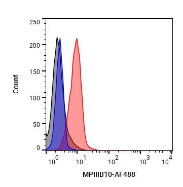 Flow cytometric analysis of 1X10⁶ A549 cells with Anti-Mouse/Rat/Human-SPP1 Antibody (MPIIIB10) (HY-P990117, red). Cells were fixed with 4% paraformaldehyde. Then stained with the primary antibody at 1/200 dilution for an hour at 4℃. AF 488-conjugated AffiniPure Goat Anti-Mouse IgG H&L (HY-P8005) was used as the secondary antibody at 1/1,000 dilution for 30 minutes at 4℃. Mouse IgG1 kappa (HY-P99977, blue) was used as the isotype control, cells without incubation with primary antibody were used as the unlabeled control (black).


Chemical Information
-
Appearance Liquid
-
Molecular Weight 150 kDa
-
Color Colorless to light yellow
-
SMILES
[Anti-Mouse/Rat/Human Osteopontin/SPP1 Antibody (MPIIIB10)]
-
Formulation
Please refer to the lot-specific COA for specific buffer information.
-
Storage
Please store the product under the recommended conditions in the Certificate of Analysis.

Protocols
-
EdU Incorporation Assay (Click Chemistry-Based DNA Synthesis Measurement)
The EdU incorporation assay measures DNA synthesis by adding the thymidine analog 5-ethynyl-2′-deoxyuridine to cells or tissues, where it is incorporated into newly synthesized DNA during S phase. Incorporated EdU is detected by copper-catalyzed azide-alkyne cycloaddition, in which a fluorescent azide covalently reacts with the ethynyl group on EdU, allowing S-phase cells to be detected by fluorescence microscopy, flow cytometry, or high-content imaging. EdU detection does not require DNA denaturation or anti-BrdU antibody access, which preserves sample structure and improves compatibility with immunostaining and multiparameter cytometry compared with BrdU-based detection. EdU can be cytotoxic in a cell-type- and exposure-dependent manner, so pulse duration, concentration, and continuous-labeling designs should be validated for each cell type.
-
BrdU Incorporation Assay
Bromodeoxyuridine (BrdU) incorporation assay is based on the principle that BrdU, a thymidine analog, is incorporated into newly synthesized DNA during the S phase of the cell cycle, thereby serving as a marker of DNA replication and cellular proliferation. Incorporated BrdU can be detected using anti-BrdU antibodies following DNA denaturation, enabling visualization or quantification of proliferating cells through immunochemical detection methods such as immunofluorescence or immunohistochemistry.
-
Patient-Derived Xenograft (PDX)
Patient-derived xenograft (PDX) models are generated by engrafting primary human tumor tissue directly into immunodeficient mice, allowing in vivo propagation of patient tumor biology without initial in vitro adaptation. These models are used to preserve key histopathological and molecular characteristics of the original tumor and enable assessment of tumor growth dynamics and therapeutic response in a living organism. The biological readout is tumor engraftment and subsequent growth in the murine host, which reflects the ability of human tumor cells to survive, vascularize, and expand in an immunocompromised microenvironment.
-
Patient-Derived Orthotopic Xenograft (PDOX)
Patient-derived orthotopic xenograft (PDOX) modeling implants fresh patient tumor tissue or patient-derived tumor cells into the anatomically corresponding organ or tissue site of immunodeficient mice, usually by surgical orthotopic implantation, to preserve patient tumor histology, local microenvironmental context, invasion, metastatic behavior, and treatment-response features better than subcutaneous implantation. PDOX readouts include tumor engraftment, orthotopic tumor growth, local invasion, metastasis, recurrence after resection, histologic similarity to the donor tumor, biomarker retention, molecular concordance, survival, and response or resistance to therapy. PDOX models are used for preclinical drug testing and individualized therapy evaluation, but engraftment success varies by tumor type and specimen quality.
-
Subcutaneous Cell-Line-Derived Xenograft
Subcutaneous cell-line-derived xenograft (CDX) models are established by implanting cultured human cancer cell lines into immunodeficient mice, where the injected cells form localized tumors that can be monitored in vivo as a measure of tumorigenic potential, growth kinetics, and treatment response. These models are widely used in oncology research because they allow reproducible tumor formation and enable comparative assessment of tumor growth between different cell lines or genetic manipulations in a controlled in vivo microenvironment. Subcutaneous implantation of cancer cells in immunodeficient mice is a standard approach for evaluating tumor growth behavior and therapeutic response across multiple cancer types, including prostate, esophageal, pancreatic, and colon cancer models.
-
Orthotopic Cell-Line Xenograft
Orthotopic cell-line xenograft models involve implantation of human cancer cell lines into the anatomically corresponding organ of immunodeficient mice to reproduce tumor growth within a native microenvironment, enabling more clinically relevant tumor behavior compared with subcutaneous models. These models are widely used because orthotopic placement better recapitulates tumor progression, including invasion and metastatic spread, which are often underrepresented in heterotopic implantation systems. Compared with conventional xenografts, orthotopic implantation is described as more technically complex but provides improved simulation of tumor-microenvironment interactions and metastatic behavior, making it particularly valuable for translational oncology research. Surgical orthotopic implantation approaches have been emphasized as enabling faithful reproduction of clinical cancer features, including metastasis and disease progression patterns that align with the tumor’s organ of origi


Purity & Documentation
-
Data Sheet (271 KB)
-
SDS (251 KB)
- English - EN (251 KB)
- Français - FR (251 KB)
- Deutsch - DE (251 KB)
- Norwegian - NO (251 KB)
- Español - ES (251 KB)
- Swedish - SV (251 KB)
- Italian - IT (251 KB)
- Korean - KR (251 KB)
- Portuguese - PT (251 KB)
-
Inhibitory Antibodies User Guide (603 KB)
References
[1]. Kim EK, et al. Tumor-derived osteopontin suppresses antitumor immunity by promoting extramedullary myelopoiesis. Cancer Res. 2014 Nov 15;74(22):6705-16. [Content Brief]
[2]. Ashizawa N, et al. Osteopontin is produced by rat cardiac fibroblasts and mediates A(II)-induced DNA synthesis and collagen gel contraction. J Clin Invest. 1996 Nov 15;98(10):2218-27. [Content Brief]
[3]. Kim JY, et al. Echinochrome A inhibits HMGB1-induced vascular smooth muscle cell migration by suppressing osteopontin expression. Korean J Physiol Pharmacol. 2025 Jan 1;29(1):83-92. [Content Brief]

Calculators
Concentration (start) × Volume (start) = Concentration (final) × Volume (final)


Anti-Mouse/Rat/Human Osteopontin/SPP1 Antibody (MPIIIB10) Related Classifications
HY-P990117 Related Classifications
 Powered by Bioz