



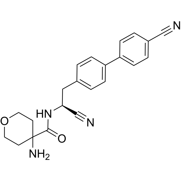
AZD5248

Based on 1 Customer Validation
AZD5248 is an orally active, selective dipeptidyl peptidase 1 (cathepsin C) inhibitor, with IC50 values of 1 nM and 17 nM against human CatC, 44 nM against human DPP1, and 67 nM against rat DPP1. It exhibits low clearance and high bioavailability in animal models. AZD5248 forms an irreversible covalent bond with the catalytic Cys234 residue of CatC, exerts reversible inhibition via its nitrile moiety, blocks CatC-dependent amyloid formation, and reduces the activation levels of neutrophil serine proteases in bone marrow and blood. AZD5248 reacts with aortic elastin aldehydes to form stable 4-imidazolinones, induces ultrastructural changes in aortic tissue, and has an α-amino acid-based backbone. AZD5248 reduces the severity of acute pancreatitis in mouse models. AZD5248 can be used in research on chronic obstructive pulmonary disease, acute pancreatitis, neurodegenerative diseases, lysosomal storage disorders, acute lung injury, cystic fibrosis, and neutrophil-mediated inflammatory diseases.
Nur für Forschungszwecke. Wir verkaufen nicht an Patienten.
- Reinheit : 98.9%
- CAS. Nr.: 1254318-44-9
- Formel: C22H22N4O2
- Molecular Weight:374.44
-
Speicherung:Powder -20°C, 3 years ; In solvent -80°C, 6 months , -20°C, 1 month

To place orders, for customer services and technical support, please contact: MedChemExpress USA
Tel: 609-228-6898 E-mail: [email protected] [email protected]


-
 Biologische Aktivität
-
Chemical Information
- Protokoll
- Reinheit & Dokumentation
- Verweise
-
 Help & FAQs


Alle Cathepsin Isoform-spezifische Produkte anzeigen
More

Biologische Aktivität
Beschreibung
In Vitro
AZD5248 (1-10 μM) potently inhibits recombinant human Cathepsin C with an enzyme IC50 of 1 nM[1].
AZD5248 exhibits fast reactivity in an in vitro propionaldehyde assay, with 15% remaining after 1 h incubation and a half-life of 0.36 h, indicating aortic binding liability[1].
AZD5248 (Serial dilutions; 3 hours) potently inhibits recombinant human Cathepsin C with an IC50 of 2.9 nM[2].
AZD5248 binds to the active site of human Cathepsin C, as evidenced by its co-crystal structure with the protein, and shares overlapping binding space with compound 54 in the enzyme's active site[3].
AZD5248 (50-100 μM; 18 h) shows high reactivity with model aldehydes in pH 7.4 phosphate buffer at 37 °C, with a half-life of 39 min for reaction with 5 mM propionaldehyde[4].
AZD5248 (100 μM; overnight preincubation, 2 h incubation) competitively inhibits 14C]AZD5248 binding to Han Wistar rat aortic homogenate, showing strong activity in this in vitro covalent binding assay[4].
AZD5248 potently inhibits DPP1, with supporting data indicating high selectivity for the target[5].
MedChemExpress (MCE) has not independently confirmed the accuracy of these methods. They are for reference only. Further protocols information, click here.
In Vivo
AZD5248 (20 mg/kg; p.o.; single dose) shows persistent, high-affinity binding to rat aortic tissue, with radioactivity retained in the aorta for at least 21 days after a single oral 20 mg/kg dose[4].
AZD5248 (20-200 mg/kg/day; p.o.; daily; 14-28 days) causes ultrastructural changes to rat aortic endothelium and smooth muscle cells, including reduced attachment to elastic laminae and increased intercellular substance[4].
AZD5248 (10 mg/kg; p.o.; twice daily; 8 days) reduces bone marrow neutrophil serine protease activity by 64-90% in rats[5].
MedChemExpress (MCE) has not independently confirmed the accuracy of these methods. They are for reference only.
-
Animal Model:C57BL/6 (6-8 weeks old, gender and weight matched, 20-25 g, caerulein-induced acute pancreatitis)[2]
-
Dosage:20 mg/kg
-
Administration:p.o.; twice daily; 6 days
-
Result:Reduced pancreatic CatC activity to ~70% of sham levels, with p < 0.05 versus caerulein+vehicle group.
Reduced pancreatic tissue damage compared to the caerulein+vehicle group.
-
Animal Model:Lister hooded rats (male)[4]
-
Dosage:20 mg/kg
-
Administration:p.o.; single dose
-
Result:Reached peak concentrations in most tissues 1 hour post-dosing.
Eliminated most tissue radioactivity by 21 days, except for residual radioactivity retained in the aortic wall, outer ear, intervertebral ligament, eye, renal outer medulla, liver, and spleen.
Showed high affinity for elastic tissues, particularly the aorta, with strong retention visible on autoradiograms even at 21 days post-administration.
-
Animal Model:Wistar Hannover rats (male, female)[4]
-
Dosage:20 mg/kg/day; 60 mg/kg/day; 200 mg/kg/day
-
Administration:p.o.; daily; 28 days (20, 60 mg/kg/day); p.o.; daily; ~14 days (200 mg/kg/day, females); p.o.; daily; 28 days (200 mg/kg/day, males)
-
Result:Caused endothelial cells to show reduced association to elastic laminae, morphological changes, and loss of flattened structure.
Induced smooth muscle cells to lose organized contact.
Increased intercellular substance in aortic samples compared to controls.
-
Animal Model:Sprague Dawley (male, 200-400 g)[5]
-
Dosage:10 mg·kg-1
-
Administration:p.o.; twice daily; 8 days
-
Result:Achieved a 90% reduction in bone marrow neutrophil elastase (NE) activity, a 64% reduction in bone marrow proteinase 3 (PR3) activity, and an 88% reduction in bone marrow cathepsin G (CatG) activity compared to vehicle-treated controls after 8 days of treatment.
Recovered bone marrow NE activity to 20% below baseline, PR3 activity to 21% below baseline, and CatG activity to 8% below baseline by day 9 post-treatment.
Reached bone marrow NE activity 10% above baseline, PR3 activity 6% above baseline, and CatG activity 4% above baseline by day 18 post-treatment.
Estimated a k_out (first-order rate constant for loss of response) of 0.0102 h-1, corresponding to a rat neutrophil turnover time of 4 days via an indirect response model fitted to the data.


Chemical Information
-
CAS. Nr. 1254318-44-9
-
Appearance Solid
-
Molecular Weight 374.44
-
Formel C22H22N4O2
-
Color White to off-white
-
SMILES
N#CC1=CC=C(C2=CC=C(C[C@H](NC(C3(N)CCOCC3)=O)C#N)C=C2)C=C1
-
Versand
Room temperature in continental US; may vary elsewhere.
-
Speicherung
Powder -20°C 3 years In solvent -80°C 6 months -20°C 1 month

Protokoll
-
Phagocytosis Functional Assay
A phagocytosis functional assay measures the ability of phagocytic cells, such as neutrophils, macrophages, monocytes, or microglia/macrophages, to bind and internalize particulate targets including bacteria, yeast particles, beads, or myelin particles. Fluorescent flow-cytometry assays detect target uptake as fluorescence associated with gated phagocytes, while pH-sensitive dyes such as pHrodo increase signal in acidic phagosomal compartments and therefore preferentially report internalized particles rather than particles remaining outside the cell. Microscopy or high-content imaging can be used to confirm intracellular localization and, in some protocols, to follow uptake kinetics.
-
Fibrosis/Collagen Morphometry
Fibrosis and collagen morphometry is based on the quantitative visualization of fibrillar collagen deposition in tissue sections using histochemical stains such as Sirius Red (Picrosirius Red) or Masson's trichrome, followed by image-based or polarization-enhanced analysis to estimate collagen proportional area as a surrogate of extracellular matrix accumulation during fibrotic remodeling. Sirius Red combined with polarized light microscopy enhances detection of collagen fibers due to birefringence properties, enabling more specific visualization of collagen type I and III fibrils compared to conventional bright-field histology, while whole-section or region-restricted digital morphometry reduces field-selection bias in fibrosis assessment. Alternative quantitative approaches include second harmonic generation (SHG) and two-photon excited fluorescence microscopy, which enable label-free detection of fibrillar collagen and have been validated against histological staining and biochemica
-
Collagen: Sirius Red Staining
Sirius Red or picrosirius red staining is a histochemical method for visualizing collagen-rich extracellular matrix in tissue sections, and collagen fibers are detected as red-stained structures under bright-field microscopy with enhanced birefringence under polarized light. Picrosirius red is useful for assessing total collagen organization, distribution, and fibrosis burden, but polarized color should not be interpreted as a definitive collagen type I versus type III readout because color is affected by fiber orientation, thickness, and packing.
-
Connective Tissue: Masson's Trichrome/Collagen Trichrome Staining
Masson’s Trichrome (collagen/trichrome staining) is a histological technique that differentially stains tissue compartments using sequential acidic dyes to distinguish collagen from muscle and cytoplasmic components based on dye affinity and tissue permeability differences, enabling visualization of fibrosis and connective tissue architecture in histological sections. The classical formulation typically uses Weigert's iron hematoxylin for nuclear staining, Biebrich scarlet-acid fuchsin for cytoplasm and muscle, and aniline blue (or light green variants) for collagen, producing a characteristic blue/green collagen signal contrasted against red cytoplasm and dark nuclei. The staining principle relies on selective displacement of smaller dye molecules by larger anionic dyes in collagen-rich regions under controlled acidified conditions, which enhances collagen-specific dye retention. This property makes the method widely used for fibrosis assessment in organs such as heart, liver, lung, a
-
Protocol for Pharmacokinetic Study
Pharmacokinetic studies quantify how an organism handles a drug over time through absorption, distribution, metabolism, and excretion, and the core experimental readout is the concentration-time profile of parent drug and, when relevant, metabolites in biological matrices such as plasma, whole blood, urine, bile, or tissue. Pharmacokinetic analysis links dose, route, exposure, clearance, half-life, distribution, bioavailability, and systemic exposure to drug efficacy and toxicity hypotheses rather than measuring a signaling pathway directly. The literature links pharmacokinetics to drug-development phenotypes by showing that drug metabolism and pharmacokinetics influence compound progression, exposure-response interpretation, safety margins, dosing strategy, and failure risk during discovery and development. DMPK science contributes to compound optimization by integrating physicochemical properties, in vitro metabolism, transporter behavior, in vivo exposure, and pharmacodynamic contex
-
Fibers: Elastic Fiber Staining
Elastic fiber staining is a histochemical technique used to selectively visualize elastin-rich structures such as elastic fibers in connective tissues (e. g. , blood vessels, lung, dermis) based on the affinity of specific dyes or oxidation products for elastin-associated amino acid residues and cross-linked elastic matrix components. Classical methods such as Verhoeff-Van Gieson (VVG), resorcin-fuchsin (Weigert-type stains), or aldehyde fuchsin rely on differential binding of dye complexes to elastic fibers, allowing them to be distinguished from collagen and other extracellular matrix components by contrast staining (typically black or deep purple elastic fibers against red collagen counterstain). These methods are widely used in histopathology to evaluate elastic fiber integrity, fragmentation, or remodeling in vascular diseases, pulmonary pathology, and connective tissue disorders.
-
Lysosome and acidic-vesicle live-cell staining
Lysosome and acidic-vesicle live-cell staining detects acidic intracellular compartments by using membrane-permeant acidotropic probes that accumulate in low-pH vesicles, including lysosomes, late endosomes, autolysosomes, and acidic phagosomes. LysoTracker staining is commonly used as an intensity-based readout of acidic lysosomal compartment abundance or enlargement, while acridine orange produces green fluorescence in less concentrated compartments and red fluorescence after concentration-dependent accumulation in acidic vesicular organelles. Loss or reduction of acridine-orange red signal can be used as a readout of lysosomal membrane permeabilization or reduced acidic-vesicle integrity. This protocol is designed for live cultured cells and can be adapted for fluorescence microscopy, high-content imaging, plate-reader readout, or flow cytometry when the selected literature supports the readout. Because these dyes report acidotropic accumulation rather than lysosome identity alone,
-
Inhalation Toxicity Study
Inhalation toxicity studies expose rodents to a controlled aerosol, vapor, gas, or smoke atmosphere and assess respiratory and systemic toxicity using exposure-atmosphere characterization, clinical observations, body and organ weights, bronchoalveolar lavage fluid, histopathology, blood chemistry, hematology, and, when included, molecular endpoints such as transcriptomics, proteomics, lipidomics, or tissue burden analysis. The primary biological readouts are airway irritation, pulmonary inflammation, cytotoxicity, altered surfactant or lipid homeostasis, impaired particle clearance, and tissue remodeling, reflected by BALF cell differentials, BALF protein, LDH, phosphatase activities, cytokines, lung weight, microscopic respiratory-tract lesions, and retained lung burden.
-
Amyloid: Congo Red Amyloid Staining
Congo red amyloid staining is a histochemical method used to detect extracellular amyloid deposits in tissue sections based on the affinity of Congo red dye for β-pleated sheet-rich protein aggregates. When bound to amyloid, Congo red produces characteristic apple-green birefringence under polarized light microscopy, which is widely regarded as a diagnostic feature of amyloid deposition in histopathology. The diagnostic principle relies on the combination of dye binding (congophilia) and optical anisotropy under polarized illumination, which distinguishes amyloid from most non-amyloid eosinophilic extracellular deposits in routine histological evaluation. Amyloid identification by Congo red staining remains a cornerstone in diagnostic pathology despite the availability of adjunct methods such as immunohistochemistry and mass spectrometry, particularly because of its ability to localize deposits directly within tissue architecture. The specificity of Congo red-positive deposits is incre


Reinheit & Dokumentation
-
Data Sheet (292 KB)
-
SDS (419 KB)
- English - EN (419 KB)
- Français - FR (419 KB)
- Deutsch - DE (419 KB)
- Norwegian - NO (419 KB)
- Español - ES (419 KB)
- Swedish - SV (419 KB)
- Italian - IT (419 KB)
- Korean - KR (419 KB)
- Portuguese - PT (419 KB)
-
Handling Instructions (2659 KB)
Verweise
[1]. Banerjee A, et al. Development of potent and selective Cathepsin C inhibitors free of aortic binding liability by application of a conformational restriction strategy. Bioorg Med Chem Lett. 2021;47:128202. [Content Brief]
[2]. Hou W, et al. Identification and Optimization of Novel Cathepsin C Inhibitors Derived from EGFR Inhibitors. J Med Chem. 2019;62(12):5901-5919. [Content Brief]
[3]. Chen X, et al. Discovery and In Vivo Anti-inflammatory Activity Evaluation of a Novel Non-peptidyl Non-covalent Cathepsin C Inhibitor. J Med Chem. 2021;64(16):11857-11885. [Content Brief]
[4]. Bragg RA, et al. Aortic Binding of AZD5248: Mechanistic Insight and Reactivity Assays To Support Lead Optimzation. Chem Res Toxicol. 2015;28(10):1991-1999. [Content Brief]
[5]. Chalmers JD, Kettritz R, Korkmaz B. Dipeptidyl peptidase 1 inhibition as a potential therapeutic approach in neutrophil-mediated inflammatory disease. Frontiers in immunology. 2023 Dec 14;14:1239151. [Content Brief]

Calculators
Konzentration (Stammlösung) × Volumen (Stammlösung) = Konzentration (Ziellösung) × Volumen (Ziellösung)


AZD5248 Related Classifications
HY-12733 Related Classifications
 Powered by Bioz
Keywords
- AZD5248
- 1254318-44-9
- AZD 5248
- AZD-5248
- Dipeptidyl Peptidase
- Cathepsin
- acute pancreatitis
- Han Wistar rat
- caerulein-induced acute pancreatitis mouse model
- neurodegenerative diseases
- neutrophil serine protease
- Cys234
- dipeptidyl peptidase 1
- chronic obstructive pulmonary disease
- cathepsin C
- THP1 cells
- Inhibitor
- inhibitor
- inhibit