Emavusertib phosphate
Based on 7 publication(s) in Google Scholar
Emavusertib phosphate (CA-4948 phosphate) is the phosphate salt form of Emavusertib (HY-135317). Emavusertib phosphate is an orally active inhibitor for IRAK4 (IC50=57 nM) and FLT3. Emavusertib phosphate inhibits NF-κB and MyD88 signaling pathways, reduces the generation of pro-inflammatory cytokines like IL-6 and IL-10, thereby exhibiting anti-inflammatory and anti-proliferative activities against cancer cells, leading to cell apoptosis. Emavusertib phosphate exhibits antitumor activity in mouse model.
For research use only. We do not sell to patients.
- CAS No.: 2376399-38-9
- Formula: C24H28N7O9P
- Molecular Weight:589.49
-
Storage:
Please store the product under the recommended conditions in the Certificate of Analysis.
Publications Citing Use of MedChemExpress (MCE) Emavusertib phosphate
More-
Cell Proliferation/Viability Assay
-
WB
-
In Vivo Efficacy Study
-
IHC
-
Bio/Physico-chemical Assay
Biological Activity
Description
Chemical Information
-
CAS No. 2376399-38-9
-
Molecular Weight 589.49
-
Formula C24H28N7O9P
-
SMILES
O=P(O)(O)O.O=C(C1=COC(C2=CC=NC(C)=C2)=N1)NC3=C(N4CC[C@H](C4)O)N=C5C(OC(N6CCOCC6)=N5)=C3
-
Synonyms
CA-4948 phosphate
-
Shipping
Room temperature in continental US; may vary elsewhere.
-
Storage
Please store the product under the recommended conditions in the Certificate of Analysis.
Publications (7)
-
Journal Impact Factor
-
Most Recent
-
Blood
2023 Sep 14;142(11):989-1007. PMID: 37172199
Emavusertib phosphate purchased from MedChemExpress. Usage Cited in: Blood. 2023 Sep 14;142(11):989-1007. [Abstract]
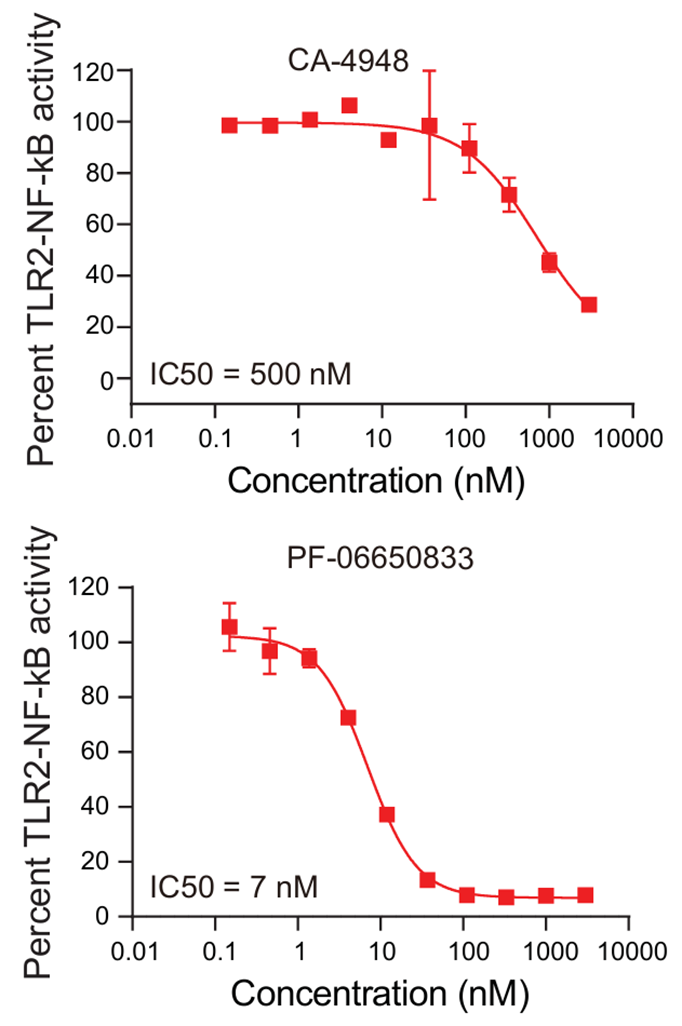
IC50 curves for CA-4948 (Emavusertib) (0.01-10000 nM; 24 h) and PF-06650833 in an assay measuring NF-kB activity upon TLR2 stimulation with PAM3CSK4 in THP1 NF-kB reporter cells.
Emavusertib phosphate purchased from MedChemExpress. Usage Cited in: Blood. 2023 Sep 14;142(11):989-1007. [Abstract]
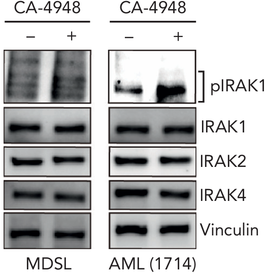
Immunoblots for phospho-IRAK1, total IRAK1, IRAK2, and IRAK4 in MDSL and AML (1714) treated for 24 hours with CA-4948 (Emavusertib) (10 μM). The results showed that treatment with CA-4948 resulted in increased IRAK1 phosphorylation.
-
Leukemia
Targeting of IRAK4 and GSPT1 enhances therapeutic efficacy in AML via c-Myc destabilization. [Abstract]2025 Sep;39(9):2163-2173. PMID: 40670672
Emavusertib phosphate purchased from MedChemExpress. Usage Cited in: Leukemia. 2025 Sep;39(9):2163-2173. [Abstract]
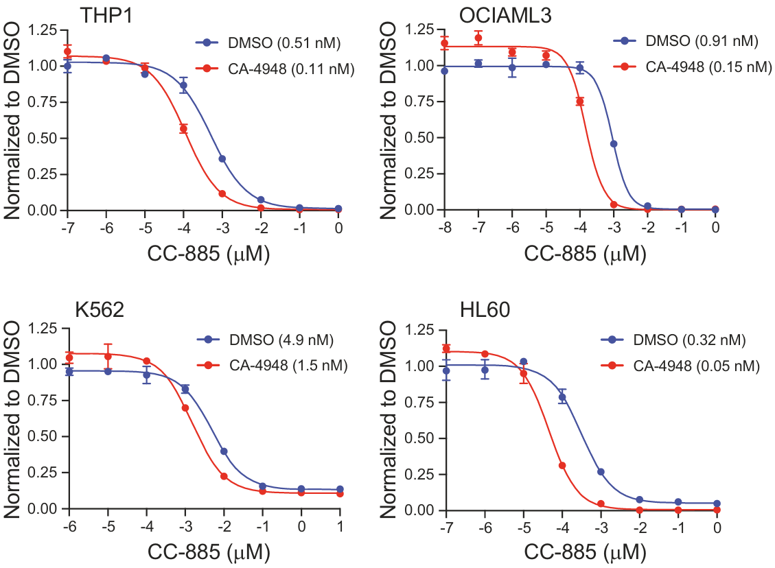
Cell viability was determined in THP1 and HL60 cells cultured with 10 µM CA-4948 (Emavusertib) or in OCI-AML3 and K562 cultured with 1 µM CA-4948 or vehicle (n = 4) for 14 days and then treated with CC-885 (n = 4).
Emavusertib phosphate purchased from MedChemExpress. Usage Cited in: Leukemia. 2025 Sep;39(9):2163-2173. [Abstract]
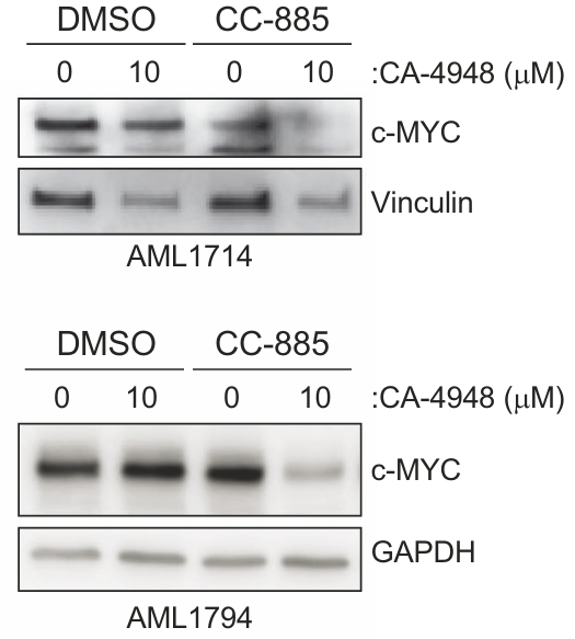
Immunoblots for c-MYC in the patient-derived AML samples (AML1794, AML1714) cultured with CA-4948 (Emavusertib) (10 µM) or vehicle for 14 days and then treated with CC-885 (50 nM) for 4 h.
-
Cell Rep
Disruption of fibroblast MYD88 signaling promotes antitumor immunity in pancreatic ductal adenocarcinoma. [Abstract]2025 Sep 24;44(10):116347. PMID: 41004339
Emavusertib phosphate purchased from MedChemExpress. Usage Cited in: Cell Rep. 2025 Sep 24;44(10):116347. [Abstract]
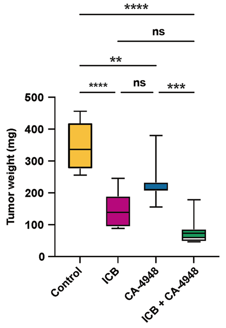
Harvested tumor weights by treatment condition (n = 8 per condition).Tumor growth studies demonstrated statistically significant inhibition with both CA-4948 (Emavusertib) (50 mg/kg; oral gavage; once daily) alone and ICB alone.
Emavusertib phosphate purchased from MedChemExpress. Usage Cited in: Cell Rep. 2025 Sep 24;44(10):116347. [Abstract]
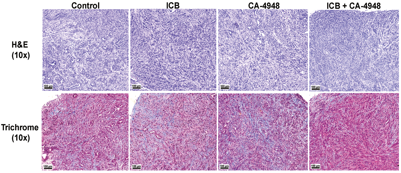
H&E and trichrome staining of tumors by treatment condition (Emavusertib (50 mg/kg; oral gavage; once daily), ect.) at 10× magnification. The scale bar represents 100 μm.
-
Front Immunol
Activation of Toll-Like Receptor 7 Signaling Pathway in Primary Sjögren's Syndrome-Associated Thrombocytopenia. [Abstract]2021 Mar 9:12:637659. PMID: 33767707 -
Curr Issues Mol Biol
FLT3 and IRAK4 Inhibitor Emavusertib in Combination with BH3-Mimetics in the Treatment of Acute Myeloid Leukemia. [Abstract]2024 Mar 29;46(4):2946-2960. PMID: 38666914 -
-
Protocols
-
Apoptosis
Apoptosis, also called programmed cell death, is generally characterized by distinct morphological characteristics.
-
TUNEL staining for apoptotic DNA fragmentation
TUNEL staining detects DNA strand breaks by using terminal deoxynucleotidyl transferase to add labeled nucleotides to exposed 3′-OH DNA termini, generating either microscopic staining in fixed cells or tissue sections, or fluorescence/cytometric signal in cell suspensions. TUNEL positivity reflects DNA fragmentation but should not be interpreted alone as definitive apoptosis, because TUNEL can also label necrotic, autolytic, mechanically damaged, or DNA-repair-associated DNA breaks.
-
Cell Cytotoxicity Assay
Cytotoxicity assays are usually based on the assessment of cell membrane damage, which can also be indirectly detected by measuring cell viability. Detection methods include MTT assay, CKK-8 assay, LDH assay and ATP assay, etc.
-
LPS-Induced Endotoxemia/Systemic Inflammation
Lipopolysaccharide (LPS)-induced endotoxemia is a widely used in vivo model of acute systemic inflammation in which LPS, a Gram-negative bacterial endotoxin, activates innate immune signaling primarily through TLR4, leading to rapid and transient induction of pro-inflammatory cytokines such as TNF-α, IL-6, and IL-1β in circulation and tissues. This cytokine surge is commonly used as a measurable readout of systemic inflammatory activation and immune dysregulation, and is typically assessed within hours after intraperitoneal LPS administration in mouse models of endotoxemia. The model captures key features of systemic inflammatory response syndrome, including cytokine release, immune cell activation, and downstream tissue responses, and has been used to evaluate anti-inflammatory interventions such as cytokine modulation, lipid mediators, and immune cell-targeting therapies.
-
Annexin V plus membrane-impermeant dye apoptosis staining
Annexin V-based apoptosis assays rely on the detection of phosphatidylserine (PS) externalization from the inner leaflet of the plasma membrane to the outer leaflet, an early biochemical hallmark of apoptosis. Fluorescently labeled Annexin V binds PS in a calcium-dependent manner, enabling identification of early apoptotic cells by flow cytometry or fluorescence microscopy. When combined with a membrane-impermeant DNA-binding dye (e. g. , propidium iodide), this approach allows discrimination between viable (Annexin V−/dye−), early apoptotic (Annexin V+/dye−), and late apoptotic or necrotic (Annexin V+/dye+) cell populations by assessing membrane integrity and PS exposure.
-
Apoptosis Solutions
Apoptosis is a regulated, generally non-lytic cell-death pathway that removes unwanted, damaged, infected, or abnormal cells through coordinated morphological changes, caspase activation, DNA fragmentation, and membrane remodeling. The intrinsic apoptosis pathway is controlled mainly by mitochondrial outer membrane permeabilization, BCL-2 family proteins, cytochrome c release, apoptosome formation, caspase-9 activation, and downstream executioner caspase-3/7 activation. The extrinsic apoptosis pathway is initiated by death receptors such as Fas, TNFR, and TRAIL receptors, which recruit adaptor proteins and activate caspase-8 before engaging executioner caspases or mitochondrial amplification through BID cleavage. Apoptosis is linked to many phenotypes, including cancer cell killing, tissue homeostasis, immune regulation, neurodegeneration, infection response, and treatment-induced cytotoxicity; unresolved questions include how apoptosis interacts with necroptosis, pyroptosis, ferroptos
-
Research Protocol for Inflammation-related Diseases
The NLRP3 inflammasome is a cytosolic innate immune signaling platform that integrates priming signals and danger-signal activation to promote caspase-1 activation, maturation of IL-1β and IL-18, and gasdermin D-mediated pyroptotic cell death. The core experimental logic is to determine whether inflammatory disease phenotypes are driven by increased NLRP3 expression, ASC-containing inflammasome assembly, caspase-1 cleavage, GSDMD cleavage, and extracellular release of IL-1β/IL-18 rather than by nonspecific cell injury alone. The pathway is strongly linked to inflammation-related disease phenotypes because monosodium urate crystals activate NALP3/NLRP3 inflammasome signaling in gout-like crystal inflammation, cholesterol crystals activate NLRP3 inflammasomes in atherogenesis models, and DSS-induced intestinal inflammation has been reported to involve NLRP3 inflammasome activity. However, experimental colitis studies also show context-dependent protective effects of NLRP3 inflammasome co
-
Subcutaneous Cell-Line-Derived Xenograft
Subcutaneous cell-line-derived xenograft (CDX) models are established by implanting cultured human cancer cell lines into immunodeficient mice, where the injected cells form localized tumors that can be monitored in vivo as a measure of tumorigenic potential, growth kinetics, and treatment response. These models are widely used in oncology research because they allow reproducible tumor formation and enable comparative assessment of tumor growth between different cell lines or genetic manipulations in a controlled in vivo microenvironment. Subcutaneous implantation of cancer cells in immunodeficient mice is a standard approach for evaluating tumor growth behavior and therapeutic response across multiple cancer types, including prostate, esophageal, pancreatic, and colon cancer models.
-
Cell Viability Determination by MTT Colorimetric Assay
The following protocol uses the MTT colorimetric assay as a classic literature-established method for assessing cell viability/metabolic activity in cultured mammalian cells. MTT[3-(4,5-dimethylthiazol-2-yl)-2,5-diphenyltetrazolium bromide] is reduced by metabolically active cells to a colored formazan product; the amount of formazan is quantified spectrophotometrically and provides an indirect measure of metabolically active viable cells. Importantly, MTT reduction reflects cellular oxidoreductase/metabolic activity rather than an absolute direct count of living cells, so changes in cellular metabolism can alter the signal independently of cell number.
-
How to Choose the Right Model Animal
Choosing the right model animal is a validity-driven decision in which the species, strain, sex, age, genetic background, disease-induction method, outcome measures, and welfare burden must match the scientific question rather than laboratory tradition or convenience. A model should be selected by judging face validity, construct validity, and predictive validity: whether it resembles the human phenotype, whether it reproduces relevant mechanisms, and whether results are likely to predict human biology or treatment response. Animal studies often fail to translate because of species differences, weak disease resemblance, poor experimental design, inadequate reporting, publication bias, and underuse of randomization, blinding, and sample-size justification. Unresolved questions include how to rank competing models objectively, how much human-disease complexity must be reproduced for a given objective, and when non-animal systems such as organoids, ex vivo tissue, or computational models
Purity & Documentation
References
[1]. Wiese MD, et al. Investigational IRAK-4 inhibitors for the treatment of rheumatoid arthritis. Expert Opin Investig Drugs. 2020 Apr 17:1-8. [Content Brief]
[2]. Guidetti F, et al. Targeting IRAK4 with Emavusertib in Lymphoma Models with Secondary Resistance to PI3K and BTK Inhibitors. J Clin Med. 2023 Jan 4;12(2):399. [Content Brief]
[3]. Parrondo RD, et al. IRAK-4 inhibition: emavusertib for the treatment of lymphoid and myeloid malignancies. Front Immunol. 2023 Oct 26;14:1239082. [Content Brief]
Calculators
Concentration (start) × Volume (start) = Concentration (final) × Volume (final)