



Isobavachalcone
Based on 10 publication(s) in Google Scholar
Isobavachalcone (Corylifolinin) is derived from Psoralea corylifolia Linn. and is a potent inhibitor of Akt signaling pathway, which induces apoptosis in human cancer cells (Inhibits OVCAR-8 cell growth with an IC50 value of 7.92 μM). Isobavachalcone also induces Reactive Oxyen Species (ROS) generation in OVCAR-8 cells and has exhibit cancer anti-promotive and anti-proliferative activity.
Nos produits utilisent uniquement pour la recherche. Nous ne vendons pas aux patients.
- Pureté : 98.99%
- CAS No.: 20784-50-3
- Formule: C20H20O4
- Masse moléculaire:324.37
-
Stockage:
4°C, protect from light
* In solvent : -80°C, 1 year; -20°C, 6 months (protect from light)

To place orders, for customer services and technical support, please contact: MedChemExpress USA
Tel: 609-228-6898 E-mail: [email protected] [email protected]


-
 Activité biologique
-
Chemical Information
-
Solvant et solubilité
- Protocole
- Pureté et documentation
- Références
-
 Help & FAQs


-
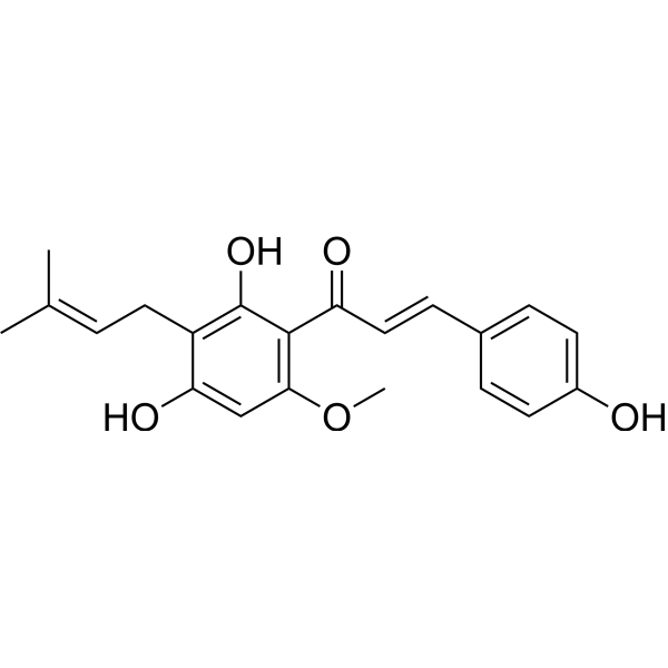
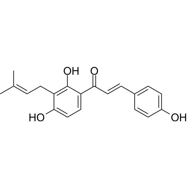
HY-N1067
XanthohumolCOX Inhibitor -
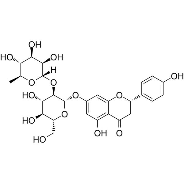
HY-N0153
NaringinCytochrome P450 Inhibitor -
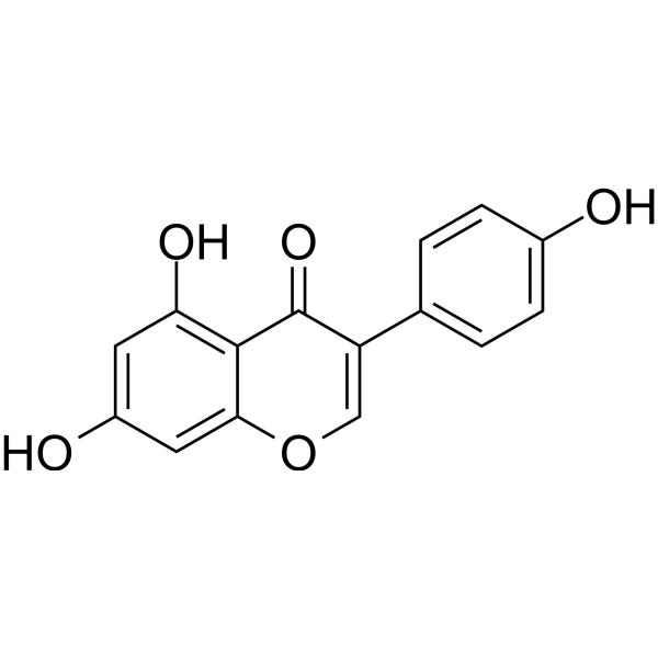
HY-14596
GenisteinAutophagy Inducer -
HY-10191
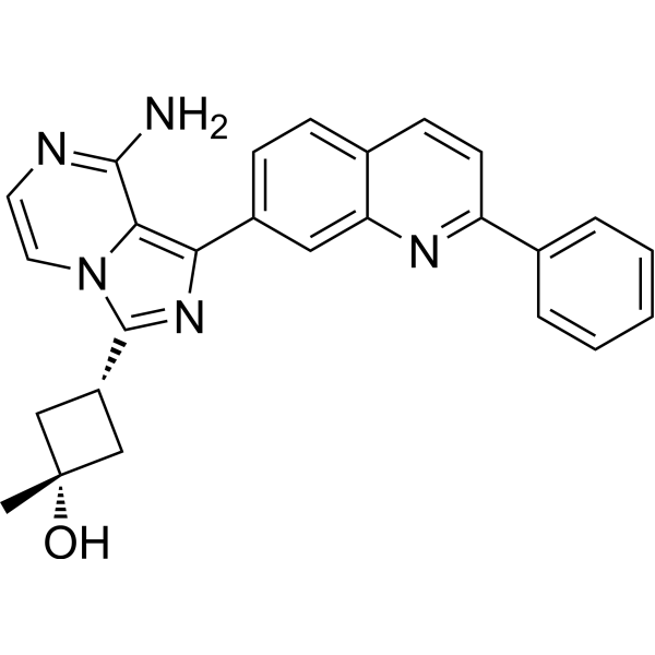
LinsitinibIGF-1R/Insulin Receptor Inhibitor -

HY-10219
RapamycinmTOR Inhibitor -
HY-N0005
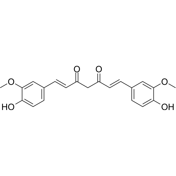
Curcuminp300 Histone Acetylatransferase Inhibitor -
HY-18085
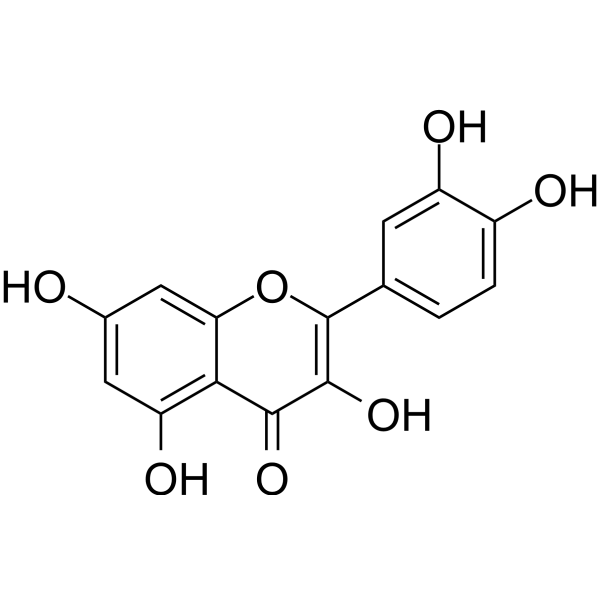
QuercetinFlavonoid Antioxidant -
HY-18258
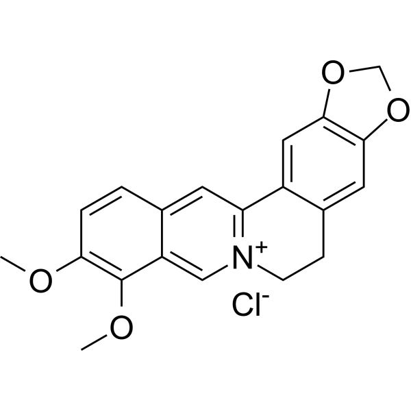
Berberine chlorideAntibiotic -
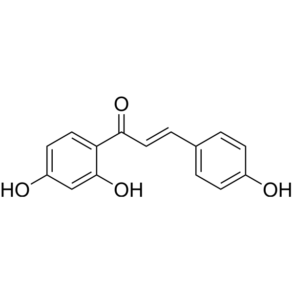
HY-N0102
IsoliquiritigeninAutophagy Inducer -
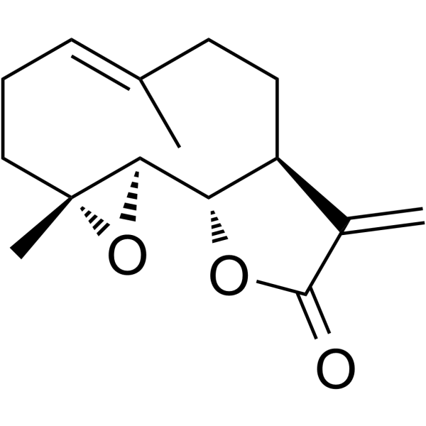
HY-N0141
ParthenolideNF-κB Inhibitor -

HY-13259
MG-132Proteasome Inhibitor -
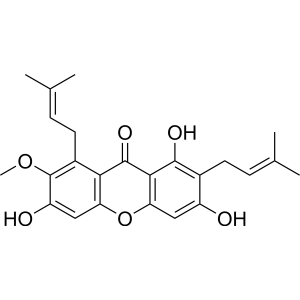
HY-N0328
alpha-MangostinIDH1-R132H Inhibitor
Publications Citing Use of MedChemExpress (MCE) Isobavachalcone
More- Acta Pharm Sin B. 2021 Jan;11(1):143-155. [Abstract]
- Food Chem. 2025 Dec 30:497:146992. [Abstract]
- Food Chem. 2025 Oct 15:489:144992. [Abstract]
- J Ethnopharmacol. 2024 May 23:326:117827. [Abstract]
- J Ethnopharmacol. 2022 Nov 15:298:115593. [Abstract]
- J Ethnopharmacol. 2022 May 4;294:115342. [Abstract]
- Food Funct. 2021 Sep 7;12(17):7749-7761. [Abstract]
- Int Immunopharmacol. 2013 Jan;15(1):38-41. [Abstract]
- Chem Biol Interact. 2024 Sep 1:400:111133. [Abstract]
- ACS Chem Neurosci. 2021 Jan 6;12(1):123-132. [Abstract]
 Customer Validation & Images

-
Histological Imaging/Staining

-
WB
-
Cell Proliferation/Viability Assay


Activité biologique
Description
IC50 & Target
IC50: 7.92 μM (OVCAR-8 cell)[1]
Cellular Effect
|
Cell Line

|
Type | Value | Description | References |
|---|---|---|---|---|
| A549 | IC50 |
>10 μM
Compound: 8
|
Cytotoxicity against human A549 cells after 48 hrs by MTT assay
Cytotoxicity against human A549 cells after 48 hrs by MTT assay
|
[PMID: 25710081] |
| HCT-116 | IC50 |
75.48 μM
Compound: 50
|
Antiproliferative activity against human HCT-116 cells assessed as reduction in cell viability
Antiproliferative activity against human HCT-116 cells assessed as reduction in cell viability
|
[PMID: 33445154] |
| HUVEC | IC50 |
>100 μM
Compound: 1c
|
Toxicity against HUVEC incubated for 48 hrs by MTT assay
Toxicity against HUVEC incubated for 48 hrs by MTT assay
|
[PMID: 25590864] |
| K562 | IC50 |
>10 μM
Compound: 8
|
Cytotoxicity against human K562 cells after 48 hrs by MTT assay
Cytotoxicity against human K562 cells after 48 hrs by MTT assay
|
[PMID: 25710081] |
| K562 | IC50 |
29.49 μM
Compound: 1c
|
Antitumor activity against human K562 cells incubated for 48 hrs by MTT assay
Antitumor activity against human K562 cells incubated for 48 hrs by MTT assay
|
[PMID: 25590864] |
| OVCAR-8 | IC50 |
7.92 μM
Compound: 60; IBC
|
Cytotoxicity against human OVCAR-8 cells assessed as cell growth inhibition measured after 72 hrs by MTT assay
Cytotoxicity against human OVCAR-8 cells assessed as cell growth inhibition measured after 72 hrs by MTT assay
|
[PMID: 35151222] |
| PC-3 | IC50 |
15.06 μM
Compound: 60; IBC
|
Cytotoxicity against human PC3 cells assessed as cell growth inhibition measured after 72 hrs by MTT assay
Cytotoxicity against human PC3 cells assessed as cell growth inhibition measured after 72 hrs by MTT assay
|
[PMID: 35151222] |
| Platelet | IC50 |
0.5 μM
Compound: 1
|
Antiplatelet activity against rabbit platelet assessed as inhibition of arachidonic acid-induced platelet aggregation preincubated for 3 mins by turbidimetric method
Antiplatelet activity against rabbit platelet assessed as inhibition of arachidonic acid-induced platelet aggregation preincubated for 3 mins by turbidimetric method
|
[PMID: 8759164] |
| Platelet | IC50 |
41.6 μM
Compound: 1
|
Antiplatelet activity against rabbit platelet assessed as inhibition of platelet-activating factor-induced platelet aggregation preincubated for 3 mins by turbidimetric method
Antiplatelet activity against rabbit platelet assessed as inhibition of platelet-activating factor-induced platelet aggregation preincubated for 3 mins by turbidimetric method
|
[PMID: 8759164] |
| Platelet | IC50 |
65.1 μM
Compound: 1
|
Antiplatelet activity against rabbit platelet assessed as inhibition of collagen-induced platelet aggregation preincubated for 3 mins by turbidimetric method
Antiplatelet activity against rabbit platelet assessed as inhibition of collagen-induced platelet aggregation preincubated for 3 mins by turbidimetric method
|
[PMID: 8759164] |
| Raji | IC50 |
320 molar ratio
Compound: 4
|
Inhibition of TPA-induced EBV-early antigen activation in human Raji cells
Inhibition of TPA-induced EBV-early antigen activation in human Raji cells
|
[PMID: 16441065] |
| RAW264.7 | IC50 |
1.6 μM
Compound: Table 4, R5C1
|
Inhibition of LPS-induced nitric oxide production in mouse RAW264.7 cells
Inhibition of LPS-induced nitric oxide production in mouse RAW264.7 cells
|
[PMID: 32208222] |
| RAW264.7 | IC50 |
16.4 μM
Compound: 2
|
Cytotoxicity against mouse RAW264.7 cells by MTT assay
Cytotoxicity against mouse RAW264.7 cells by MTT assay
|
[PMID: 16643064] |
| RAW264.7 | IC50 |
3.5 μg/mL
Compound: 133
|
Antiinflammatory activity in mouse RAW264.7 cells assessed as inhibition of LPS-induced nitric oxide production
Antiinflammatory activity in mouse RAW264.7 cells assessed as inhibition of LPS-induced nitric oxide production
|
[PMID: 31255927] |
| RAW264.7 | IC50 |
3.5 μg/mL
Compound: 157
|
Anti-inflammatory activity in mouse RAW264.7 cells assessed as reduction in LPS-induced NO production pre-treated with compounds for 2 hrs followed by LPS-stimulation measured for 22 hrs by Griess reagent based analysis
Anti-inflammatory activity in mouse RAW264.7 cells assessed as reduction in LPS-induced NO production pre-treated with compounds for 2 hrs followed by LPS-stimulation measured for 22 hrs by Griess reagent based analysis
|
[PMID: 37683361] |
| RAW264.7 | IC50 |
6.4 μM
Compound: 2
|
Inhibition of LPS-induced nitric oxide production in mouse RAW264.7 cells assessed as nitrite accumulation after 20 hrs
Inhibition of LPS-induced nitric oxide production in mouse RAW264.7 cells assessed as nitrite accumulation after 20 hrs
|
[PMID: 16643064] |
| SW480 | IC50 |
44.07 μM
Compound: 50
|
Antiproliferative activity against human SW480 cells assessed as reduction in cell viability
Antiproliferative activity against human SW480 cells assessed as reduction in cell viability
|
[PMID: 33445154] |
| THP-1 | CC50 |
35.95 μM
Compound: 1
|
Cytotoxicity against human THP1 cells assessed as cell viability after 72 hrs by MTT assay
Cytotoxicity against human THP1 cells assessed as cell viability after 72 hrs by MTT assay
|
[PMID: 26906638] |
| Vero | CC50 |
726.82 μg/mL
Compound: 14, 4-hydroxy-isocordoin
|
Cytotoxicity against african green monkey Vero cells assessed as cell death after 72 hrs by MTT assay
Cytotoxicity against african green monkey Vero cells assessed as cell death after 72 hrs by MTT assay
|
[PMID: 22169259] |
| Vero | IC50 |
31.25 μg/mL
Compound: 14, 4-hydroxy-isocordoin
|
Cytotoxicity against African green monkey Vero cells assessed as inhibition of cell growth after 72 hrs by MTT assay
Cytotoxicity against African green monkey Vero cells assessed as inhibition of cell growth after 72 hrs by MTT assay
|
[PMID: 22169259] |
In Vitro
Isobavachalcone (6.0-48.0 μM; 72 hours; OVCAR-8, PC3, A549, MCF-7, L-02 and HUVEC cells) treatment inhibits the proliferation of human cancer cells. Isobavachalcone inhibits PC3, A549, MCF-7, L-02 and HUVEC cells growth with IC50 values of 15.06 μM, 32.2 μM, 28.29 μM, 31.61 μM and 31.3 μM, respectively[1].
Isobavachalcone (0-18 μM; 6 hours; OVCAR-8 and PC3 cells) treatment results in a concentration-and time-dependent down-regulation of the Ser-473 phosphorylation of Akt and GSK3b phosphorylation[1].
Isobavachalcone (0-18 μM; 72 hours; OVCAR-8 and PC3 cells) treatment causes apoptosis via caspase- and ROS-involved mitochondrial pathway[1].
MedChemExpress (MCE) has not independently confirmed the accuracy of these methods. They are for reference only.
-
Cell Line:OVCAR-8, PC3, A549, MCF-7, L-02 and HUVEC cells
-
Concentration:6.0-48.0 μM
-
Incubation Time:72 hours
-
Result:Inhibited the proliferation of human cancer cells.
-
Cell Line:OVCAR-8 or PC3 cells
-
Concentration:0 μM, 6 μM, 12 μM, and 18 μM
-
Incubation Time:6 hours
-
Result:A concentration-and time-dependent down-regulation of the Ser-473 phosphorylation of Akt. GSK3b phosphorylation was also inhibited in a concentration- and time-dependent manner.
-
Cell Line:OVCAR-8 cells and PC3 cells
-
Concentration:0 μM, 6 μM, 12 μM, and 18 μM
-
Incubation Time:72 hours
-
Result:Led to dose dependent increase of apoptosis.
In Vivo
MedChemExpress (MCE) has not independently confirmed the accuracy of these methods. They are for reference only.
-
Animal Model:Seven-week-old specific pathogen-free female Kunming mice (18-22 g)[1]
-
Dosage:20 mg/kg
-
Administration:Intraperitoneal injection; for 0.5, 1, 2, 4, 6, 8, 12, 24 hours
-
Result:Increased in blood glucose levels.
- Detection of Apoptosis
- TUNEL staining for apoptotic DNA fragmentation
- Annexin V plus membrane-impermeant dye apoptosis staining
- Protocol for Cell Counting and Cell Density Analysis
- ROS/oxidative-stress fluorescent staining
- Somatic Cell Culture
- Apoptosis Solutions
- CCK-8/WST-8 Cell Proliferation Assay
- Cell Counting-Based Growth Curve Assay
- MTT Cell Proliferation Assay
- Cell Viability Determination by MTT Colorimetric Assay


Chemical Information
-
CAS No. 20784-50-3
-
Appearance Solid
-
Masse moléculaire 324.37
-
Formule C20H20O4
-
Color Light yellow to yellow
-
SMILES
O=C(/C=C/C1=CC=C(C=C1)O)C2=CC=C(C(C/C=C(C)\C)=C2O)O
-
Synonyms
Corylifolinin; Isobacachalcone
-
Structure Classification
-
Livraison
Room temperature in continental US; may vary elsewhere.
-
Stockage
4°C, protect from light
* In solvent : -80°C, 1 year; -20°C, 6 months (protect from light)


Publications (10)
-
Journal Impact Factor
-
Most Recent
-
Acta Pharm Sin B
Chrysin serves as a novel inhibitor of DGK α/FAK interaction to suppress the malignancy of esophageal squamous cell carcinoma (ESCC). [Abstract]2021 Jan;11(1):143-155. PMID: 33532186 -
Food Chem
Effects of sun drying combined with baking processes on the flavor quality of Chongqing Tuocha raw tea. [Abstract]2025 Dec 30:497:146992. PMID: 41285060 -
Food Chem
Flavonoid-mediated metabolic underpinning quality variation in red bud-sport pear mutants. [Abstract]2025 Oct 15:489:144992. PMID: 40466530 -
J Ethnopharmacol
An integrated network pharmacology, molecular docking and experiment validation study to investigate the potential mechanism of Isobavachalcone in the treatment of osteoarthritis. [Abstract]2024 May 23:326:117827. PMID: 38310989 -
J Ethnopharmacol
2022 Nov 15:298:115593. PMID: 35973629 -
J Ethnopharmacol
Investigation of the mechanism of Isobavachalcone in treating rheumatoid arthritis through a combination strategy of network pharmacology and experimental verification. [Abstract]2022 May 4;294:115342. PMID: 35525528 -
Food Funct
Isobavachalcone ameliorates cognitive deficits, and Aβ and tau pathologies in triple-transgenic mice with Alzheimer's disease. [Abstract]2021 Sep 7;12(17):7749-7761. PMID: 34269361
Isobavachalcone purchased from MedChemExpress. Usage Cited in: Food Funct. 2021 Sep 7;12(17):7749-7761. [Abstract]
Representative images of the cortex by silver staining treated with Isobavachalcone (ISO) (5, 10 μg/mL).
-
Int Immunopharmacol
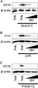
Isobavachalcone suppresses expression of inducible nitric oxide synthase induced by Toll-like receptor agonists. [Abstract]2013 Jan;15(1):38-41. PMID: 23164691
Isobavachalcone purchased from MedChemExpress. Usage Cited in: Int Immunopharmacol. 2013 Jan;15(1):38-41. [Abstract]
IBC inhibits iNOS protein induced by TLRs agonists. A–C) RAW264.7 cells are pretreated with 20 or 50 μM IBC for 1 h and then further stimulated with MALP-2 (10 μg/mL) (A), LPS (10 μg/mL) (B), or poly[I:C] (10 μg/mL) (C) for an additional 8 h. Cell lysates are analyzed for iNOS and β-actin protein by immunoblots. Veh, vehicle; IBC, isobavachalcone.
Isobavachalcone purchased from MedChemExpress. Usage Cited in: Int Immunopharmacol. 2013 Jan;15(1):38-41. [Abstract]
RAW264.7 cells were treated with Isobavachalcone (IBC) (20, 50, 100 μM) for 4 h. Twenty microliters of the CellTiter 96 AQueous One Solution Reagent was added directly to culture wells.
-
Chem Biol Interact
Bavachinin, a main compound of Psoraleae Fructus, facilitates GSDMD-mediated pyroptosis and causes hepatotoxicity in mice. [Abstract]2024 Sep 1:400:111133. PMID: 38969277 -
ACS Chem Neurosci
Inhibitory Effects of Isobavachalcone on Tau Protein Aggregation, Tau Phosphorylation, and Oligomeric Tau-Induced Apoptosis. [Abstract]2021 Jan 6;12(1):123-132. PMID: 33320518


Solvant et solubilité
In Vitro:
DMSO : 100 mg/mL (308.29 mM; Need ultrasonic; Hygroscopic DMSO has a significant impact on the solubility of product, please use newly opened DMSO)
Please refer to the solubility information to select the appropriate solvent. Once prepared, please aliquot and store the solution to prevent product inactivation from repeated freeze-thaw cycles.
Storage method and period of stock solution: -80°C, 1 year; -20°C, 6 months (protect from light). When stored at -80°C, please use it within 1 year. When stored at -20°C, please use it within 6 months.
Please refer to the solubility information to select the appropriate solvent. Once prepared, please aliquot and store the solution to prevent product inactivation from repeated freeze-thaw cycles.
Storage method and period of stock solution: -80°C, 1 year; -20°C, 6 months (protect from light). When stored at -80°C, please use it within 1 year. When stored at -20°C, please use it within 6 months.
Concentration (start) × Volume (start) = Concentration (final) × Volume (final)
In Vivo:
Select the appropriate dissolution method based on your experimental animal and administration route.
- For the following dissolution methods, please ensure to first prepare a clear stock solution using an In Vitro approach and then sequentially add co-solvents:
- To ensure reliable experimental results, the clarified stock solution can be appropriately stored based on storage conditions. As for the working solution for In Vivo experiments, it is recommended to prepare freshly and use it on the same day.
- The percentages shown for the solvents indicate their volumetric ratio in the final prepared solution. If precipitation or phase separation occurs during preparation, heat and/or sonication can be used to aid dissolution.
Add each solvent one by one: 10% DMSO 90% (20% SBE-β-CD in Saline)
Solubility: ≥ 2.08 mg/mL (6.41 mM); Clear solution
This protocol yields a clear solution of ≥ 2.08 mg/mL (saturation unknown).
Taking 1 mL working solution as an example, add 100 μL DMSO stock solution (20.8 mg/mL) to 900 μL 20% SBE-β-CD in Saline, and mix evenly.
Preparation of 20% SBE-β-CD in Saline (4°C, storage for one week): 2 g SBE-β-CD powder is dissolved in 10 mL Saline, completely dissolve until clear.
In Vivo Dissolution Calculator
Please enter the basic information of animal experiments:
-
-
-
-
Recommended: Prepare an additional quantity of animals to account for potential losses during experiments.
Please enter your animal formula composition:
-
%DMSO +
Recommended: Keep the proportion of DMSO in working solution below 2% if your animal is weak.
-
%+
-
+%Tween-80 + +
-
%Saline +
The co-solvents required include: DMSO, . All of co-solvents are available by MedChemExpress (MCE). , Tween 80. All of co-solvents are available by MedChemExpress (MCE).
Working solution concentration: 0.22 mg/mL
Method for preparing stock solution: mg drug dissolved in μL DMSO. Stock solution concentration: mg/mL. * In solvent : -80°C, 1 year; -20°C, 6 months (protect from light)
1. Take μL DMSO stock solution;
2. Add μL .
μL , mix evenly;
3. Then add μL Tween 80, mix evenly;
4. Then add μL
Please ensure that the stock solution in the first step is dissolved to a clear state, and add co-solvents in sequence. You can use ultrasonic heating (ultrasonic cleaner, recommended frequency 20-40 kHz), vortexing, etc. to assist dissolution.


Pureté et documentation
-
Fiche technique (276 KB)
-
SDS (393 KB)
- English - EN (393 KB)
- Français - FR (393 KB)
- Deutsch - DE (393 KB)
- Norwegian - NO (393 KB)
- Español - ES (393 KB)
- Swedish - SV (393 KB)
- Italian - IT (393 KB)
- Korean - KR (393 KB)
- Portuguese - PT (393 KB)
-
Instruction de manipulation (2659 KB)
Références


Complete Stock Solution Preparation Table
Please refer to the solubility information to select the appropriate solvent. Once prepared, please aliquot and store the solution to prevent product inactivation from repeated freeze-thaw cycles.
Storage method and period of stock solution: -80°C, 1 year; -20°C, 6 months (protect from light). When stored at -80°C, please use it within 1 year. When stored at -20°C, please use it within 6 months.
| Optional Solvent | Concentration Solvent Mass | 1 mg | 5 mg | 10 mg | 25 mg |
|---|---|---|---|---|---|
| DMSO | 1 mM | 3.0829 mL | 15.4145 mL | 30.8290 mL | 77.0725 mL |
| 5 mM | 0.6166 mL | 3.0829 mL | 6.1658 mL | 15.4145 mL | |
| 10 mM | 0.3083 mL | 1.5414 mL | 3.0829 mL | 7.7072 mL | |
| 15 mM | 0.2055 mL | 1.0276 mL | 2.0553 mL | 5.1382 mL | |
| 20 mM | 0.1541 mL | 0.7707 mL | 1.5414 mL | 3.8536 mL | |
| 25 mM | 0.1233 mL | 0.6166 mL | 1.2332 mL | 3.0829 mL | |
| 30 mM | 0.1028 mL | 0.5138 mL | 1.0276 mL | 2.5691 mL | |
| 40 mM | 0.0771 mL | 0.3854 mL | 0.7707 mL | 1.9268 mL | |
| 50 mM | 0.0617 mL | 0.3083 mL | 0.6166 mL | 1.5414 mL | |
| 60 mM | 0.0514 mL | 0.2569 mL | 0.5138 mL | 1.2845 mL | |
| 80 mM | 0.0385 mL | 0.1927 mL | 0.3854 mL | 0.9634 mL | |
| 100 mM | 0.0308 mL | 0.1541 mL | 0.3083 mL | 0.7707 mL |


Isobavachalcone Related Classifications
HY-13065 Related Classifications
 Powered by Bioz