



R243
Based on 3 publication(s) in Google Scholar
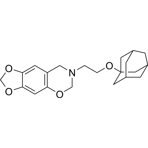
R243 is a potent and selective CCR8 antagonist. R243 inhibits CCL1/CCR8 interaction and inhibits CCR8 signaling and chemotaxis. R243 has antinociceptive and anti-inflammatory effects.
Nos produits utilisent uniquement pour la recherche. Nous ne vendons pas aux patients.
- Pureté : 98.09%
- CAS No.: 688352-84-3
- Formule: C21H27NO4
- Masse moléculaire:357.44
-
Stockage:Powder -20°C, 3 years , 4°C, 2 years ; In solvent -80°C, 6 months , -20°C, 1 month

To place orders, for customer services and technical support, please contact: MedChemExpress USA
Tel: 609-228-6898 E-mail: [email protected] [email protected]


-
 Activité biologique
-
Chemical Information
-
Solvant et solubilité
- Protocole
- Pureté et documentation
- Références
-
 Help & FAQs


-
HY-13004
MaravirocCCR5 Antagonist -
HY-15724
VercirnonCCR9 Antagonist -
HY-124416
ML604086CCR8 Inhibitor -
HY-12080
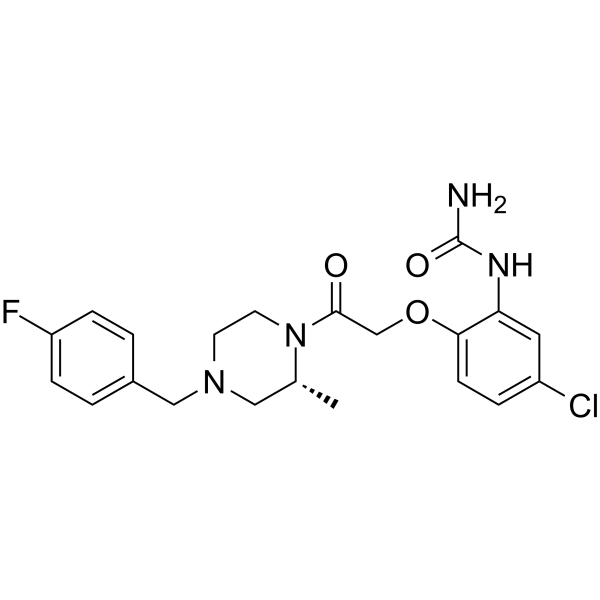
BX471CCR1 Antagonist -
HY-121885
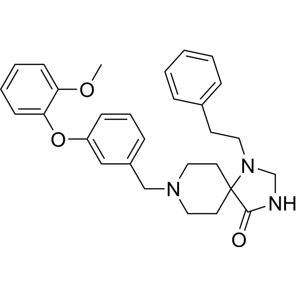
LMD-009CCR8 Nonpeptide Agonist -
HY-101038A
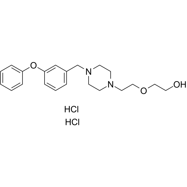
ZK756326 dihydrochlorideCCR8 Agonist -
HY-103364
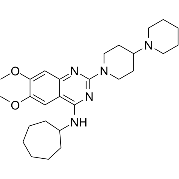
C-021CCR4 Antagonist -
HY-116835
BI-6901CCR10 Antagonist -
HY-112701
CCR6 inhibitor 1CCR Inhibitor -
HY-17450A
Aplaviroc hydrochlorideCCR5 Antagonist -
HY-15418
RS 504393CCR2 Antagonist -
HY-14882
CenicrivirocCCR2/5 Antagonist
Publications Citing Use of MedChemExpress (MCE) R243
More

Activité biologique
Description
IC50 & Target
[1]|
CCR8 |
In Vitro
R243 has CCR8-antagonistic effects on CCL1-induced Ca2+ flux and CCL1-driven peritoneal macrophages aggregation[1].
R243 attenuates secretion of TNF-α, IL-6, and most strikingly IL-10 from wild-type peritoneal macrophages (WT PMφ)[1].
R243-treated WT PMφ shows suppressed c-jun N-terminal kinase activity and NF-κB signaling after lipopolysaccharide (LPS) treatment when compared with WT PMφ[1].
MedChemExpress (MCE) has not independently confirmed the accuracy of these methods. They are for reference only.
In Vivo
MedChemExpress (MCE) has not independently confirmed the accuracy of these methods. They are for reference only.
-
Animal Model:Male Swiss mice (7-9 weeks old) injected with CCL1[2]
-
Dosage:0.1 mg/kg, 0.3mg/kg, 1 mg/kg
-
Administration:Intraperitoneal injection; once
-
Result:The analgesic effect evoked by CCL1 (10 μg/kg; 1 h; s.c.) was dose-dependently inhibited.


Chemical Information
-
CAS No. 688352-84-3
-
Appearance Solid
-
Masse moléculaire 357.44
-
Formule C21H27NO4
-
Color White to off-white
-
SMILES
[C@@H]12CC(C[C@H](C3)C1)(C[C@@H]3C2)OCCN4CC(C=C(OCO5)C5=C6)=C6OC4
-
Livraison
Room temperature in continental US; may vary elsewhere.
-
Stockage
Powder -20°C 3 years 4°C 2 years In solvent -80°C 6 months -20°C 1 month


Publications (3)
-
Journal Impact Factor
-
Most Recent
-
Sci Adv
Earlier onset of chemotherapy-induced neuropathic pain in females by ICAM-1-mediated accumulation of perivascular macrophages. [Abstract]2025 Apr 18;11(16):eadu2159. PMID: 40238872 -
J Immunother Cancer
2024 Dec 4;12(12):e009629. PMID: 39631847 -
JCI Insight
ANGPTL8 links refeeding to monocyte dynamics and metabolic inflammation via the CCL5-CCR5 axis. [Abstract]2025 Nov 25:e196605. PMID: 41289013


Solvant et solubilité
In Vitro:
DMSO : 100 mg/mL (279.77 mM; ultrasonic and warming and heat to 60°C; Hygroscopic DMSO has a significant impact on the solubility of product, please use newly opened DMSO)
Please refer to the solubility information to select the appropriate solvent. Once prepared, please aliquot and store the solution to prevent product inactivation from repeated freeze-thaw cycles.
Storage method and period of stock solution: -80°C, 6 months; -20°C, 1 month. When stored at -80°C, please use it within 6 months. When stored at -20°C, please use it within 1 month.
Please refer to the solubility information to select the appropriate solvent. Once prepared, please aliquot and store the solution to prevent product inactivation from repeated freeze-thaw cycles.
Storage method and period of stock solution: -80°C, 6 months; -20°C, 1 month. When stored at -80°C, please use it within 6 months. When stored at -20°C, please use it within 1 month.
Concentration (start) × Volume (start) = Concentration (final) × Volume (final)
In Vivo:
Select the appropriate dissolution method based on your experimental animal and administration route.
- For the following dissolution methods, please ensure to first prepare a clear stock solution using an In Vitro approach and then sequentially add co-solvents:
- To ensure reliable experimental results, the clarified stock solution can be appropriately stored based on storage conditions. As for the working solution for In Vivo experiments, it is recommended to prepare freshly and use it on the same day.
- The percentages shown for the solvents indicate their volumetric ratio in the final prepared solution. If precipitation or phase separation occurs during preparation, heat and/or sonication can be used to aid dissolution.
Add each solvent one by one: 10% DMSO 90% (20% SBE-β-CD in Saline)
Solubility: ≥ 2.08 mg/mL (5.82 mM); Clear solution
This protocol yields a clear solution of ≥ 2.08 mg/mL (saturation unknown).
Taking 1 mL working solution as an example, add 100 μL DMSO stock solution (20.8 mg/mL) to 900 μL 20% SBE-β-CD in Saline, and mix evenly.
Preparation of 20% SBE-β-CD in Saline (4°C, storage for one week): 2 g SBE-β-CD powder is dissolved in 10 mL Saline, completely dissolve until clear.
In Vivo Dissolution Calculator
Please enter the basic information of animal experiments:
-
-
-
-
Recommended: Prepare an additional quantity of animals to account for potential losses during experiments.
Please enter your animal formula composition:
-
%DMSO +
Recommended: Keep the proportion of DMSO in working solution below 2% if your animal is weak.
-
%+
-
+%Tween-80 + +
-
%Saline +
The co-solvents required include: DMSO, . All of co-solvents are available by MedChemExpress (MCE). , Tween 80. All of co-solvents are available by MedChemExpress (MCE).
Working solution concentration: 0.22 mg/mL
Method for preparing stock solution: mg drug dissolved in μL DMSO. Stock solution concentration: mg/mL.
1. Take μL DMSO stock solution;
2. Add μL .
μL , mix evenly;
3. Then add μL Tween 80, mix evenly;
4. Then add μL
Please ensure that the stock solution in the first step is dissolved to a clear state, and add co-solvents in sequence. You can use ultrasonic heating (ultrasonic cleaner, recommended frequency 20-40 kHz), vortexing, etc. to assist dissolution.


Pureté et documentation
-
Fiche technique (275 KB)
-
SDS (251 KB)
- English - EN (251 KB)
- Français - FR (251 KB)
- Deutsch - DE (251 KB)
- Norwegian - NO (251 KB)
- Español - ES (251 KB)
- Swedish - SV (251 KB)
- Italian - IT (251 KB)
- Korean - KR (251 KB)
- Portuguese - PT (251 KB)
-
Instruction de manipulation (2659 KB)
Références
[1]. Tomoyuki Oshio, et al. Chemokine Receptor CCR8 Is Required for Lipopolysaccharide-Triggered Cytokine Production in Mouse Peritoneal Macrophages. PLoS One. 2014 Apr 8;9(4):e94445. [Content Brief]
[2]. Mario García-Domínguez, et al. The Systemic Administration of the Chemokine CCL1 Evokes Thermal Analgesia in Mice Through the Activation of the Endocannabinoid System. Cell Mol Neurobiol. 2019 Nov;39(8):1115-1124. [Content Brief]


Complete Stock Solution Preparation Table
Please refer to the solubility information to select the appropriate solvent. Once prepared, please aliquot and store the solution to prevent product inactivation from repeated freeze-thaw cycles.
Storage method and period of stock solution: -80°C, 6 months; -20°C, 1 month. When stored at -80°C, please use it within 6 months. When stored at -20°C, please use it within 1 month.
| Optional Solvent | Concentration Solvent Mass | 1 mg | 5 mg | 10 mg | 25 mg |
|---|---|---|---|---|---|
| DMSO | 1 mM | 2.7977 mL | 13.9884 mL | 27.9767 mL | 69.9418 mL |
| 5 mM | 0.5595 mL | 2.7977 mL | 5.5953 mL | 13.9884 mL | |
| 10 mM | 0.2798 mL | 1.3988 mL | 2.7977 mL | 6.9942 mL | |
| 15 mM | 0.1865 mL | 0.9326 mL | 1.8651 mL | 4.6628 mL | |
| 20 mM | 0.1399 mL | 0.6994 mL | 1.3988 mL | 3.4971 mL | |
| 25 mM | 0.1119 mL | 0.5595 mL | 1.1191 mL | 2.7977 mL | |
| 30 mM | 0.0933 mL | 0.4663 mL | 0.9326 mL | 2.3314 mL | |
| 40 mM | 0.0699 mL | 0.3497 mL | 0.6994 mL | 1.7485 mL | |
| 50 mM | 0.0560 mL | 0.2798 mL | 0.5595 mL | 1.3988 mL | |
| 60 mM | 0.0466 mL | 0.2331 mL | 0.4663 mL | 1.1657 mL | |
| 80 mM | 0.0350 mL | 0.1749 mL | 0.3497 mL | 0.8743 mL | |
| 100 mM | 0.0280 mL | 0.1399 mL | 0.2798 mL | 0.6994 mL |


R243 Related Classifications
HY-122219 Related Classifications
 Powered by Bioz