



Calenduloside E
Based on 1 publication(s) in Google Scholar
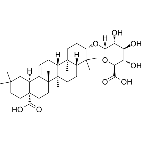
Calenduloside E is a pentacyclic triterpenoid saponin that can be extracted from the bark and roots of Aralia ovata, and has anti-inflammatory and anti-apoptotic activities. Calenduloside E alleviates atherosclerosis by regulating macrophage polarization, improves mitochondrial function by regulating the AMPK-SIRT3 pathway, and alleviates acute liver injury. In addition, Calenduloside E promotes the interaction between L-type calcium channels and Bcl-2 related apoptosis genes, inhibits calcium overload, and alleviates myocardial ischemia/reperfusion injury. Calenduloside E also improves non-alcoholic fatty liver disease by regulating heat shock-dependent pathways, and inhibits ROS mediated JAK1-STAT3 pathways to reduce cellular inflammatory responses.
For research use only. We do not sell to patients.
- Purity : 98.92%
- CAS No.: 26020-14-4
- Formula: C36H56O9
- Molecular Weight:632.82
-
Storage:Powder -20°C, 3 years , 4°C, 2 years ; In solvent -80°C, 6 months , -20°C, 1 month

To place orders, for customer services and technical support, please contact: MedChemExpress USA
Tel: 609-228-6898 E-mail: [email protected] [email protected]


-
 Biological Activity
-
Chemical Information
-
Solvent & Solubility
- Protocols
- Purity & Documentation
- References
-
 Help & FAQs


-
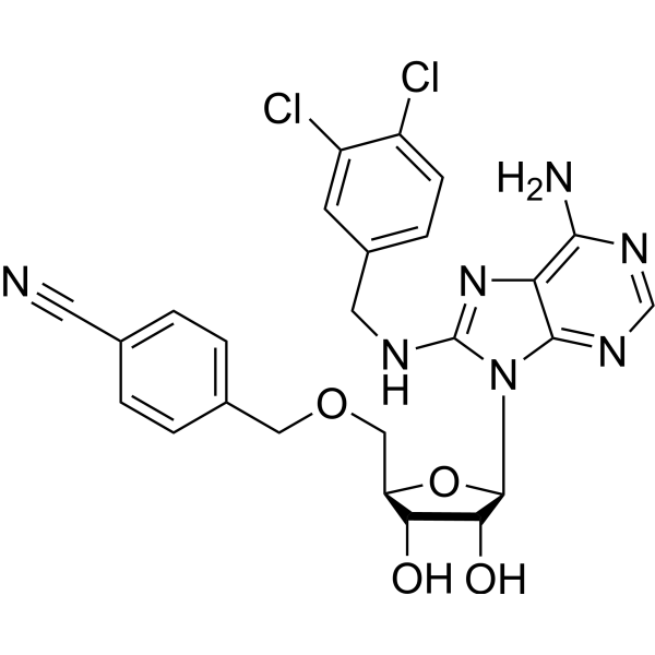
HY-10941
VER-155008Hsp70 Inhibitor -
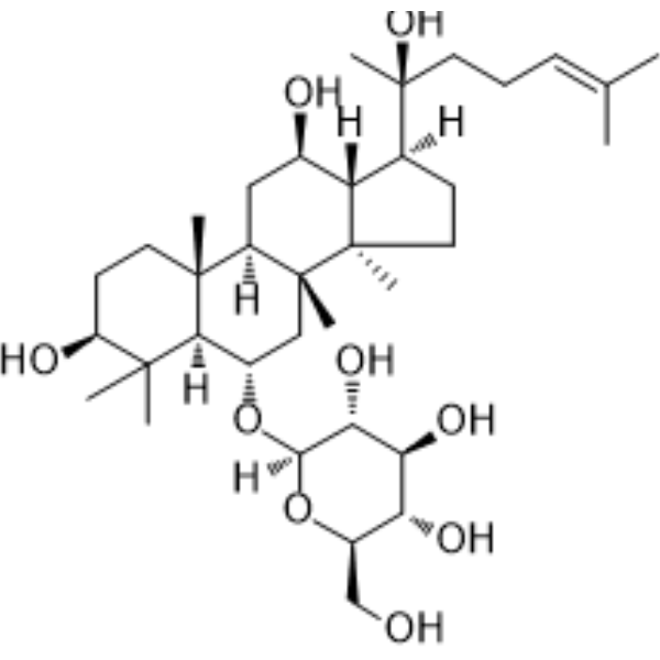
HY-N0604
Ginsenoside Rh1Endogenous Metabolite -
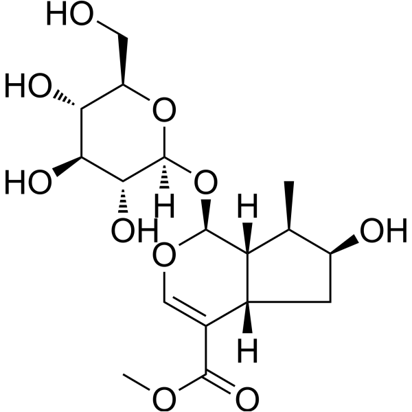
HY-N0512
LoganinIiridoid Glycoside -
HY-A0059
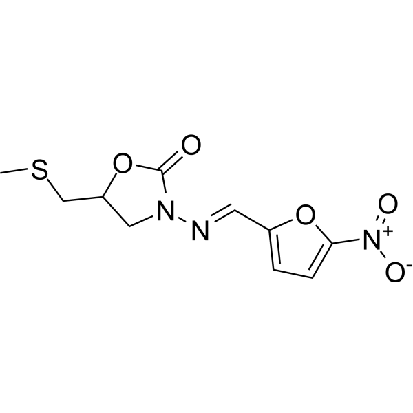
NifuratelBroad-Spectrum Antibiotic -
HY-103000
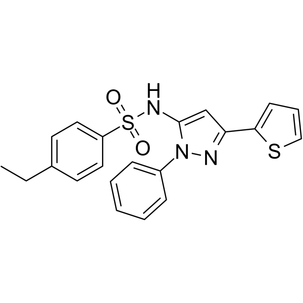
HSF1AHSF1 Activator -
HY-N0604R
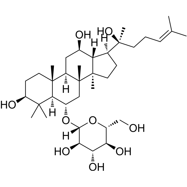
Ginsenoside Rh1 (Standard)Endogenous Metabolite -

HY-N0512R
Loganin (Standard)Natural Product -

HY-A0059R
Nifuratel (Standard)Bacterial Inhibitor -
HY-B0809B
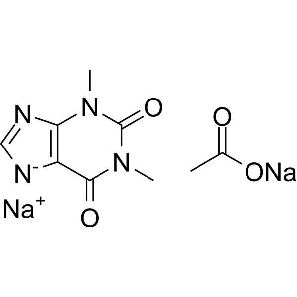
Theophylline sodium acetatePDE Inhibitor -

HY-103000R
HSF1A (Standard)Reference Standard
Publications Citing Use of MedChemExpress (MCE) Calenduloside E
MoreAll AMPK Isoforms
MoreAll Calcium Channel Isoforms
More

Biological Activity
Description
Cellular Effect
|
Cell Line

|
Type | Value | Description | References |
|---|---|---|---|---|
| HCT-116 | IC50 |
18 μM
Compound: OAG
|
Cytotoxicity against human HCT116 cells assessed as reduction in cell viability after 72 hrs by MTT assay
Cytotoxicity against human HCT116 cells assessed as reduction in cell viability after 72 hrs by MTT assay
|
10.1039/C5MD00502G |
| MCF7 | IC50 |
11.33 μM
Compound: OAG
|
Cytotoxicity against human MCF7 cells assessed as reduction in cell viability after 72 hrs by MTT assay
Cytotoxicity against human MCF7 cells assessed as reduction in cell viability after 72 hrs by MTT assay
|
10.1039/C5MD00502G |
| MOLT-4 | IC50 |
13.34 μM
Compound: OAG
|
Cytotoxicity against human MOLT4 cells assessed as reduction in cell viability after 72 hrs by MTT assay
Cytotoxicity against human MOLT4 cells assessed as reduction in cell viability after 72 hrs by MTT assay
|
10.1039/C5MD00502G |
| NCI-H460 | IC50 |
6 μM
Compound: 18
|
Cytotoxicity against human NCI-H460 cells incubated for 72 hrs by cell-titer 96 aqueous non-radioactive cell proliferation assay
Cytotoxicity against human NCI-H460 cells incubated for 72 hrs by cell-titer 96 aqueous non-radioactive cell proliferation assay
|
[PMID: 26287548] |
| SK-BR-3 | IC50 |
11.88 μM
Compound: OAG
|
Cytotoxicity against human SKBR3 cells assessed as reduction in cell viability after 72 hrs by MTT assay
Cytotoxicity against human SKBR3 cells assessed as reduction in cell viability after 72 hrs by MTT assay
|
10.1039/C5MD00502G |
In Vitro
Calenduloside E (1.25 μg/mL; 24 h) inhibits glycolysis-mediated M1 macrophage polarization[2]. Calenduloside E (1 μM; 2 h) alleviates LPS (HY-D1056)/D-galn-induced AML12 and LX2 cell damage and AMPK-SIRT3 signaling pathway protein expression[3]. Calenduloside E (0-16 μM; 24 h) inhibits inflammasome activation and pyroptosis in AML-12 cells stimulated by lipid mixture[5].
MedChemExpress (MCE) has not independently confirmed the accuracy of these methods. They are for reference only.
-
Cell Line:ox-LDL-induced M1 macrophages
-
Concentration:1.25 μg/mL
-
Incubation Time:24 h
-
Result:Reduced the levels of IL-1 β, IL-6, PFKFB3, GLUT1 and LDHA.
-
Cell Line:LPS/ d-galn-induced AML12 and LX2 cells
-
Concentration:1 μM
-
Incubation Time:2 h
-
Result:Reduced ROS and JC-1 levels, as well as cell apoptosis.
-
Cell Line:AML-12 cells stimulated with lipid mixture
-
Concentration:2, 4, 8 and 16 μM
-
Incubation Time:24 h
-
Result:Inhibited the increased expression of NLRP3, Caspase-1 p20, and IL-1β.
In Vivo
MedChemExpress (MCE) has not independently confirmed the accuracy of these methods. They are for reference only.
-
Animal Model:HFD-fed ApoE-/- mice[2]
-
Dosage:11 mg/kg
-
Administration:i.g.;Once a day for 16 weeks
-
Result:Reduced the levels of IL-1β, IL-6, TNF-α, and monocyte chemoattractant protein-1 (MCP-1) in the serum of ApoE-/- mice.
-
Animal Model:LPS (HY-D1056)/dGalN-induced acute liver injury in mice[3]
-
Dosage:15 and 30 mg/kg
-
Administration:i.g.; 1 time per day for 7 consecutive days
-
Result:Improved hepatocyte infiltration and reduced hepatocyte necrosis and shrinkage.
Reduced hepatocyte ROS levels and serum MDA levels, and increased GSH-Px and SOD levels.
-
Animal Model:Rat Model of MI/R Injury[4]
-
Dosage:7.5, 15 and 30 mg/kg
-
Administration:i.g.; 1 time per day for 3 consecutive days
-
Result:Myocardial infarction area/risk area decreased to 53%, 38% and 43% respectively.
Restored the expression of calcium-regulating proteins, including calcium transporters (SERCA, a1C, RyR2, and NCX) to normal levels.
-
Animal Model:Establishment of NAFLD model in apoE-/- mice by western diet[5]
-
Dosage:5 and 10 mg/kg
-
Administration:i.g.; 1 time per day for 4 weeks
-
Result:Reduced the expression of TNF-α, MCP-1, CCL2, Ly6c and cd68 in the liver.
Reversed the upregulation of lipogenic genes FASN, Srebpf, ACC and PPARγ and lipid uptake gene cd36.
Reduced the expression of NLRP3, pNLRC4, NLRC4, cleaved GSDMD, cleaved Caspase1 and IL-1β.
- Phagocytosis Functional Assay
- Detection of Apoptosis
- Mitochondrial membrane-potential fluorescent assay
- TUNEL staining for apoptotic DNA fragmentation
- Annexin V plus membrane-impermeant dye apoptosis staining
- ROS/oxidative-stress fluorescent staining
- Mitochondrial membrane-potential and mitochondrial mass staining
- Apoptosis Solutions
- Primary monocyte-to-macrophage differentiation
- Research Protocol for Cardiovascular Diseases
- Research Protocol for Inflammation-related Diseases
- Research Protocol for Metabolic Diseases


Chemical Information
-
CAS No. 26020-14-4
-
Appearance Solid
-
Molecular Weight 632.82
-
Formula C36H56O9
-
Color White to off-white
-
SMILES
C[C@]12[C@]3(C([C@@]4([H])[C@](C(O)=O)(CCC(C)(C)C4)CC3)=CC[C@]1([H])[C@@]5([C@@](C(C)([C@@H](O[C@]6([H])O[C@@H]([C@@H](O)[C@H](O)[C@H]6O)C(O)=O)CC5)C)([H])CC2)C)C
-
Structure Classification
-
Shipping
Room temperature in continental US; may vary elsewhere.
-
Storage
Powder -20°C 3 years 4°C 2 years In solvent -80°C 6 months -20°C 1 month


Publications (1)
-
Journal Impact Factor
-
Most Recent
-
J Mol Histol
Calenduloside E alleviates cerebral ischemia/reperfusion injury by preserving mitochondrial function. [Abstract]2022 Aug;53(4):713-727. PMID: 35819738


Solvent & Solubility
In Vitro:
DMSO : 100 mg/mL (158.02 mM; Need ultrasonic; Hygroscopic DMSO has a significant impact on the solubility of product, please use newly opened DMSO)
Please refer to the solubility information to select the appropriate solvent. Once prepared, please aliquot and store the solution to prevent product inactivation from repeated freeze-thaw cycles.
Storage method and period of stock solution: -80°C, 6 months; -20°C, 1 month. When stored at -80°C, please use it within 6 months. When stored at -20°C, please use it within 1 month.
Please refer to the solubility information to select the appropriate solvent. Once prepared, please aliquot and store the solution to prevent product inactivation from repeated freeze-thaw cycles.
Storage method and period of stock solution: -80°C, 6 months; -20°C, 1 month. When stored at -80°C, please use it within 6 months. When stored at -20°C, please use it within 1 month.
Concentration (start) × Volume (start) = Concentration (final) × Volume (final)
In Vivo:
Select the appropriate dissolution method based on your experimental animal and administration route.
- For the following dissolution methods, please ensure to first prepare a clear stock solution using an In Vitro approach and then sequentially add co-solvents:
- To ensure reliable experimental results, the clarified stock solution can be appropriately stored based on storage conditions. As for the working solution for In Vivo experiments, it is recommended to prepare freshly and use it on the same day.
- The percentages shown for the solvents indicate their volumetric ratio in the final prepared solution. If precipitation or phase separation occurs during preparation, heat and/or sonication can be used to aid dissolution.
Add each solvent one by one: 10% DMSO 90% (20% SBE-β-CD in Saline)
Solubility: ≥ 2.5 mg/mL (3.95 mM); Clear solution
This protocol yields a clear solution of ≥ 2.5 mg/mL (saturation unknown).
Taking 1 mL working solution as an example, add 100 μL DMSO stock solution (25.0 mg/mL) to 900 μL 20% SBE-β-CD in Saline, and mix evenly.
Preparation of 20% SBE-β-CD in Saline (4°C, storage for one week): 2 g SBE-β-CD powder is dissolved in 10 mL Saline, completely dissolve until clear.
In Vivo Dissolution Calculator
Please enter the basic information of animal experiments:
-
-
-
-
Recommended: Prepare an additional quantity of animals to account for potential losses during experiments.
Please enter your animal formula composition:
-
%DMSO +
Recommended: Keep the proportion of DMSO in working solution below 2% if your animal is weak.
-
%+
-
+%Tween-80 + +
-
%Saline +
The co-solvents required include: DMSO, . All of co-solvents are available by MedChemExpress (MCE). , Tween 80. All of co-solvents are available by MedChemExpress (MCE).
Working solution concentration: 0.22 mg/mL
Method for preparing stock solution: mg drug dissolved in μL DMSO. Stock solution concentration: mg/mL.
1. Take μL DMSO stock solution;
2. Add μL .
μL , mix evenly;
3. Then add μL Tween 80, mix evenly;
4. Then add μL
Please ensure that the stock solution in the first step is dissolved to a clear state, and add co-solvents in sequence. You can use ultrasonic heating (ultrasonic cleaner, recommended frequency 20-40 kHz), vortexing, etc. to assist dissolution.


Purity & Documentation
-
Data Sheet (288 KB)
-
SDS (254 KB)
- English - EN (254 KB)
- Français - FR (254 KB)
- Deutsch - DE (254 KB)
- Norwegian - NO (254 KB)
- Español - ES (254 KB)
- Swedish - SV (254 KB)
- Italian - IT (254 KB)
- Korean - KR (254 KB)
- Portuguese - PT (254 KB)
-
Handling Instructions (2659 KB)
References
[1]. Tian Y, et al. The clickable activity-based probe of anti-apoptotic calenduloside E. Pharm Biol. 2019 Dec;57(1):133-139. [Content Brief]
[2]. Lanfang Li, et al. "Calenduloside e modulates macrophage polarization via KLF2-regulated glycolysis, contributing to attenuates atherosclerosis." International Immunopharmacology 117 (2023): 109730. [Content Brief]
[3]. Pengli Guo, et al. "Isolation of Calenduloside E from achyranthes bidentata blume and its effects on LPS/D-GalN-induced acute liver injury in mice by regulating the AMPK-SIRT3 signaling pathway." Phytomedicine 125 (2024): 155353. [Content Brief]
[4]. Ruiying Wang, et al. "Calenduloside E suppresses calcium overload by promoting the interaction between L-type calcium channels and Bcl2-associated athanogene 3 to alleviate myocardial ischemia/reperfusion injury." Journal of Advanced Research 34 (2021): 173-186. [Content Brief]
[5]. Yifei Le, et al. "Calenduloside E ameliorates non-alcoholic fatty liver disease via modulating a pyroptosis-dependent pathway." Journal of Ethnopharmacology 319 (2024): 117239. [Content Brief]
[6]. Min Wang, et al. "Calenduloside E ameliorates myocardial ischemia‐reperfusion injury through regulation of AMPK and mitochondrial OPA1." Oxidative Medicine and Cellular Longevity 2020.1 (2020): 2415269. [Content Brief]


Complete Stock Solution Preparation Table
Please refer to the solubility information to select the appropriate solvent. Once prepared, please aliquot and store the solution to prevent product inactivation from repeated freeze-thaw cycles.
Storage method and period of stock solution: -80°C, 6 months; -20°C, 1 month. When stored at -80°C, please use it within 6 months. When stored at -20°C, please use it within 1 month.
| Optional Solvent | Concentration Solvent Mass | 1 mg | 5 mg | 10 mg | 25 mg |
|---|---|---|---|---|---|
| DMSO | 1 mM | 1.5802 mL | 7.9011 mL | 15.8023 mL | 39.5057 mL |
| 5 mM | 0.3160 mL | 1.5802 mL | 3.1605 mL | 7.9011 mL | |
| 10 mM | 0.1580 mL | 0.7901 mL | 1.5802 mL | 3.9506 mL | |
| 15 mM | 0.1053 mL | 0.5267 mL | 1.0535 mL | 2.6337 mL | |
| 20 mM | 0.0790 mL | 0.3951 mL | 0.7901 mL | 1.9753 mL | |
| 25 mM | 0.0632 mL | 0.3160 mL | 0.6321 mL | 1.5802 mL | |
| 30 mM | 0.0527 mL | 0.2634 mL | 0.5267 mL | 1.3169 mL | |
| 40 mM | 0.0395 mL | 0.1975 mL | 0.3951 mL | 0.9876 mL | |
| 50 mM | 0.0316 mL | 0.1580 mL | 0.3160 mL | 0.7901 mL | |
| 60 mM | 0.0263 mL | 0.1317 mL | 0.2634 mL | 0.6584 mL | |
| 80 mM | 0.0198 mL | 0.0988 mL | 0.1975 mL | 0.4938 mL | |
| 100 mM | 0.0158 mL | 0.0790 mL | 0.1580 mL | 0.3951 mL |


Calenduloside E Related Classifications
HY-N6850 Related Classifications
 Powered by Bioz