



FC131

Based on 1 Customer Validation
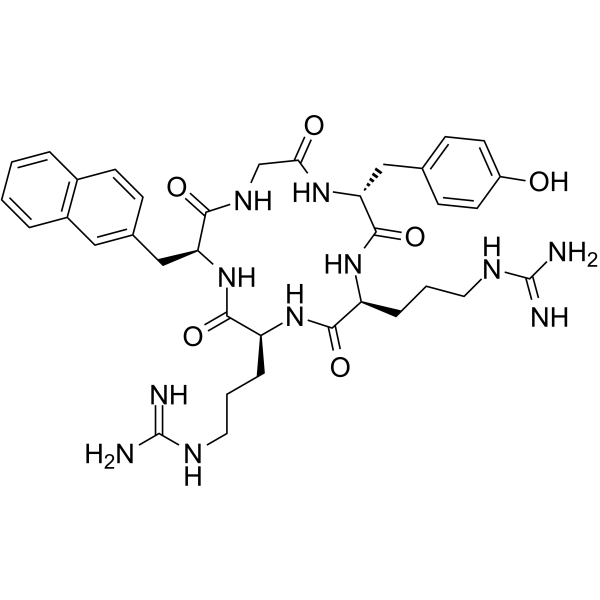
FC131 is a CXCR4 antagonist that inhibits AKT kinase activity in AML OCI-AML3 cells. D-ArgFC131, a derivative of FC131, inhibits the phosphorylation of downstream ERK1/2 and Akt, induces caspase-3 pathway-mediated apoptosis, and causes cell cycle arrest. FC131 exerts cytotoxic effects in cancer cells such as AML cells. FC131 is applicable for cancer-related research.
Nur für Forschungszwecke. Wir verkaufen nicht an Patienten.
- Reinheit : 98.13%
- CAS. Nr.: 606968-52-9
- Formel: C36H47N11O6
- Molecular Weight:729.83
-
Speicherung:
Sealed storage, away from moisture.
Powder -80°C, 2 years , -20°C, 1 year* In solvent : -80°C, 6 months; -20°C, 1 month (sealed storage, away from moisture)

To place orders, for customer services and technical support, please contact: MedChemExpress USA
Tel: 609-228-6898 E-mail: [email protected] [email protected]


-
 Biologische Aktivität
-
Chemical Information
-
Lösungsmittel & Löslichkeit
- Protokoll
- Reinheit & Dokumentation
- Verweise
-
 Help & FAQs


-
HY-P1103
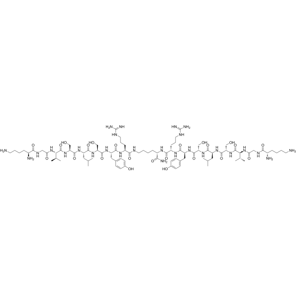
CTCE-9908CXCR4 Antagonist -
HY-P1034
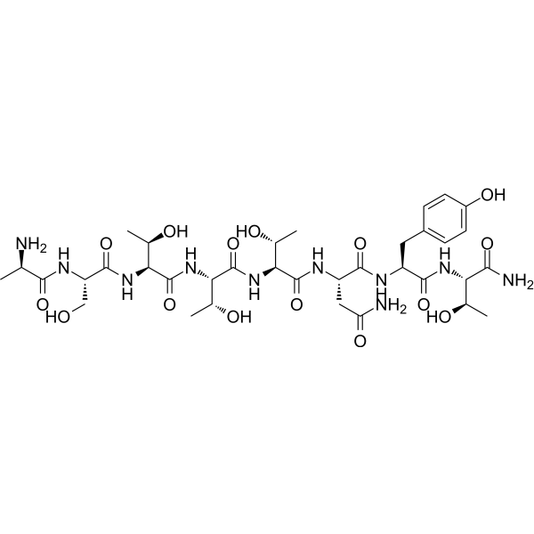
DAPTACCR5 Inhibitor -
HY-50688
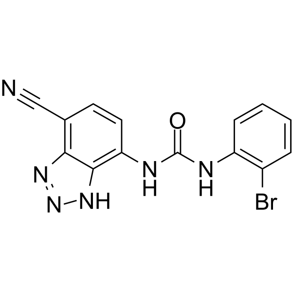
SB-265610CXCR2 Antagonist -
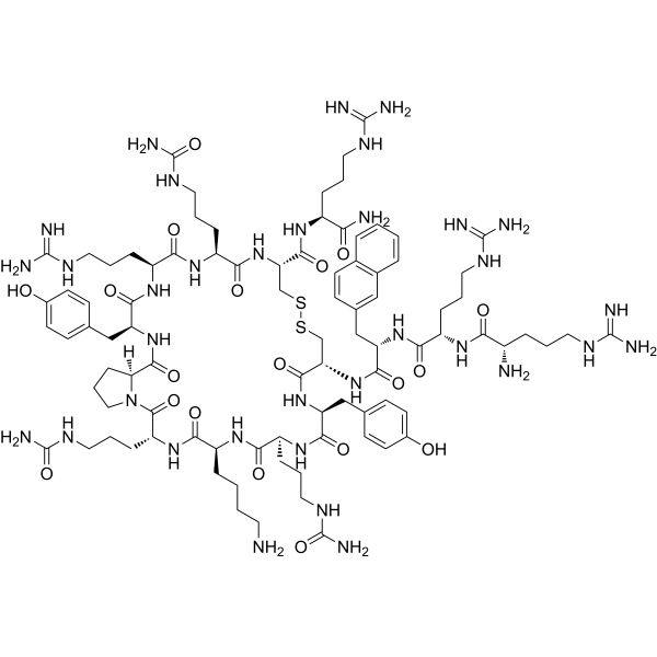
HY-P1102
TC14012CXCR4 Antagonist -
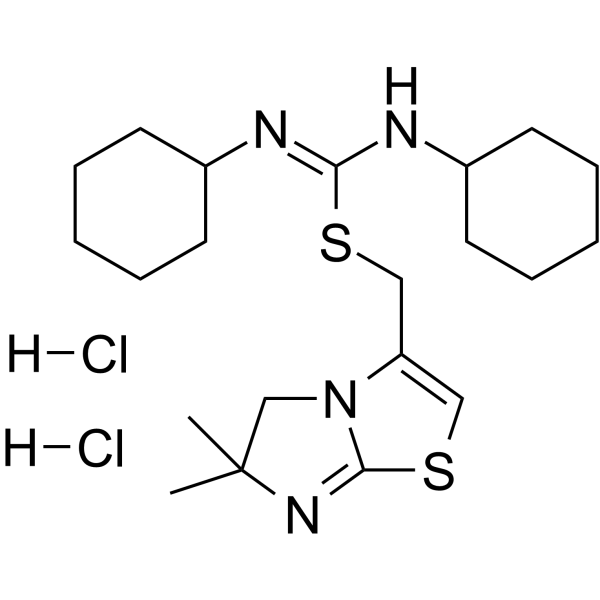
HY-101458A
IT1t dihydrochlorideCXCR4 Antagonist -
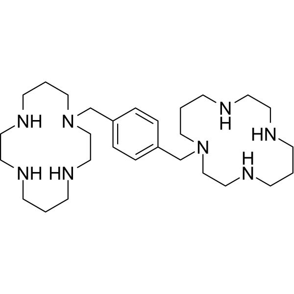
HY-10046
PlerixaforCXCR4 Antagonist -
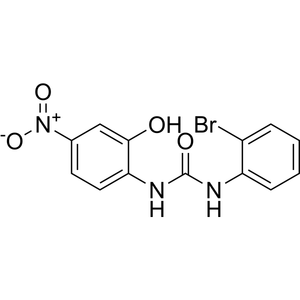
HY-16711
SB225002CXCR2 Antagonist -
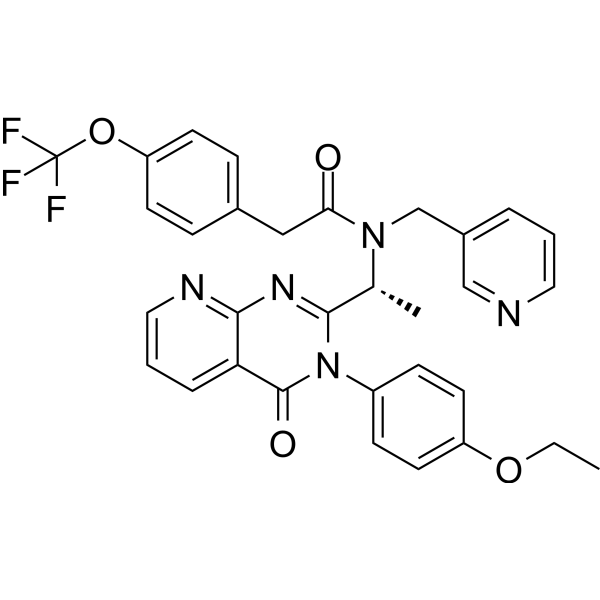
HY-15319
AMG 487CXCR3 Antagonist -
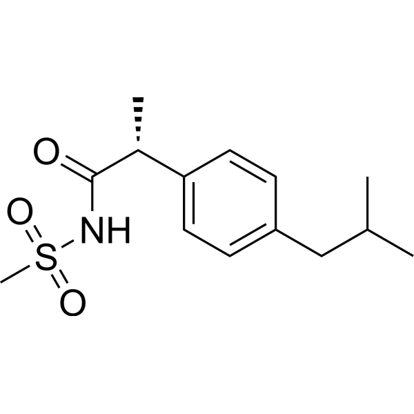
HY-15251
ReparixinCXCR1/2 Inhibitor -
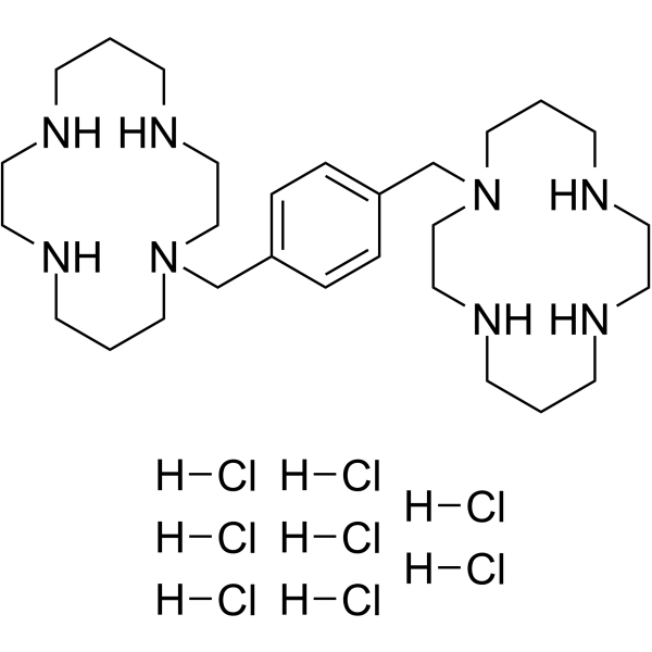
HY-50912
Plerixafor octahydrochlorideCXCR4 Antagonist -
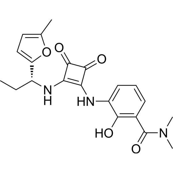
HY-10198
NavarixinCXCR1/2 Antagonist -
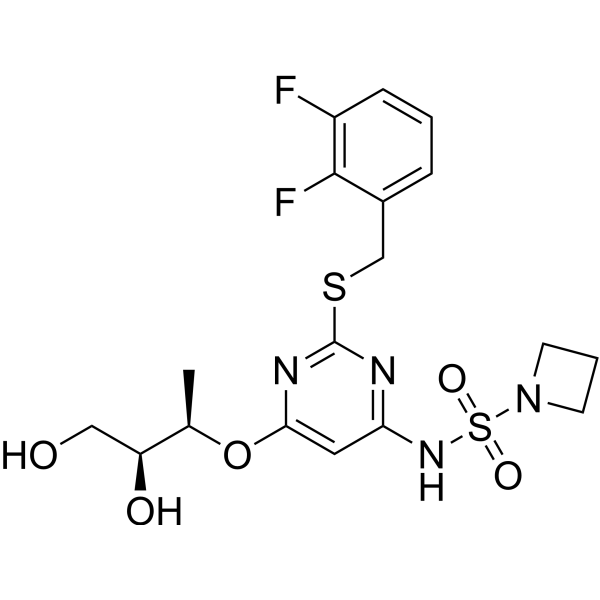
HY-19855
AZD-5069CXCR2 Antagonist


Biologische Aktivität
Beschreibung
|
CXCR4 |
Akt |
Cellular Effect
|
Cell Line

|
Type | Value | Description | References |
|---|---|---|---|---|
| CHO | IC50 |
>30 μM
Compound: 1a
|
Inhibition of [125I]SDF-1alpha binding to CXCR7 (unknown origin) expressed in CHO cell membranes incubated for 1 hr by radioligand displacement assay
Inhibition of [125I]SDF-1alpha binding to CXCR7 (unknown origin) expressed in CHO cell membranes incubated for 1 hr by radioligand displacement assay
|
[PMID: 26042340] |
| CHO | IC50 |
0.0084 μM
Compound: 3
|
Inhibition of [125I]SDF-1 binding to C-X-C chemokine receptor type 4 (CXCR4) expressed in CHO cells
Inhibition of [125I]SDF-1 binding to C-X-C chemokine receptor type 4 (CXCR4) expressed in CHO cells
|
[PMID: 15857134] |
| CHO | IC50 |
0.035 μM
Compound: 2
|
Displacement of [125I]SDF1 from human CXCR4 expressed in CHO cells
Displacement of [125I]SDF1 from human CXCR4 expressed in CHO cells
|
[PMID: 18539453] |
| HEK293 | IC50 |
0.084 μM
Compound: FC131
|
Displacement of [125I]SDF-1alpha from CXCR4 expressed in HEK293 cell membrane after 1 hr
Displacement of [125I]SDF-1alpha from CXCR4 expressed in HEK293 cell membrane after 1 hr
|
[PMID: 22352868] |
| HEK293 | IC50 |
1.2 μM
Compound: 1a
|
Inhibition of [125I]SDF-1alpha binding to CXCR4 (unknown origin) expressed in HEK293 cell membranes incubated for 1 hr by radioligand displacement assay
Inhibition of [125I]SDF-1alpha binding to CXCR4 (unknown origin) expressed in HEK293 cell membranes incubated for 1 hr by radioligand displacement assay
|
[PMID: 26042340] |
| HEK293 | IC50 |
126 nM
Compound: 2, FC131
|
Displacement of [125I]-SDF-1alpha from CXCR4 receptor expressed in HEK293 cells after 1 hr by scintillation counting
Displacement of [125I]-SDF-1alpha from CXCR4 receptor expressed in HEK293 cells after 1 hr by scintillation counting
|
[PMID: 24900333] |
| HeLa | EC50 |
21 nM
Compound: 2, FC131
|
Antiviral activity against Human immunodeficiency virus 1 3B infected in human HeLa cells assessed as inhibition of viral replication after 48 hrs by MAGI assay
Antiviral activity against Human immunodeficiency virus 1 3B infected in human HeLa cells assessed as inhibition of viral replication after 48 hrs by MAGI assay
|
[PMID: 24900333] |
| HeLa | EC50 |
21 nM
Compound: 2, FC131
|
Antiviral activity against Human immunodeficiency virus 1 NL4.3 infected in human HeLa cells assessed as inhibition of viral replication after 48 hrs by MAGI assay
Antiviral activity against Human immunodeficiency virus 1 NL4.3 infected in human HeLa cells assessed as inhibition of viral replication after 48 hrs by MAGI assay
|
[PMID: 24900333] |
| MT4 | CC50 |
>10 μM
Compound: FC131
|
Cytotoxicity against human MT4 cells assessed as reduction of cell viability
Cytotoxicity against human MT4 cells assessed as reduction of cell viability
|
[PMID: 22579418] |
| MT4 | EC50 |
0.16 μM
Compound: FC131
|
Antiviral activity against X4-tropic HIV1 NL4.3 infected in human MT4 cells assessed as protection from virus-induced cytopathogenicity
Antiviral activity against X4-tropic HIV1 NL4.3 infected in human MT4 cells assessed as protection from virus-induced cytopathogenicity
|
[PMID: 22579418] |
In Vitro
FC131 (0.5-4.0 μM; 48 h) combined with Panobinostat (HY-10224) synergistically induces apoptosis in human AML OCI-AML3 cells, and the corresponding combination index value is less than 1.0[1].
FC131 (2 μM plus 50 nM Panobinostat; 48 h) induces significantly higher lethality in primary human AML cells than in normal human CD34+ bone marrow progenitor cells[1].
FC131 (1 μM; 24 h) partially inhibits AKT kinase activity in human AML OCI-AML3 cells, and combined treatment with 50 nM Panobinostat enhances this AKT kinase inhibitory effect without altering the Panobinostat-mediated reduction in the levels of CXCR4, GRK3 and downstream signaling proteins[1].
FC131 (100 nM; 6 h) inhibits the activity of the GH promoter in GH3 rat pituitary tumor cells, reducing luciferase activity to 0.3-fold that of the control group[2].
FC131 (10-100 nM; 6 days) slightly inhibits the proliferation of GH3 rat pituitary tumor cells[2].
FC131 (1 nM-100 μM; 10 min pre-incubation, 90 min co-incubation with CXCL12) potently inhibits CXCL12-mediated IP accumulation in COS-7 cells expressing wild-type CXCR4, with an IC50 of 0.40 μM; the H113A, D171N and D262N mutations significantly reduce its potency, while the W94A and D97A mutations enhance its potency[3].
FC131 (1 nM-100 μM; 3 h) binds to wild-type CXCR4 expressed in COS-7 cells, with an IC50 of 0.76 μM; the H113A, Y116A, D171N and D262N mutations significantly reduce its binding affinity, the H281A, D187A and E288A mutations moderately reduce its binding affinity, while the W94A and D97A mutations increase its binding affinity[3].
MedChemExpress (MCE) has not independently confirmed the accuracy of these methods. They are for reference only.
-
Cell Line:human AML OCI-AML3 cell line
-
Concentration:1 μM
50 nM Panobinostat -
Incubation Time:24 h
-
Result:Inhibied AKT kinase activity in human AML OCI-AML3 cells, and combined treatment with 50 nM Panobinostat enhances this AKT kinase inhibitory effect without altering the Panobinostat-mediated reduction in the levels of CXCR4, GRK3 and downstream signaling proteins.
-
Cell Line:GH3 rat pituitary tumor cells
-
Concentration:10, 100 nM
-
Incubation Time:6 days (twice-daily treatment)
-
Result:Reduced GH3 cell number to ~90% of the control at both 10 nM and 100 nM.
- Kinase activity and phosphorylation assays
- Western Blot
- Detection of Apoptosis
- TUNEL staining for apoptotic DNA fragmentation
- Flow cytometric DNA-content cell-cycle staining
- Annexin V plus membrane-impermeant dye apoptosis staining
- BrdU Incorporation Assay
- Apoptosis Solutions
- Protocol for Cell Cycle
- Protocol for Kinase activity and phosphorylation assays
- Cell Viability Determination by MTT Colorimetric Assay


Chemical Information
-
CAS. Nr. 606968-52-9
-
Appearance Solid
-
Molecular Weight 729.83
-
Formel C36H47N11O6
-
Color White to off-white
-
Versand
Room temperature in continental US; may vary elsewhere.
-
Speicherung
Sealed storage, away from moisture
Powder -80°C 2 years -20°C 1 year * In solvent : -80°C, 6 months; -20°C, 1 month (sealed storage, away from moisture)


Lösungsmittel & Löslichkeit
In Vitro:
DMSO : 100 mg/mL (137.02 mM; Need ultrasonic; Hygroscopic DMSO has a significant impact on the solubility of product, please use newly opened DMSO)
Please refer to the solubility information to select the appropriate solvent. Once prepared, please aliquot and store the solution to prevent product inactivation from repeated freeze-thaw cycles.
Storage method and period of stock solution: -80°C, 6 months; -20°C, 1 month (sealed storage, away from moisture). When stored at -80°C, please use it within 6 months. When stored at -20°C, please use it within 1 month.
Please refer to the solubility information to select the appropriate solvent. Once prepared, please aliquot and store the solution to prevent product inactivation from repeated freeze-thaw cycles.
Storage method and period of stock solution: -80°C, 6 months; -20°C, 1 month (sealed storage, away from moisture). When stored at -80°C, please use it within 6 months. When stored at -20°C, please use it within 1 month.
Konzentration (Stammlösung) × Volumen (Stammlösung) = Konzentration (Ziellösung) × Volumen (Ziellösung)


Reinheit & Dokumentation
-
Data Sheet (280 KB)
-
SDS (252 KB)
- English - EN (252 KB)
- Français - FR (252 KB)
- Deutsch - DE (252 KB)
- Norwegian - NO (252 KB)
- Español - ES (252 KB)
- Swedish - SV (252 KB)
- Italian - IT (252 KB)
- Korean - KR (252 KB)
- Portuguese - PT (252 KB)
-
Handling Instructions (2659 KB)
Verweise
[1]. Mandawat A, et al. Pan-histone deacetylase inhibitor panobinostat depletes CXCR4 levels and signaling and exerts synergistic antimyeloid activity in combination with CXCR4 antagonists. Blood. 2010 Dec 09;116(24):5306-15. [Content Brief]
[2]. Kim JM, et al. The cyclic pentapeptide d-Arg3FC131, a CXCR4 antagonist, induces apoptosis of somatotrope tumor and inhibits tumor growth in nude mice. Endocrinology. 2011 Feb;152(2):536-44. [Content Brief]
[3]. Thiele S, et al. Determination of the binding mode for the cyclopentapeptide CXCR4 antagonist FC131 using a dual approach of ligand modifications and receptor mutagenesis. British journal of pharmacology. 2014 Dec;171(23):5313-29. [Content Brief]


Complete Stock Solution Preparation Table
Please refer to the solubility information to select the appropriate solvent. Once prepared, please aliquot and store the solution to prevent product inactivation from repeated freeze-thaw cycles.
Storage method and period of stock solution: -80°C, 6 months; -20°C, 1 month (sealed storage, away from moisture). When stored at -80°C, please use it within 6 months. When stored at -20°C, please use it within 1 month.
| Optional Solvent | Concentration Solvent Mass | 1 mg | 5 mg | 10 mg | 25 mg |
|---|---|---|---|---|---|
| DMSO | 1 mM | 1.3702 mL | 6.8509 mL | 13.7018 mL | 34.2546 mL |
| 5 mM | 0.2740 mL | 1.3702 mL | 2.7404 mL | 6.8509 mL | |
| 10 mM | 0.1370 mL | 0.6851 mL | 1.3702 mL | 3.4255 mL | |
| 15 mM | 0.0913 mL | 0.4567 mL | 0.9135 mL | 2.2836 mL | |
| 20 mM | 0.0685 mL | 0.3425 mL | 0.6851 mL | 1.7127 mL | |
| 25 mM | 0.0548 mL | 0.2740 mL | 0.5481 mL | 1.3702 mL | |
| 30 mM | 0.0457 mL | 0.2284 mL | 0.4567 mL | 1.1418 mL | |
| 40 mM | 0.0343 mL | 0.1713 mL | 0.3425 mL | 0.8564 mL | |
| 50 mM | 0.0274 mL | 0.1370 mL | 0.2740 mL | 0.6851 mL | |
| 60 mM | 0.0228 mL | 0.1142 mL | 0.2284 mL | 0.5709 mL | |
| 80 mM | 0.0171 mL | 0.0856 mL | 0.1713 mL | 0.4282 mL | |
| 100 mM | 0.0137 mL | 0.0685 mL | 0.1370 mL | 0.3425 mL |


FC131 Related Classifications
HY-P1104 Related Classifications
 Powered by Bioz