



Higenamine
Based on 12 publication(s) in Google Scholar
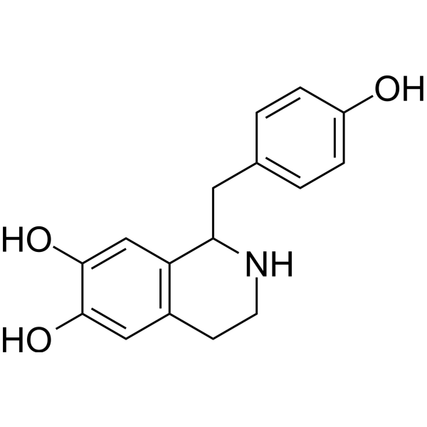
Higenamine (Norcoclaurine), a β2-AR agonist with antioxidant capability, is a key component of the Chinese herb aconite root that prescribes for treating symptoms of heart failure in the oriental Asian countries. Higenamine is also a α1-adrenergic receptor antagonist with hypotensive effect. is a selective LSD1 inhibitor (IC50=1.47 μM) that can be isolated from aconite. Higenamine hydrochloride has anti-inflammatory and antibacterial activity. Higenamine protects myocyte Apoptosis and ischemia/reperfusion (I/R) injury through selective activation of beta2-adrenergic receptor (β2-AR). Higenamine also reduces I/R-induced myocardial infarction in mice. Higenamine can attenuate IL-1β-induced Apoptosis through ROS-mediated PI3K/Akt signaling pathway. Higenamine protects brain cells from oxygen deprivation. Higenamine can promote bone formation in osteoporosis through the SMAD2/3 pathway. Higenamine can be used to study cancer, inflammation, cardiorenal syndrome and other diseases.
For research use only. We do not sell to patients.
- Purity : 99.31%
- CAS No.: 5843-65-2
- Formula: C16H17NO3
- Molecular Weight:271.31
-
Storage:
4°C, sealed storage, away from moisture
* In solvent : -80°C, 6 months; -20°C, 1 month (sealed storage, away from moisture)

To place orders, for customer services and technical support, please contact: MedChemExpress USA
Tel: 609-228-6898 E-mail: [email protected] [email protected]


-
 Biological Activity
-
Chemical Information
-
Solvent & Solubility
- Protocols
- Purity & Documentation
- References
-
 Help & FAQs


-
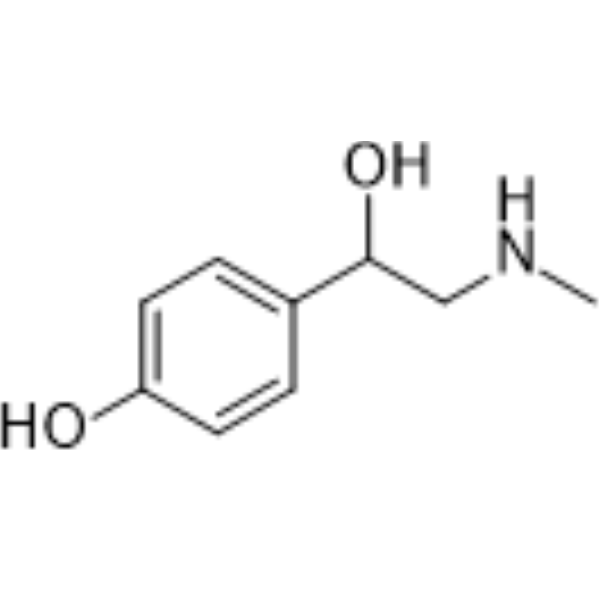
HY-N0132
SynephrineAdrenergic Receptor Agonist -
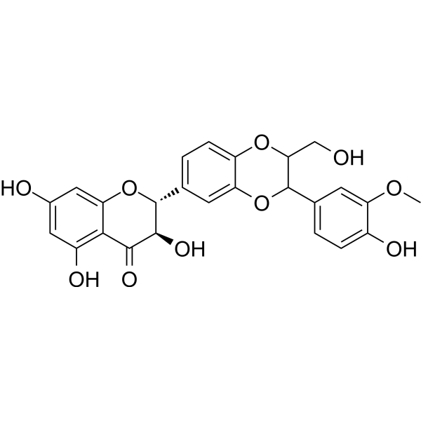
HY-N0779A
SilybinApoptosis Inducer -
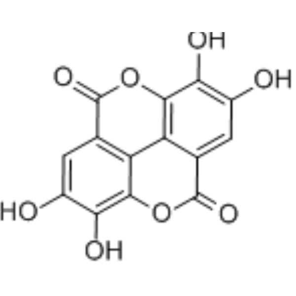
HY-B0183
Ellagic acidSHP2 Inhibitor -
HY-N3610
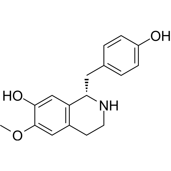
CoclaurinenAChR Antagonist -
HY-14383
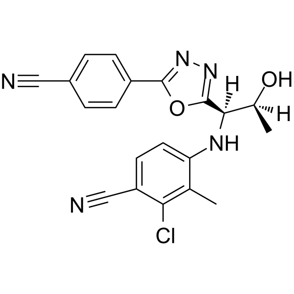
VosilasarmAndrogen Receptor Modulator -
HY-N0028
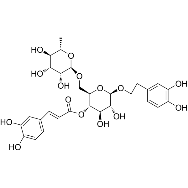
Forsythiaside AAnti-Inflammatory Agent -

HY-W206168
Tetrahydropapaveroline hydrobromideDA Uptake Inhibitor -

HY-N2037R
Higenamine (Standard)Adrenergic Receptor Agonist, LSD1 Inhibitor
Publications Citing Use of MedChemExpress (MCE) Higenamine
More- Cell. 2025 Sep 18;188(19):5142-5156.e23. [Abstract]
- Arch Toxicol. 2025 May;99(5):1999-2021. [Abstract]
- Food Sci Hum Wellness. 2025.
- Ind Crops Prod. 2025 Dec 2;238:122414.
- Nutrients. 2024 May 22;16(11):1567. [Abstract]
- Eur J Pharmacol. 2026 Jun 10:1026:178966. [Abstract]
- Toxicology. 2026 Mar 6:154442. [Abstract]
- J Inflamm Res. 2024 Oct 14:17:7295-7310. [Abstract]
- J Pharm Biomed Anal. 2021 Feb 20:195:113870. [Abstract]
- Cardiovasc Toxicol. 2026 Apr 4;26(4):36. [Abstract]
- Fitoterapia. 2026 Sep:193:107388.
- SSRN. 2026 Jul 3.
All MAP3K Isoforms
MoreAll Adrenergic Receptor Isoforms
More

Biological Activity
Description
IC50 & Target
|
β adrenergic receptor |
In Vitro
Higenamine (3-100 μM; 72 h) can inhibit the differentiation of MV4-11 and MOLM-13 cells by inhibiting the activity of LSD1[1].
Higenamine (1-100μM; 8 h) can enhance the activity of HO-1 in C6 cells and protect brain cells from cell hypoxia damage [2].
Higenamine (10-50 μM; 8 h) can inhibit apoptosis in C6 cells[2].
Higenamine (10-40 μM; 24 h) can inhibit the production of IL-1β-induced ROS and activate the ROS-mediated PI3K/Akt signaling pathway, which has anti-apoptotic activity in HNPCs[3].
Higenamine (0.08-250 μM; 0.5-24 h) promotes phosphorylation of SMAD2/3 in a time- and dose-dependent manner in BMSCs[6].
Higenamine (0-120 μM, 24 h) reverses H2O2 (250 μM, 24 h) induced cell death and apoptosis in neonatal rat ventricular myocytes (NRVMs)[7].
MedChemExpress (MCE) has not independently confirmed the accuracy of these methods. They are for reference only.
-
Cell Line:MV4-11, MOLM-13
-
Concentration:3 μM, 10 μM, 30 μM, 100 μM
-
Incubation Time:72 h
-
Result:Could up-regulate the expression levels of LSD1 substrate H3K4me1 and H3K4me2 in a dose-dependent manner, but did not affect the expression levels of H3K4me3, H3 and LSD1.
Promoted P53 expression in a dose-dependent manner.
-
Cell Line:C6
-
Concentration:1 μM, 5μM, 10 μM, 50 μM,100 μM
-
Incubation Time:8 h
-
Result:Increased HO-1 expression in a concentration-dependent manner under hypoxia and normoxia conditions.
-
Cell Line:MV4-11, MOLM-13
-
Concentration:3 μM, 10 μM, 30 μM, 100 μM
-
Incubation Time:72 h
-
Result:Significantly down-regulated the expression levels of HoxA9 and Meis1 in leukemia cells in a dose-dependent manner.
-
Cell Line:NRVMs
-
Concentration:0-120 μM
-
Incubation Time:24 h
-
Result:Dose dependently attenuated H2O2-stimulated early and late apoptosis of NRVMs.
In Vivo
Higenamine hydrochloride (0.5-4.5 mg/kg; Single dose) improves cardiac and renal function in rats with cardio-renal syndrome (CRS) and alleviates cardiac and renal fibrosis by targeting ASK1/MAPK (ERK, P38)/NF-kB signaling pathway in Sprague-Dawley rats[4].
Higenamine hydrochloride (20 mg/kg-30 mg/kg; Intraperitoneal injection; Once daily for 60 days) promotes bone formation and prevents accelerated bone loss in SAMP6 mice[6].
Higenamine (10 mg/kg, i.p., 2 h prior to the surgery) protects against I/R-induced myocardial infarction in mice bearing ischemia/reperfusion injury[7].
MedChemExpress (MCE) has not independently confirmed the accuracy of these methods. They are for reference only.
-
Animal Model:Spontaneous osteoporosis SAMP6 mice model[6]
-
Dosage:10 mg/kg, 20 mg/kg
-
Administration:Intraperitoneal injection (i.p.);Once daily for 60 days
-
Result:Significantly increased the expression of P1NP and OCN (P1NP and OCN are markers of bone formation).
-
Animal Model:Mice bearing ischemia/reperfusion injury[7]
-
Dosage:10 mg/kg
-
Administration:i.p., 2 h prior to the surgery
-
Result:Decreased the myocardial infarct areas (MI) and increased myocyte survival in mice bearing ischemia/reperfusion injury.
- Research Protocol for Infectious Diseases
- Detection of Apoptosis
- TUNEL staining for apoptotic DNA fragmentation
- Annexin V plus membrane-impermeant dye apoptosis staining
- ROS/oxidative-stress fluorescent staining
- Apoptosis Solutions
- Research Protocol for Cardiovascular Diseases
- Research Protocol for Inflammation-related Diseases
- Bacterial live/dead nucleic-acid viability staining
- Pyroptosis Solutions


Chemical Information
-
CAS No. 5843-65-2
-
Appearance Solid
-
Molecular Weight 271.31
-
Formula C16H17NO3
-
Color White to off-white
-
SMILES
OC1=CC2=C(C(CC3=CC=C(O)C=C3)NCC2)C=C1O
-
Synonyms
Norcoclaurine; Demethyl-Coclaurine
-
Structure Classification
-
Initial Source
-
Shipping
Room temperature in continental US; may vary elsewhere.
-
Storage
4°C, sealed storage, away from moisture
* In solvent : -80°C, 6 months; -20°C, 1 month (sealed storage, away from moisture)


Publications (12)
-
Journal Impact Factor
-
Most Recent
-
Cell
2025 Sep 18;188(19):5142-5156.e23. PMID: 40555230 -
Arch Toxicol
Quantitative in vitro-to-in vivo extrapolation of human adrenergic and trace amine-associated receptor 1 potencies of pre-workout supplement ingredients using physiologically based kinetic modelling-based reverse dosimetry. [Abstract]2025 May;99(5):1999-2021. PMID: 40178592 -
-
-
Nutrients
In Vitro Activation of Human Adrenergic Receptors and Trace Amine-Associated Receptor 1 by Phenethylamine Analogues Present in Food Supplements. [Abstract]2024 May 22;16(11):1567. PMID: 38892500 -
Eur J Pharmacol
Phenethylamines in pre-workout supplements alter arterial pressure, heart rate, and body temperature in rats. [Abstract]2026 Jun 10:1026:178966. PMID: 42128191 -
Toxicology
In vitro inhibition of monoamine transport by amphetamine-like pre-workout supplement ingredients. [Abstract]2026 Mar 6:154442. PMID: 41796714 -
J Inflamm Res
Shenfu Injection Mediated NLRP3/Caspase 1 Through (R)-Norcoclaurinee Alleviates Sepsis-Induced Cognitive Dysfunction. [Abstract]2024 Oct 14:17:7295-7310. PMID: 39429846 -
J Pharm Biomed Anal
Screening method of mildronate and over 300 doping agents by reversed-phase liquid chromatography-high resolution mass spectrometry. [Abstract]2021 Feb 20:195:113870. PMID: 33453569 -
Cardiovasc Toxicol
Potential Cardiovascular Risks of Phenethylamine Analogues in Food Supplements: Evidence from Vasocontraction of Rat Artery Segments. [Abstract]2026 Apr 4;26(4):36. PMID: 41934574 -
-


Solvent & Solubility
In Vitro:
DMSO : ≥ 100 mg/mL (368.58 mM; Hygroscopic DMSO has a significant impact on the solubility of product, please use newly opened DMSO)
H2O : < 0.1 mg/mL (insoluble)
* "≥" means soluble, but saturation unknown.
Please refer to the solubility information to select the appropriate solvent. Once prepared, please aliquot and store the solution to prevent product inactivation from repeated freeze-thaw cycles.
Storage method and period of stock solution: -80°C, 6 months; -20°C, 1 month (sealed storage, away from moisture). When stored at -80°C, please use it within 6 months. When stored at -20°C, please use it within 1 month.
Please refer to the solubility information to select the appropriate solvent. Once prepared, please aliquot and store the solution to prevent product inactivation from repeated freeze-thaw cycles.
Storage method and period of stock solution: -80°C, 6 months; -20°C, 1 month (sealed storage, away from moisture). When stored at -80°C, please use it within 6 months. When stored at -20°C, please use it within 1 month.
Concentration (start) × Volume (start) = Concentration (final) × Volume (final)
In Vivo:
Select the appropriate dissolution method based on your experimental animal and administration route.
- For the following dissolution methods, please ensure to first prepare a clear stock solution using an In Vitro approach and then sequentially add co-solvents:
- To ensure reliable experimental results, the clarified stock solution can be appropriately stored based on storage conditions. As for the working solution for In Vivo experiments, it is recommended to prepare freshly and use it on the same day.
- The percentages shown for the solvents indicate their volumetric ratio in the final prepared solution. If precipitation or phase separation occurs during preparation, heat and/or sonication can be used to aid dissolution.
Add each solvent one by one: 10% DMSO 40% PEG300 5% Tween-80 45% Saline
Solubility: ≥ 2.5 mg/mL (9.21 mM); Clear solution
This protocol yields a clear solution of ≥ 2.5 mg/mL (saturation unknown).
Taking 1 mL working solution as an example, add 100 μL DMSO stock solution (25.0 mg/mL) to 400 μL PEG300, and mix evenly; then add 50 μL Tween-80 and mix evenly; then add 450 μL Saline to adjust the volume to 1 mL.
Preparation of Saline: Dissolve 0.9 g sodium chloride in ddH₂O and dilute to 100 mL to obtain a clear Saline solution.
Add each solvent one by one: 10% DMSO 90% (20% SBE-β-CD in Saline)
Solubility: ≥ 2.5 mg/mL (9.21 mM); Clear solution
This protocol yields a clear solution of ≥ 2.5 mg/mL (saturation unknown).
Taking 1 mL working solution as an example, add 100 μL DMSO stock solution (25.0 mg/mL) to 900 μL 20% SBE-β-CD in Saline, and mix evenly.
Preparation of 20% SBE-β-CD in Saline (4°C, storage for one week): 2 g SBE-β-CD powder is dissolved in 10 mL Saline, completely dissolve until clear.
In Vivo Dissolution Calculator
Please enter the basic information of animal experiments:
-
-
-
-
Recommended: Prepare an additional quantity of animals to account for potential losses during experiments.
Please enter your animal formula composition:
-
%DMSO +
Recommended: Keep the proportion of DMSO in working solution below 2% if your animal is weak.
-
%+
-
+%Tween-80 + +
-
%Saline +
The co-solvents required include: DMSO, . All of co-solvents are available by MedChemExpress (MCE). , Tween 80. All of co-solvents are available by MedChemExpress (MCE).
Working solution concentration: 0.22 mg/mL
Method for preparing stock solution: mg drug dissolved in μL DMSO. Stock solution concentration: mg/mL. * In solvent : -80°C, 6 months; -20°C, 1 month (sealed storage, away from moisture)
1. Take μL DMSO stock solution;
2. Add μL .
μL , mix evenly;
3. Then add μL Tween 80, mix evenly;
4. Then add μL
Please ensure that the stock solution in the first step is dissolved to a clear state, and add co-solvents in sequence. You can use ultrasonic heating (ultrasonic cleaner, recommended frequency 20-40 kHz), vortexing, etc. to assist dissolution.


Purity & Documentation
-
Data Sheet (286 KB)
-
SDS (393 KB)
- English - EN (393 KB)
- Français - FR (393 KB)
- Deutsch - DE (393 KB)
- Norwegian - NO (393 KB)
- Español - ES (393 KB)
- Swedish - SV (393 KB)
- Italian - IT (393 KB)
- Korean - KR (393 KB)
- Portuguese - PT (393 KB)
-
Handling Instructions (2659 KB)
References
[1]. Fang Y, et al. Discovery of higenamine as a potent, selective and cellular active natural LSD1 inhibitor for MLL-rearranged leukemia therapy. Bioorg Chem. 2021 Apr;109:104723. [Content Brief]
[2]. Ha YM, et al. Higenamine reduces HMGB1 during hypoxia-induced brain injury by induction of heme oxygenase-1 through PI3K/Akt/Nrf-2 signal pathways. Apoptosis. 2012 May;17(5):463-74. [Content Brief]
[3]. Zhu X, et al. Higenamine mitigates interleukin-1β-induced human nucleus pulposus cell apoptosis by ROS-mediated PI3K/Akt signaling. Mol Cell Biochem. 2021 Nov;476(11):3889-3897. [Content Brief]
[4]. Deng T, et al. Higenamine Improves Cardiac and Renal Fibrosis in Rats With Cardiorenal Syndrome via ASK1 Signaling Pathway. J Cardiovasc Pharmacol. 2020 Jun;75(6):535-544. [Content Brief]
[5]. Erasto, Paul et al. Evaluation of Antimycobacterial Activity of Higenamine Using Galleria mellonella as an In Vivo Infection Model. Natural products and bioprospecting vol. 8,1 (2018): 63-69. [Content Brief]
[6]. Dong, Hui et al. Higenamine Promotes Osteogenesis Via IQGAP1/SMAD4 Signaling Pathway and Prevents Age- and Estrogen-Dependent Bone Loss in Mice. Journal of bone and mineral research : the official journal of the American Society for Bone and Mineral Research vol. 38,5 (2023): 775-791. [Content Brief]
[7]. Wu MP, et al. Higenamine protects ischemia/reperfusion induced cardiac injury and myocyte apoptosis through activation of β2-AR/PI3K/AKT signaling pathway. Pharmacol Res. 2016 Feb;104:115-23. [Content Brief]
[8]. Lee SR, et al. Acute oral intake of a higenamine-based dietary supplement increases circulating free fatty acidsand energy expenditure in human subjects. Lipids Health Dis. 2013 Oct 21;12:148. [Content Brief]
[9]. Wen J, et al. Role of Higenamine in Heart Diseases: A Mini-Review. Front Pharmacol. 2022 Jan 10;12:798495. [Content Brief]
[10]. Chen DT, et al. Pharmacological effects of higenamine based on signalling pathways and mechanism of action. Front Pharmacol. 2022 Sep 15;13:981048. [Content Brief]
[11]. Romeo I, et al. The Antioxidant Capability of Higenamine: Insights from Theory. Antioxidants (Basel). 2020 Apr 25;9(5):358. [Content Brief]


Complete Stock Solution Preparation Table
Please refer to the solubility information to select the appropriate solvent. Once prepared, please aliquot and store the solution to prevent product inactivation from repeated freeze-thaw cycles.
Storage method and period of stock solution: -80°C, 6 months; -20°C, 1 month (sealed storage, away from moisture). When stored at -80°C, please use it within 6 months. When stored at -20°C, please use it within 1 month.
| Optional Solvent | Concentration Solvent Mass | 1 mg | 5 mg | 10 mg | 25 mg |
|---|---|---|---|---|---|
| DMSO | 1 mM | 3.6858 mL | 18.4291 mL | 36.8582 mL | 92.1455 mL |
| 5 mM | 0.7372 mL | 3.6858 mL | 7.3716 mL | 18.4291 mL | |
| 10 mM | 0.3686 mL | 1.8429 mL | 3.6858 mL | 9.2146 mL | |
| 15 mM | 0.2457 mL | 1.2286 mL | 2.4572 mL | 6.1430 mL | |
| 20 mM | 0.1843 mL | 0.9215 mL | 1.8429 mL | 4.6073 mL | |
| 25 mM | 0.1474 mL | 0.7372 mL | 1.4743 mL | 3.6858 mL | |
| 30 mM | 0.1229 mL | 0.6143 mL | 1.2286 mL | 3.0715 mL | |
| 40 mM | 0.0921 mL | 0.4607 mL | 0.9215 mL | 2.3036 mL | |
| 50 mM | 0.0737 mL | 0.3686 mL | 0.7372 mL | 1.8429 mL | |
| 60 mM | 0.0614 mL | 0.3072 mL | 0.6143 mL | 1.5358 mL | |
| 80 mM | 0.0461 mL | 0.2304 mL | 0.4607 mL | 1.1518 mL | |
| 100 mM | 0.0369 mL | 0.1843 mL | 0.3686 mL | 0.9215 mL |


Higenamine Related Classifications
HY-N2037 Related Classifications
 Powered by Bioz