



FTI-277 TFA
Based on 2 publication(s) in Google Scholar
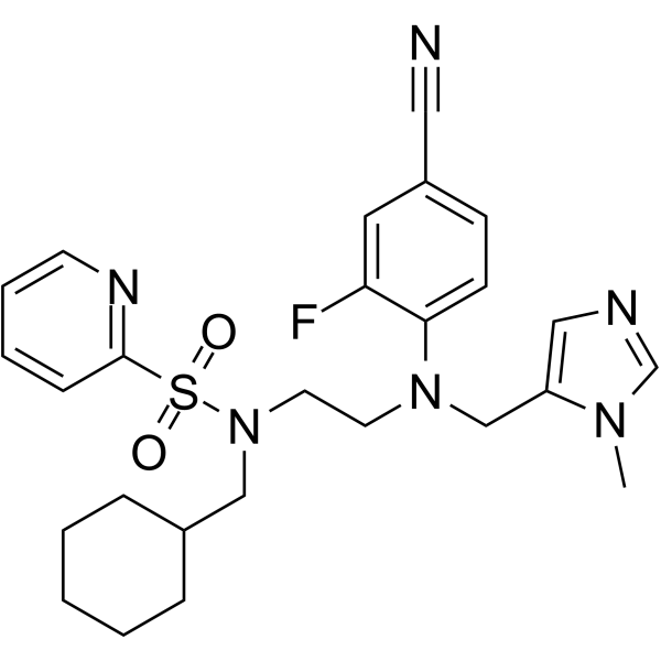
FTI-277 TFA is a farnesyltransferase (FTase) inhibitor. FTI-277 TFA inhibits Ras farnesylation, blocks the phosphorylation of downstream ERK1/2 and mTOR, and reduces membrane-bound active N-ras protein. FTI-277 TFA activates caspase 3, upregulates Bim expression, induces cell apoptosis, suppresses regulatory T cell expansion, enhances macrophage phagocytosis, and improves bacterial clearance. FTI-277 TFA activates the PI3K/Akt signaling pathway, inhibits osteoblast differentiation, and reduces the proliferation ability of neuroblastoma cells. FTI-277 TFA can be used in research related to head and neck squamous cell carcinoma, neuroblastoma, sepsis, and vascular calcification.
Nos produits utilisent uniquement pour la recherche. Nous ne vendons pas aux patients.
- CAS No.: 1217447-06-7
- Formule: C24H30F3N3O5S2
- Masse moléculaire:561.64
-
Stockage:
Please store the product under the recommended conditions in the Certificate of Analysis.


To place orders, for customer services and technical support, please contact: MedChemExpress USA
Tel: 609-228-6898 E-mail: [email protected] [email protected]


-
 Activité biologique
-
Chemical Information
- Pureté et documentation
- Références
-
 Help & FAQs




Publications Citing Use of MedChemExpress (MCE) FTI-277 TFA
More Customer Validation & Images

-
WB
Voir tous les produits spécifiques à Isoform Caspase
More

Activité biologique
FTI-277 (1-40 μM; 24-72 h) TFA decreases the viability of HEp-2 head and neck squamous cell carcinoma cells in a concentration-dependent manner, with a 58.5% reduction in viability at the highest tested concentration (40 μM) after 72 h of treatment[1].
FTI-277 (0.5-10 μM; 24-72 h) TFA decreases the viability of HSC-3 head and neck squamous cell carcinoma cells in a concentration-dependent manner, with a 77.0% reduction in viability at the highest tested concentration (10 μM) after 72 h of treatment, and exhibits greater potency against HSC-3 cells than HEp-2 cells[1].
FTI-277 (40 μM; 36 h) TFA significantly increases caspase-3 activity in HEp-2 head and neck squamous cell carcinoma cells[1].
FTI-277 (5 μM; 36 h) TFA significantly increases caspase-3 activity in HSC-3 head and neck squamous cell carcinoma cells, demonstrating greater potency than against HEp-2 cells[1].
FTI-277 (40 μM; 36 h) TFA significantly increases the percentage of annexin V-positive HEp-2 head and neck squamous cell carcinoma cells, indicating induction of apoptosis[1].
FTI-277 (5 μM; 36 h) TFA significantly increases the percentage of annexin V-positive HSC-3 head and neck squamous cell carcinoma cells, indicating induction of apoptosis with greater potency than against HEp-2 cells[1].
FTI-277 (40 μM; 48 h) TFA suppresses Ras membrane localization, reduces phosphorylation of ERK1/2 and mTOR, and increases Bim protein expression in HEp-2 head and neck squamous cell carcinoma cells[1].
FTI-277 (5 μM; 48 h) TFA suppresses Ras membrane localization, reduces phosphorylation of ERK1/2 and mTOR, and increases Bim protein expression in HSC-3 head and neck squamous cell carcinoma cells, demonstrating greater potency than against HEp-2 cells[1].
FTI-277 (5 μM; 72 h) TFA significantly decreases the viability of NW7 v-H-Ras-transfected NIH3T3 cells but has no significant effect on NV20 empty vector-transfected NIH3T3 cells, indicating selective activity against Ras-overexpressing cells[1].
FTI-277 (1-20 μM; 8-10 days) TFA inhibits osteogenic differentiation and βGP-induced mineralization in primary bovine vascular smooth muscle cells, with significant effects observed at concentrations ≥10 μM, and even when added up to 6 days post-βGP initiation[3].
FTI-277 (10 μM; 77 h) TFA enhances Akt phosphorylation in primary bovine vascular smooth muscle cells when followed by serum starvation and short-term serum stimulation[3].
FTI-277 (10-20 μM; 9-10 days) TFA maintains Akt phosphorylation during βGP-induced mineralization in primary bovine vascular smooth muscle cells, and this Akt activation is required for FTI-277's inhibitory effect on mineralization[3].
FTI-277 (10 μM) TFA inhibits phosphate-induced apoptosis in human coronary artery vascular smooth muscle cells, and this effect depends on Akt activation[3].
FTI-277 (10 μM; 96 h) TFA reduces farnesylated N-Ras protein levels by approximately 50% in IMR-5 neuroblastoma cells[4].
FTI-277 (10 μM; 96 h) TFA reduces basal MAP-kinase activity by ~47% and blunts BDNF-induced MAP-kinase activation to 35% of control levels in IMR-5 neuroblastoma cells[4].
FTI-277 (10 μM; 96 h) TFA reduces basal N-myc expression to 70% of controls and blunts BDNF-induced N-myc expression to 53% of control-stimulated levels in IMR-5 neuroblastoma cells[4].
FTI-277 (10 μM; 96 h) TFA significantly reduces cell proliferation across complete, serum-free, and BDNF-supplemented media, and abolishes BDNF-induced proliferation in IMR-5 neuroblastoma cells[4].
MedChemExpress (MCE) has not independently confirmed the accuracy of these methods. They are for reference only.
-
Cell Line:HEp-2 head and neck squamous cell carcinoma cells
-
Concentration:1 μM, 5 μM, 10 μM, 20 μM, 40 μM
-
Incubation Time:24 h, 48 h, 72 h
-
Result:Decreased HEp-2 cell viability in a concentration-dependent manner.
Reduced cell viability to 97.9% with 1 μM, 98.9% with 5 μM, 89.1% with 10 μM, 78.3% with 20 μM, and 41.5% with 40 μM after 72 h treatment.
Induced statistically significant viability reductions with 20 μM and 40 μM at 48 h, and with 10 μM, 20 μM, and 40 μM at 72 h relative to controls.
-
Cell Line:HSC-3 head and neck squamous cell carcinoma cells
-
Concentration:1 μM, 5 μM, 10 μM, 20 μM, 40 μM
-
Incubation Time:24 h, 48 h, 72 h
-
Result:Decreased HSC-3 cell viability in a concentration-dependent manner, with greater potency than in HEp-2 cells.
Reduced cell viability to 89.7% with 0.5 μM, 69.1% with 1 μM, 52.9% with 2.5 μM, 33.4% with 5 μM, and 23.0% with 10 μM after 72 h treatment.
Induced statistically significant viability reductions with all tested concentrations at 24, 48, and 72 h relative to controls.
-
Cell Line:HEp-2 head and neck squamous cell carcinoma cells
-
Concentration:40 μM
-
Incubation Time:36 h
-
Result:Increased the percentage of annexin V-positive HEp-2 cells to approximately 42%, relative to control cells with approximately 7% annexin V-positive cells.
Induced statistically significant elevation of annexin V-positive cells, indicating apoptosis induction.
-
Cell Line:HSC-3 head and neck squamous cell carcinoma cells
-
Concentration:5 μM
-
Incubation Time:36 h
-
Result:Increased the percentage of annexin V-positive HSC-3 cells to approximately 47%, relative to control cells with approximately 9% annexin V-positive cells.
Induced statistically significant elevation of annexin V-positive cells, indicating apoptosis induction with greater potency than against HEp-2 cells.
-
Cell Line:HEp-2 head and neck squamous cell carcinoma cells
-
Concentration:40 μM
-
Incubation Time:48 h
-
Result:Inhibited Ras membrane localization and increased Ras cytoplasmic localization in HEp-2 cells.
Decreased the expression of phosphorylated ERK1/2 and phosphorylated mTOR.
Increased the expression of Bim protein, relative to control cells.
-
Cell Line:HSC-3 head and neck squamous cell carcinoma cells
-
Concentration:5 μM
-
Incubation Time:48 h
-
Result:Inhibited Ras membrane localization and increased Ras cytoplasmic localization in HSC-3 cells.
Decreased the expression of phosphorylated ERK1/2 and phosphorylated mTOR.
Increased the expression of Bim protein, relative to control cells.
Demonstrated greater potency than against HEp-2 cells.
-
Cell Line:NV20 empty vector-transfected NIH3T3 cells, NW7 v-H-Ras-transfected NIH3T3 cells
-
Concentration:5 μM
-
Incubation Time:72 h
-
Result:Had no significant effect on the viability of NV20 empty vector-transfected NIH3T3 cells.
Significantly reduced the viability of NW7 v-H-Ras-transfected NIH3T3 cells to approximately 35%, relative to untreated NW7 cells.
Exhibited selective activity against Ras-overexpressing cells.
-
Cell Line:IMR-5 neuroblastoma cells
-
Concentration:10 μM
-
Incubation Time:96 h
-
Result:Reduced farnesylated N-Ras protein levels by 47%.\nReduced basal MAP-kinase activity by 47% compared to untreated controls.
Blunted BDNF-induced MAP-kinase activation to 35% of control levels.
-
Cell Line:IMR-5 neuroblastoma cells
-
Concentration:10 μM
-
Incubation Time:96 h
-
Result:Reduced basal N-myc expression to 70% of untreated control levels.
Blunted BDNF-induced N-myc expression to 53% of control-stimulated levels.
-
Cell Line:IMR-5 neuroblastoma cells
-
Concentration:10 μM
-
Incubation Time:96 h
-
Result:Decreased cell numbers by 10% in complete medium over 48 hours, compared to an ~80% increase in untreated controls.
Resulted in significantly lower cell numbers than controls in serum-free medium.
Abolished BDNF-induced 22% proliferation increase seen in control cells in serum-free medium.
Reduced cell proliferation significantly across all three media conditions.
MedChemExpress (MCE) has not independently confirmed the accuracy of these methods. They are for reference only.
-
Animal Model:C57BL/6 (male, 7 weeks of age, sepsis induced by cecal ligation and puncture)[2]
-
Dosage:25 mg/kg
-
Administration:i.p.; single dose
-
Result:Increased survival rate of septic mice to 67% (10/15 survived to 7 days) compared to 7% (1/15) in vehicle-treated group.
Significantly reduced bacterial colony-forming units in blood and peritoneal cavity at 16 hours post-CLP.
Almost completely blocked sepsis-induced increase in serum HMGB1 concentration at 16 hours post-CLP.
Reduced percentage of TUNEL-positive apoptotic cells in spleen from ~7% to ~2% and in thymus from ~9% to ~1% at 16 hours post-CLP.
Reversed sepsis-induced increase in splenic farnesylated protein levels and farnesyltransferase activity to sham-equivalent levels at 16 hours post-CLP.
Partially reversed sepsis-induced increase in splenic CD4+Foxp3+ regulatory T cells from ~22% to ~15% of total CD4+ cells without altering total CD4+ splenocyte counts.
Restored IFN-γ secretion from ~30 pg/mL to ~100 pg/mL and splenocyte proliferative response to sham-equivalent levels, and reversed sepsis-induced decrease in IFN-γ to IL-4 ratio.
Reduced sepsis-induced increases in PD-L1 and PD-1 expression on splenic CD4+ T cells and macrophages.
Increased phagocytotic activity of peritoneal F4/80+ macrophages, with percentage of fluorescent microsphere-positive cells rising from ~45% to ~65% and mean fluorescence intensity per cell rising from ~220 to ~300.


Chemical Information
-
CAS No. 1217447-06-7
-
Masse moléculaire 561.64
-
Formule C24H30F3N3O5S2
-
SMILES
OC(C(F)(F)F)=O.CSCC[C@@H](C(OC)=O)NC(C1=C(C2=CC=CC=C2)C=C(C=C1)NC[C@@H](N)CS)=O
-
Livraison
Room temperature in continental US; may vary elsewhere.
-
Stockage
Please store the product under the recommended conditions in the Certificate of Analysis.


Publications (2)
-
Journal Impact Factor
-
Most Recent
-
Fish Shellfish Immunol
Involvement of H-Ras in the adaptive immunity of Nile tilapia by regulating lymphocyte activation. [Abstract]2019 Jun:89:281-289. PMID: 30953781 -
Oncotarget
2017 Nov 22;8(65):109135-109150. PMID: 29312596
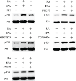
FTI-277 TFA purchased from MedChemExpress. Usage Cited in: Oncotarget. 2017 Nov 22;8(65):109135-109150. [Abstract]
MCF-7 cells are pretreated with the indicated chemical inhibitors for 30min, followed by 15 min treatment with RA (20 μM) + EPA (80 μM).Cell extracts are prepared and subjected to western blotting analysis.


Pureté et documentation
Références
[1]. Tateishi K, et al. FTI-277 and GGTI-289 induce apoptosis via inhibition of the Ras/ERK and Ras/mTOR pathway in head and neck carcinoma HEp-2 and HSC-3 cells. J BUON. 2021;26(2):606-612. [Content Brief]
[2]. Yang W, et al. Farnesyltransferase inhibitor FTI-277 reduces mortality of septic mice along with improved bacterial clearance. J Pharmacol Exp Ther. 2011;339(3):832-841. [Content Brief]
[3]. Ponnusamy A, et al. FTI-277 inhibits smooth muscle cell calcification by up-regulating PI3K/Akt signaling and inhibiting apoptosis. PLoS One. 2018;13(4):e0196232. Published 2018 Apr 24. [Content Brief]
[4]. Girgert R, et al. Farnesyltransferase inhibitor FTI-277 prevents autocrine growth stimulation of neuroblastoma by BDNF. J Cancer Res Clin Oncol. 2003;129(4):227-233. [Content Brief]

Calculators
Concentration (start) × Volume (start) = Concentration (final) × Volume (final)


FTI-277 TFA Related Classifications
HY-110038 Related Classifications
 Powered by Bioz