



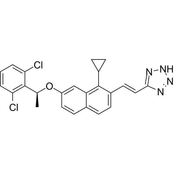
MsbA-IN-6
MsbA-IN-6 is a potent inhibitor of MsbA. MsbA-IN-6 is an antibiotic. Gram-negative ATP-binding cassette (ABC) transporter MsbA, an essential inner membrane protein, transports lipopolysaccharide from the inner leaflet to the periplasmic face of the inner membrane. MsbA-IN-6 kills Escherichia coli through inhibition of its ATPase and transport activity, with no loss of activity against clinical multidrug-resistant strains.
Nos produits utilisent uniquement pour la recherche. Nous ne vendons pas aux patients.
- CAS No.: 2909443-13-4
- Formule: C24H20Cl2N4O
- Masse moléculaire:451.35
-
Stockage:
Please store the product under the recommended conditions in the Certificate of Analysis.

To place orders, for customer services and technical support, please contact: MedChemExpress USA
Tel: 609-228-6898 E-mail: [email protected] [email protected]


-
 Activité biologique
-
Chemical Information
- Protocole
- Pureté et documentation
- Références
-
 Help & FAQs


Voir tous les produits spécifiques à Isoform Antibiotic
More

Activité biologique
Description
IC50 & Target
MsbA[1]


Chemical Information
-
CAS No. 2909443-13-4
-
Masse moléculaire 451.35
-
Formule C24H20Cl2N4O
-
SMILES
C[C@H](OC1=CC2=C(C(/C=C/C3=NNN=N3)=CC=C2C=C1)C4CC4)C5=C(C=CC=C5Cl)Cl
-
Livraison
Room temperature in continental US; may vary elsewhere.
-
Stockage
Please store the product under the recommended conditions in the Certificate of Analysis.

Protocole
-
Membrane Protein Extraction Using Detergents and Chaotropes
Membrane protein extraction with detergents and chaotropes solubilizes lipid-bilayer-associated proteins by disrupting protein-lipid and protein-protein interactions while maintaining proteins in a soluble state for downstream electrophoresis, purification, or mass spectrometry. Chaotropes such as urea and thiourea improve solubilization of difficult proteins, while nonionic and zwitterionic detergents such as CHAPS, ASB-14, SB 3-10, MEGA-10, dodecyl maltoside, and Triton X-100 differ in extraction efficiency depending on sample type and membrane protein properties.
-
Gram Staining of Tissue Sections
Gram staining of tissue sections is a histochemical technique used to differentiate Gram-positive and Gram-negative bacteria within histological specimens based on differences in bacterial cell wall structure and dye retention, adapted from classical bacteriological Gram staining into tissue-compatible “histological Gram stain” variants. In tissue applications, modifications of the Brown-Hopps and Brown-Brenn methods are commonly used to improve differentiation of microorganisms embedded within host connective tissue and to reduce overstaining or loss of Gram-negative signal, which are known limitations of earlier approaches. The principle relies on crystal violet-iodine complex retention in Gram-positive organisms and subsequent decolorization and counterstaining steps that allow contrast visualization of Gram-negative organisms against tissue background.


Pureté et documentation
Références

Calculators
Concentration (start) × Volume (start) = Concentration (final) × Volume (final)


MsbA-IN-6 Related Classifications
HY-130004 Related Classifications
 Powered by Bioz