



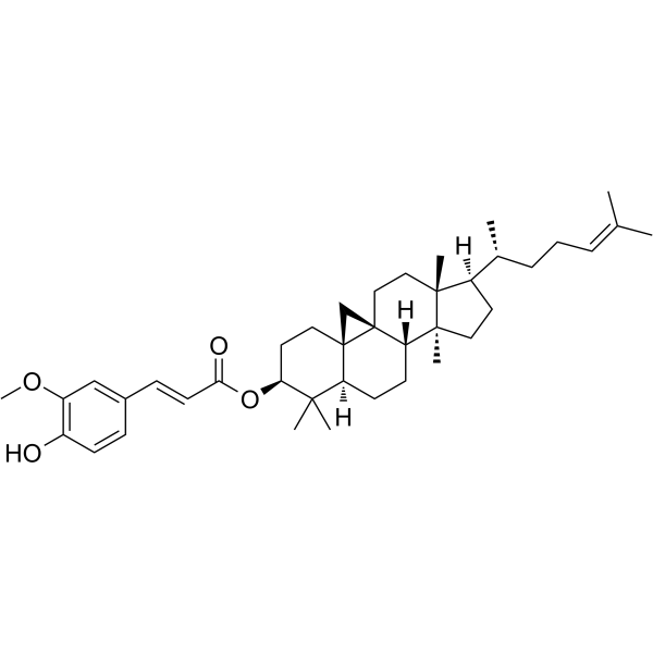
Cycloartenyl ferulate
Based on 1 publication(s) in Google Scholar
Cycloartenyl ferulate (Cycloartenol ferulate; Cycloartenol ferulic acid ester) is a derivative of γ-oryzanol (HY-B2194) with multiple biological activities including antioxidant, anti-inflammatory, and anti-tumor properties. Cycloartenyl ferulate selectively binds to IFNγR1 (binding affinity Kd = 0.5 μM) to activate the canonical JAK1/2-STAT1 signaling pathway. Cycloartenyl ferulate inhibits paraquat (PQ)-triggered apoptosis and ROS in HK2 cells. Cycloartenyl ferulate enhances the activation and cytolytic activity of natural killer (NK) cells by upregulating the expression of NK cell activation receptors (NKG2D, NKp30, NKp44) and the release of cytotoxic molecules and cytokine IFNγ. Cycloartenyl ferulate exerts anti-cancer effects in tumor mice models. Cycloartenyl ferulate can be used for the study of cancer and allergic inflammation intervention.
For research use only. We do not sell to patients.
- Purity : 99.15%
- CAS No.: 21238-33-5
- Formula: C40H58O4
- Molecular Weight:602.89
-
Storage:
4°C, protect from light
* In solvent : -80°C, 6 months; -20°C, 1 month (protect from light)

To place orders, for customer services and technical support, please contact: MedChemExpress USA
Tel: 609-228-6898 E-mail: [email protected] [email protected]


-
 Biological Activity
-
Chemical Information
-
Solvent & Solubility
- Protocols
- Purity & Documentation
- References
-
 Help & FAQs


Publications Citing Use of MedChemExpress (MCE) Cycloartenyl ferulate
More

Biological Activity
Description
Cellular Effect
|
Cell Line

|
Type | Value | Description | References |
|---|---|---|---|---|
| MCF7 | IC50 |
80 μM
Compound: 4
|
Cytotoxicity against human MCF7 cells after 72 hrs by MTT assay
Cytotoxicity against human MCF7 cells after 72 hrs by MTT assay
|
[PMID: 15679326] |
| Raji | IC50 |
15.9 nM
Compound: 17
|
Inhibition of TPA-induced Epstein-Barr virus early antigen activation in Raji cells after 48 hrs
Inhibition of TPA-induced Epstein-Barr virus early antigen activation in Raji cells after 48 hrs
|
[PMID: 17503850] |
In Vitro
Cycloartenyl ferulate (0-10 μM, 72 h) enhances cytolytic activity of NK92 cells against K562, A2058, A549 cancer cells in a dose-dependent manner, upregulates CD107a expression, activates receptors (NKG2D, NKp30, NKp44) and increases granzyme B, perforin, IFNγ release without impairing cell viability[1].
Cycloartenyl ferulate (0-10 μM, 24 h) activates NK cells via the IFNγR-JAK1/2-STAT1-dependent signaling pathway[1].
Cycloartenyl ferulate (20-80 μM, 24 h) significantly reverses the decrease in HK-2 cell viability induced by paraquat (PQ)[2].
Cycloartenyl ferulate (20-80 μM, 24 h) significantly reduces the proportion of HK-2 cells in sub-G1 phase, decreases DNA fragmentation and chromatin condensation, inhibits phosphatidylserine externalization, and reverses the apoptotic characteristics of cells caused by paraquat[2].
Cycloartenyl ferulate (20-80 μM, 0-120 min) dose-dependently and time-dependently inhibits the production of intracellular ROS and superoxide in HK-2 cells induced by PQ[2].
Cycloartenyl ferulate (80 μM, 24 h) reverses the inhibition of Nrf2 and its downstream effectors HO1 and NQO1 expression in HK-2 cells by PQ[2].
Cycloartenyl ferulate (60 min) inhibits degranulation of DNP-IgE-sensitized RBL-2H3 mast cells stimulated with DNP-HSA in a concentration-dependent manner[3].
Cycloartenyl ferulate (1-30 μM, 60 min) decreases the detected concentration of IgE by ELISA in a concentration-dependent manner[3].
MedChemExpress (MCE) has not independently confirmed the accuracy of these methods. They are for reference only.
-
Cell Line:NK92 cells
-
Concentration:0, 0.1, 1, 10 μM
-
Incubation Time:24 h
-
Result:Upregulated p-JAK1/p-JAK2/p-STAT1 without affecting p-JAK3/p-STAT3/p-STAT5/p-mTOR/p-AKT/p-ERK.
Promoted IFNγR1/IFNγR2 dimer formation in NK92 cells.
-
Cell Line:HK-2 cells
-
Concentration:20, 40, 80 μM
-
Incubation Time:24 h
-
Result:Inhibited the increase in caspase-3, -8, and -9 activities induced by PQ.
Inhibited the up-regulation of Bax protein and down-regulation of Bcl-2 protein caused by PQ.
In Vivo
Cycloartenyl ferulate (12.5-50 mg/kg, i.g., once daily, 21 days) reduces dose-dependent formation of lung melanoma nodules in C57BL/6J mice[1].
Cycloartenyl ferulate (50 mg/kg, i.g., once daily, 19 days) combined with anti-PD1 antibody inhibits tumor growth more effectively than alone in C57BL/6J mice harboring Lewis cells[1].
Cycloartenyl ferulate (0-30 μM, intradermal injection, 60 min) attenuates DNP-HSA-induced passive cutaneous anaphylaxis reaction in 8-week-old male Sprague-Dawley rats[3].
MedChemExpress (MCE) has not independently confirmed the accuracy of these methods. They are for reference only.
-
Animal Model:RMA/RMAS cells (1 × 10⁶) were intraperitoneally injected into 6-8-week-old female C57BL/6J mice (18-20 g)[1]
-
Dosage:12.5, 25, 50 mg/kg
-
Administration:i.g., once daily for 3 days
-
Result:Enhanced the clearance of MHC-I-deficient RMAS cells in a dose-dependent manner.
Induced significant elimination of MHC-I-normal RMA cells at the dose of 50 mg/kg.
-
Animal Model:B16-F10 melanoma cells (2 × 10⁵) were intravenously injected into 6-8-week-old female C57BL/6J mice (18-20 g) to establish a pulmonary metastasis model[1]
-
Dosage:12.5, 25, 50 mg/kg
-
Administration:i.g., once daily, 21 days
-
Result:Reduced the formation of lung melanoma nodules in a dose-dependent manner.
Increased the percentages of splenic CD3- NK1.1+ (Pan-NK) and CD3- NKp46+ (activated NK) cells.
-
Animal Model:Lewis lung cancer cells (2 × 10⁶) were subcutaneously implanted into the right flanks of 6-8-week-old female C57BL/6J mice (18-20 g)[1]
-
Dosage:50 mg/kg combined with anti-PD1 antibody (0.2 mg, i.p. every 3 days)
-
Administration:i.g., once daily, 19 days
-
Result:Inhibited Lewis lung tumor growth.
Reduced the populations of myeloid-derived suppressor cells (MDSCs, CD45+ CD11B+ LY6G+) and Foxp3+ Regulatory T cells (Tregs, CD3+CD4+CD25+Foxp3+) in the spleen and tumor tissue.
Increased the infiltration of NK cells (NK1.1+) and CD8+ T cells in tumor tissue.
Promoted the release of granzyme B.
- RNA extraction experimental
- Detection of Apoptosis
- TUNEL staining for apoptotic DNA fragmentation
- Annexin V plus membrane-impermeant dye apoptosis staining
- ROS/oxidative-stress fluorescent staining
- Apoptosis Solutions
- Research Protocol for Inflammation-related Diseases
- Cell Viability Determination by MTT Colorimetric Assay


Chemical Information
-
CAS No. 21238-33-5
-
Appearance Solid
-
Molecular Weight 602.89
-
Formula C40H58O4
-
Color White to off-white
-
SMILES
C[C@]12[C@@]3([H])[C@@]4(CC[C@@]1([C@@](CC2)([H])[C@H](C)CC/C=C(C)\C)C)[C@@]5([C@](CC3)([H])C(C)([C@H](CC5)OC(/C=C/C6=CC(OC)=C(C=C6)O)=O)C)C4
-
Synonyms
Cycloartenol ferulate; Cycloartenol ferulic acid ester
-
Structure Classification
-
Initial Source
-
Shipping
Room temperature in continental US; may vary elsewhere.
-
Storage
4°C, protect from light
* In solvent : -80°C, 6 months; -20°C, 1 month (protect from light)


Publications (1)
-
Journal Impact Factor
-
Most Recent
-
J Biol Chem
Cycloartenyl ferulate improves natural killer (NK) cell immunity against cancer by binding to IFNγ receptor 1. [Abstract]2023 Aug;299(8):104990. PMID: 37392850


Solvent & Solubility
In Vitro:
DMSO : 5 mg/mL (8.29 mM; ultrasonic and warming and heat to 60°C; Hygroscopic DMSO has a significant impact on the solubility of product, please use newly opened DMSO)
Please refer to the solubility information to select the appropriate solvent. Once prepared, please aliquot and store the solution to prevent product inactivation from repeated freeze-thaw cycles.
Storage method and period of stock solution: -80°C, 6 months; -20°C, 1 month (protect from light). When stored at -80°C, please use it within 6 months. When stored at -20°C, please use it within 1 month.
Please refer to the solubility information to select the appropriate solvent. Once prepared, please aliquot and store the solution to prevent product inactivation from repeated freeze-thaw cycles.
Storage method and period of stock solution: -80°C, 6 months; -20°C, 1 month (protect from light). When stored at -80°C, please use it within 6 months. When stored at -20°C, please use it within 1 month.
Concentration (start) × Volume (start) = Concentration (final) × Volume (final)
In Vivo:
Select the appropriate dissolution method based on your experimental animal and administration route.
- For the following dissolution methods, please ensure to first prepare a clear stock solution using an In Vitro approach and then sequentially add co-solvents:
- To ensure reliable experimental results, the clarified stock solution can be appropriately stored based on storage conditions. As for the working solution for In Vivo experiments, it is recommended to prepare freshly and use it on the same day.
- The percentages shown for the solvents indicate their volumetric ratio in the final prepared solution. If precipitation or phase separation occurs during preparation, heat and/or sonication can be used to aid dissolution.
In Vivo Dissolution Calculator
Please enter the basic information of animal experiments:
-
-
-
-
Recommended: Prepare an additional quantity of animals to account for potential losses during experiments.
Please enter your animal formula composition:
-
%DMSO +
Recommended: Keep the proportion of DMSO in working solution below 2% if your animal is weak.
-
%+
-
+%Tween-80 + +
-
%Saline +
The co-solvents required include: DMSO, . All of co-solvents are available by MedChemExpress (MCE). , Tween 80. All of co-solvents are available by MedChemExpress (MCE).
Working solution concentration: 0.22 mg/mL
Method for preparing stock solution: mg drug dissolved in μL DMSO. Stock solution concentration: mg/mL. * In solvent : -80°C, 6 months; -20°C, 1 month (protect from light)
1. Take μL DMSO stock solution;
2. Add μL .
μL , mix evenly;
3. Then add μL Tween 80, mix evenly;
4. Then add μL
Please ensure that the stock solution in the first step is dissolved to a clear state, and add co-solvents in sequence. You can use ultrasonic heating (ultrasonic cleaner, recommended frequency 20-40 kHz), vortexing, etc. to assist dissolution.


Purity & Documentation
-
Data Sheet (280 KB)
-
SDS (252 KB)
- English - EN (252 KB)
- Français - FR (252 KB)
- Deutsch - DE (252 KB)
- Norwegian - NO (252 KB)
- Español - ES (252 KB)
- Swedish - SV (252 KB)
- Italian - IT (252 KB)
- Korean - KR (252 KB)
- Portuguese - PT (252 KB)
-
Handling Instructions (2659 KB)
References
[1]. Li M, et al. Cycloartenyl ferulate improves natural killer (NK) cell immunity against cancer by binding to IFNγ receptor 1. J Biol Chem. 2023 Aug;299(8):104990. [Content Brief]
[2]. Guang-Liang Hong, et al. The reversal of paraquat-induced mitochondria-mediated apoptosis by cycloartenyl ferulate, the important role of Nrf2 pathway. Exp Cell Res. 2013 Nov 1;319(18):2845-55. [Content Brief]
[3]. T Oka, et al. Cycloartenyl ferulate, a component of rice bran oil-derived gamma-oryzanol, attenuates mast cell degranulation.Phytomedicine. 2010 Feb;17(2):152-6. [Content Brief]


Complete Stock Solution Preparation Table
Please refer to the solubility information to select the appropriate solvent. Once prepared, please aliquot and store the solution to prevent product inactivation from repeated freeze-thaw cycles.
Storage method and period of stock solution: -80°C, 6 months; -20°C, 1 month (protect from light). When stored at -80°C, please use it within 6 months. When stored at -20°C, please use it within 1 month.
| Optional Solvent | Concentration Solvent Mass | 1 mg | 5 mg | 10 mg | 25 mg |
|---|---|---|---|---|---|
| DMSO | 1 mM | 1.6587 mL | 8.2934 mL | 16.5868 mL | 41.4669 mL |
| 5 mM | 0.3317 mL | 1.6587 mL | 3.3174 mL | 8.2934 mL |


Cycloartenyl ferulate Related Classifications
HY-125938 Related Classifications
 Powered by Bioz