



D-Psicose

Based on 1 Customer Validation
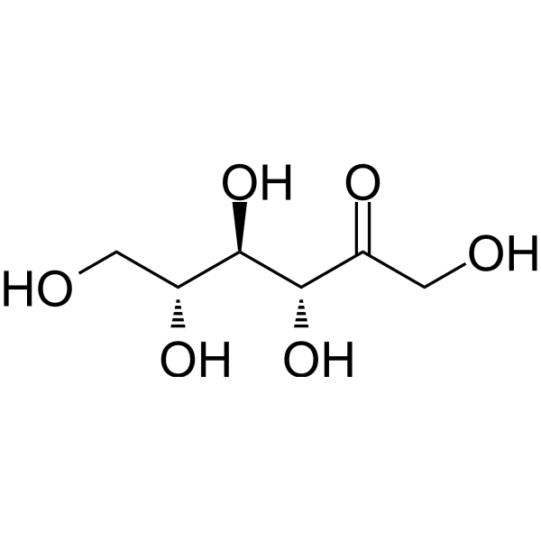
D-psicose is an orally active rare sugar. D-psicose inhibits p38-MAPK phosphorylation and MCP-1 expression. D-psicose inhibits the AGEs/RAGE/NF-κB pathway. D-psicose protects pancreatic β-islets, improves hyperglycemia and high-fat diet-induced non-alcoholic fatty liver disease.
Nur für Forschungszwecke. Wir verkaufen nicht an Patienten.
- Reinheit : 99.99%
- CAS. Nr.: 551-68-8
- Formel: C6H12O6
- Molecular Weight:180.16
-
Speicherung:
4°C, protect from light
* In solvent : -80°C, 6 months; -20°C, 1 month (protect from light)

To place orders, for customer services and technical support, please contact: MedChemExpress USA
Tel: 609-228-6898 E-mail: [email protected] [email protected]


-
 Biologische Aktivität
-
Chemical Information
-
Lösungsmittel & Löslichkeit
- Protokoll
- Reinheit & Dokumentation
- Verweise
-
 Help & FAQs


-
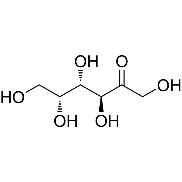
HY-42680
D-TagatoseMonosaccharide -
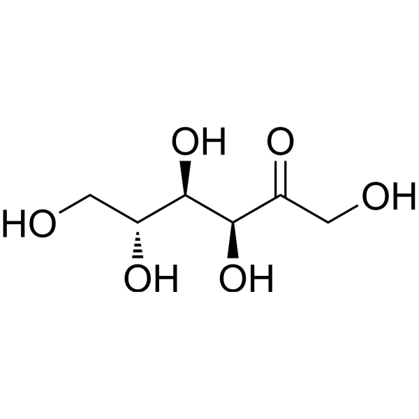
HY-N7092
D-FructoseEndogenous Metabolite -
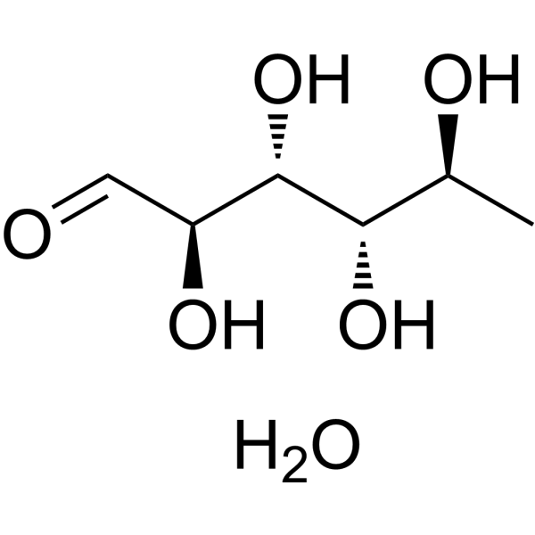
HY-N1420A
Rhamnose monohydrateMonosaccharide -

HY-N9914R
D-Psicose (Standard)Standard Of D-Psicose -
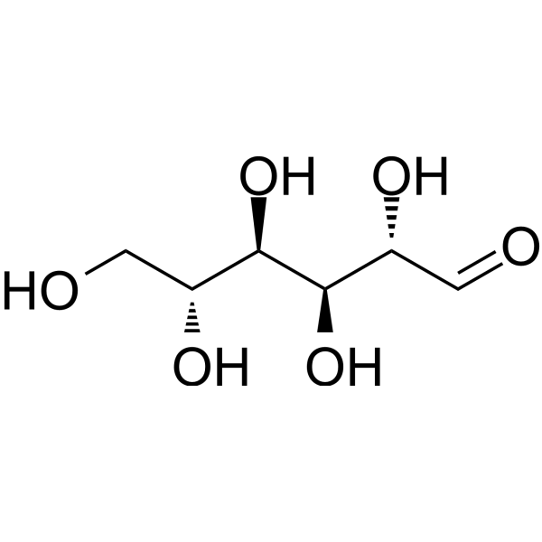
HY-N0379
D-MannoseEndogenous Metabolite -
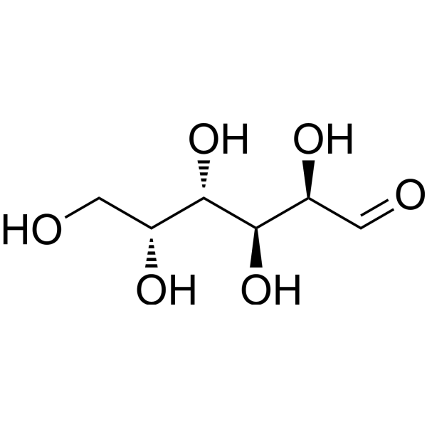
HY-N0210
D-GalactoseEndogenous Metabolite -

HY-N0379R
D-Mannose (Standard)Endogenous Metabolite -
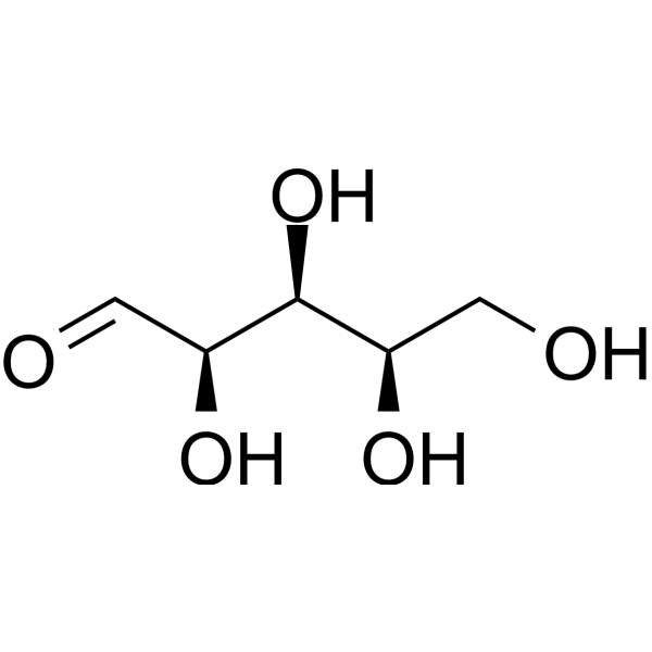
HY-N0537
XyloseEndogenous Metabolite -

HY-N7092R
D-Fructose (Standard)Endogenous Metabolite -
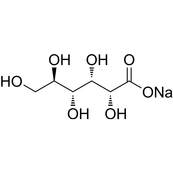
HY-B1092A
Gluconate sodiumGlucose Derivative -
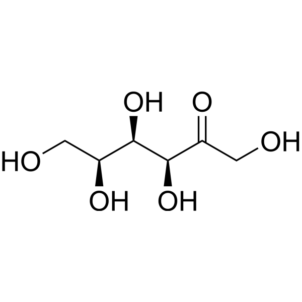
HY-W040240
L-(-)-SorboseCellulase Production Inducer -
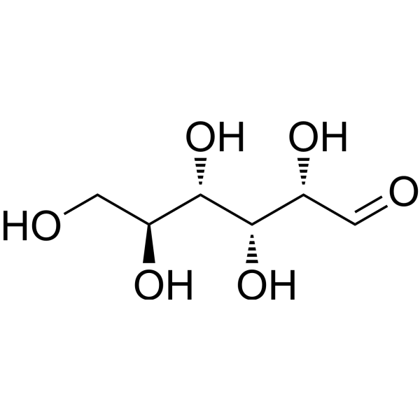
HY-W010042
L-GlucoseD-glucose Stereoisomer


Biologische Aktivität
Beschreibung
In Vitro
D-Psicose (5.6-22.4 mM; 3-5 days) inhibits high-glucose-induced MCP-1 mRNA and protein expression in HUVECs by suppressing p38-MAPK phosphorylation[1].
MedChemExpress (MCE) has not independently confirmed the accuracy of these methods. They are for reference only.
-
Cell Line:Human Umbilical Vein Endothelial Cells (HUVECs)
-
Concentration:2.8 mM, 5.6 mM, 11.2 mM, 16.8 mM, 22.4 mM
-
Incubation Time:up to 5 days
-
Result:Dose-dependently reduced MCP-1 mRNA.
Showed no significant effect at 2.8 mM.
In Vivo
D-Psicose (200 mg/kg BW; p.o.; 28 days) ameliorates hyperglycemia, improves glucose tolerance, reduces hepatic triglyceride and total cholesterol levels by 37.88% and 62.89% respectively, and slows weight gain in C57BL/6J db/db mice[3].
D-Psicose (2.5%-5%; p.o. via drinking water; 12 weeks) reduces hepatic lipid accumulation and inflammatory damage, improves serum lipid profiles and oxidative stress, modulates gut microbiota (increases Akkermansia abundance) and serum metabolomics (enhances anti-inflammatory metabolites), and inhibits the AGEs/RAGE/NF-κB pathway in high-fat diet-induced NAFLD ICR mice[4].
D-Psicose (0.01%-1%; p.o. via diet; 6 weeks) shows no promoting or preventive effects on Diethylnitrosamine (HY-N7434)-induced hepatocarcinogenesis in F344 male rats[5].
MedChemExpress (MCE) has not independently confirmed the accuracy of these methods. They are for reference only.
-
Animal Model:Male Otsuka Long-Evans Tokushima Fatty (OLETF) rats (type 2 diabetes mellitus model)[2]
-
Dosage:5% D-psicose
-
Administration:p.o. via drinking water; 13 weeks.
-
Result:Attenuated pancreatic β-islet fibrosis.
Preserved islet structure.
Reduced body weight and abdominal fat deposition.
Improves oral glucose tolerance and insulin resistance.
Clinical Trial
| NCT Number | Sponsor | Condition | Start Date |
Phase

|
|---|---|---|---|---|
| NCT01329991 | 2011-05 | PHASE1 |


Chemical Information
-
CAS. Nr. 551-68-8
-
Appearance Solid
-
Molecular Weight 180.16
-
Formel C6H12O6
-
Color White to off-white
-
SMILES
OC[C@H]([C@H]([C@H](C(CO)=O)O)O)O
-
Synonyms
D-Allulose
-
Versand
Room temperature in continental US; may vary elsewhere.
-
Speicherung
4°C, protect from light
* In solvent : -80°C, 6 months; -20°C, 1 month (protect from light)


Lösungsmittel & Löslichkeit
In Vitro:
H2O : 100 mg/mL (555.06 mM; Need ultrasonic)
DMSO : 100 mg/mL (555.06 mM; Need ultrasonic; Hygroscopic DMSO has a significant impact on the solubility of product, please use newly opened DMSO)
Please refer to the solubility information to select the appropriate solvent. Once prepared, please aliquot and store the solution to prevent product inactivation from repeated freeze-thaw cycles.
Storage method and period of stock solution: -80°C, 6 months; -20°C, 1 month (protect from light). When stored at -80°C, please use it within 6 months. When stored at -20°C, please use it within 1 month.
* Note: If you choose water as the stock solution, please dilute it to the working solution, then filter and sterilize it with a 0.22 μm filter before use.
Please refer to the solubility information to select the appropriate solvent. Once prepared, please aliquot and store the solution to prevent product inactivation from repeated freeze-thaw cycles.
Storage method and period of stock solution: -80°C, 6 months; -20°C, 1 month (protect from light). When stored at -80°C, please use it within 6 months. When stored at -20°C, please use it within 1 month.
* Note: If you choose water as the stock solution, please dilute it to the working solution, then filter and sterilize it with a 0.22 μm filter before use.
Konzentration (Stammlösung) × Volumen (Stammlösung) = Konzentration (Ziellösung) × Volumen (Ziellösung)


Reinheit & Dokumentation
-
Data Sheet (291 KB)
-
SDS (476 KB)
- English - EN (476 KB)
- Français - FR (476 KB)
- Deutsch - DE (476 KB)
- Norwegian - NO (476 KB)
- Español - ES (476 KB)
- Swedish - SV (476 KB)
- Italian - IT (476 KB)
- Korean - KR (476 KB)
- Portuguese - PT (476 KB)
-
Handling Instructions (2659 KB)
Verweise
[1]. Murao K, et al. D-Psicose inhibits the expression of MCP-1 induced by high-glucose stimulation in HUVECs. Life Sci. 2007 Jul 26;81(7):592-9. [Content Brief]
[2]. Hossain A, et al. Rare sugar D-psicose protects pancreas β-islets and thus improves insulin resistance in OLETF rats. Biochem Biophys Res Commun. 2012 Sep 7;425(4):717-23. [Content Brief]
[3]. Baek SH, et al. D-psicose, a sweet monosaccharide, ameliorate hyperglycemia, and dyslipidemia in C57BL/6J db/db mice. J Food Sci. 2010 Mar;75(2):H49-53. [Content Brief]
[4]. Tan J, et al. D-Psicose mitigates NAFLD mice induced by a high-fat diet by reducing lipid accumulation, inflammation, and oxidative stress. Front Nutr. 2025 May 27;12:1574151. [Content Brief]


Complete Stock Solution Preparation Table
Please refer to the solubility information to select the appropriate solvent. Once prepared, please aliquot and store the solution to prevent product inactivation from repeated freeze-thaw cycles.
Storage method and period of stock solution: -80°C, 6 months; -20°C, 1 month (protect from light). When stored at -80°C, please use it within 6 months. When stored at -20°C, please use it within 1 month.
| Optional Solvent | Concentration Solvent Mass | 1 mg | 5 mg | 10 mg | 25 mg |
|---|---|---|---|---|---|
| H2O / DMSO | 1 mM | 5.5506 mL | 27.7531 mL | 55.5062 mL | 138.7655 mL |
| 5 mM | 1.1101 mL | 5.5506 mL | 11.1012 mL | 27.7531 mL | |
| 10 mM | 0.5551 mL | 2.7753 mL | 5.5506 mL | 13.8766 mL | |
| 15 mM | 0.3700 mL | 1.8502 mL | 3.7004 mL | 9.2510 mL | |
| 20 mM | 0.2775 mL | 1.3877 mL | 2.7753 mL | 6.9383 mL | |
| 25 mM | 0.2220 mL | 1.1101 mL | 2.2202 mL | 5.5506 mL | |
| 30 mM | 0.1850 mL | 0.9251 mL | 1.8502 mL | 4.6255 mL | |
| 40 mM | 0.1388 mL | 0.6938 mL | 1.3877 mL | 3.4691 mL | |
| 50 mM | 0.1110 mL | 0.5551 mL | 1.1101 mL | 2.7753 mL | |
| 60 mM | 0.0925 mL | 0.4626 mL | 0.9251 mL | 2.3128 mL | |
| 80 mM | 0.0694 mL | 0.3469 mL | 0.6938 mL | 1.7346 mL | |
| 100 mM | 0.0555 mL | 0.2775 mL | 0.5551 mL | 1.3877 mL |
* Note: If you choose water as the stock solution, please dilute it to the working solution, then filter and sterilize it with a 0.22 μm filter before use.


D-Psicose Related Classifications
HY-N9914 Related Classifications
 Powered by Bioz