



G5-7

Based on 1 Customer Validation
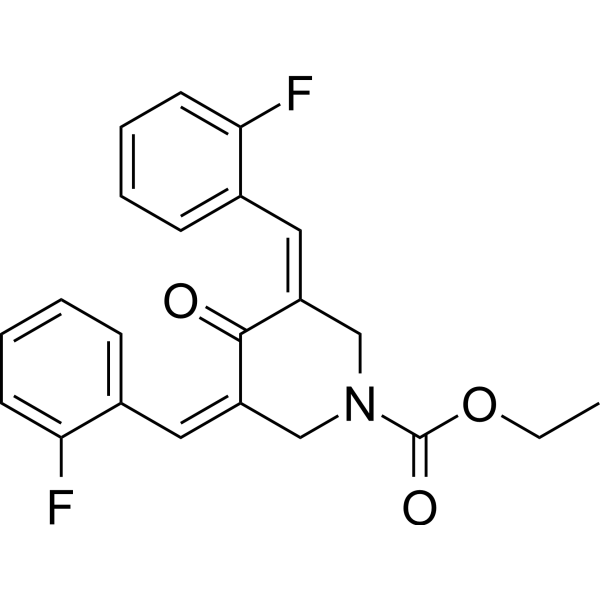
G5-7, an orally active and allosteric JAK2 inhibitor, selectively inhibits JAK2 mediated phosphorylation and activation of EGFR (Tyr1068) and STAT3 by binding to JAK2. G5-7 induces cell cycle arrest, apoptosis and possesses antiangiogenic effect. G5-7 has the potential for glioma study.
For research use only. We do not sell to patients.
- Purity : 98.45%
- CAS No.: 939681-36-4
- Formula: C22H19F2NO3
- Molecular Weight:383.39
-
Storage:Powder -20°C, 3 years ; In solvent -80°C, 6 months , -20°C, 1 month

To place orders, for customer services and technical support, please contact: MedChemExpress USA
Tel: 609-228-6898 E-mail: [email protected] [email protected]


-
 Biological Activity
-
Chemical Information
-
Solvent & Solubility
- Protocols
- Purity & Documentation
- References
-
 Help & FAQs


-
HY-50856
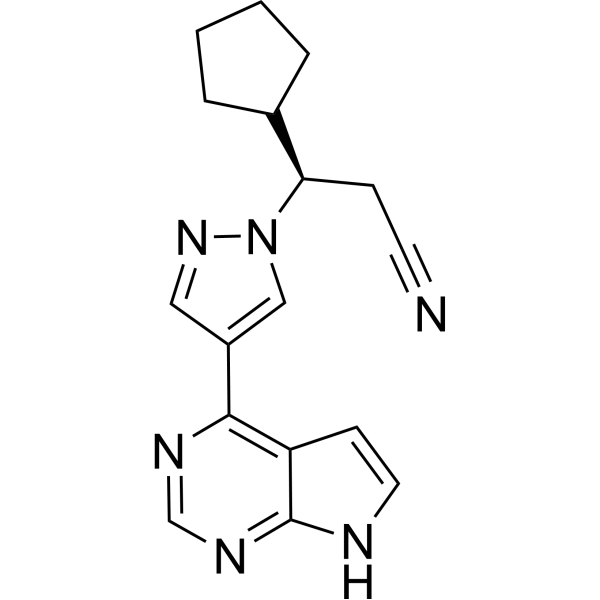
RuxolitinibJAK1/2 Inhibitor -
HY-10409
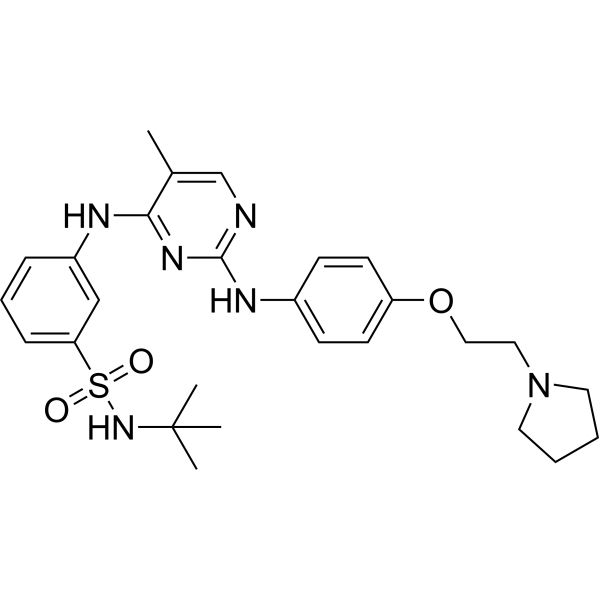
FedratinibJAK2 Inhibitor -
HY-16379
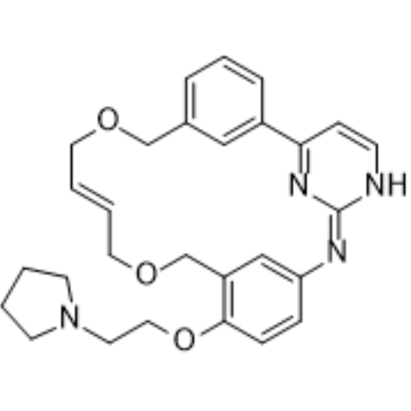
PacritinibJAK2/FLT3 Inhibitor -
HY-15312
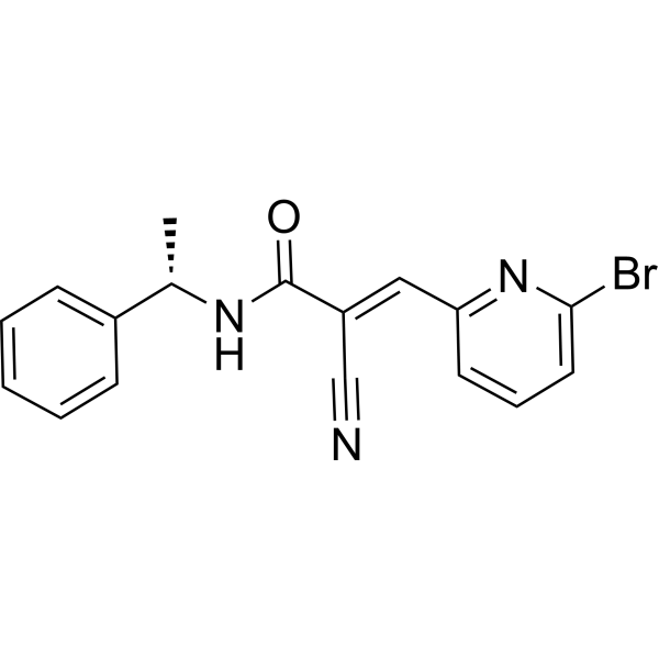
WP1066JAK2/STAT3 Inhibitor -
HY-107429
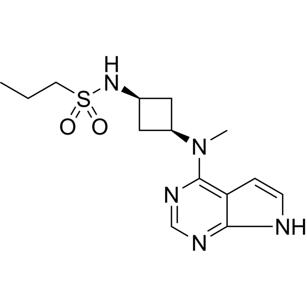
AbrocitinibJAK1 Inhibitor -
HY-113573
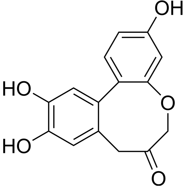
Protosappanin AJAK2/STAT3 Inhibitor -
HY-K0010
Protease Inhibitor Cocktail (EDTA-Free, 100× in DMSO) -
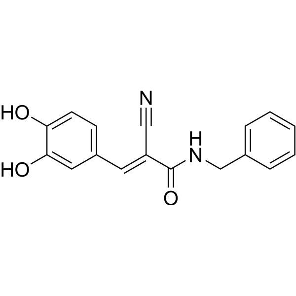
HY-12000
AG490EGFR Inhibitor -
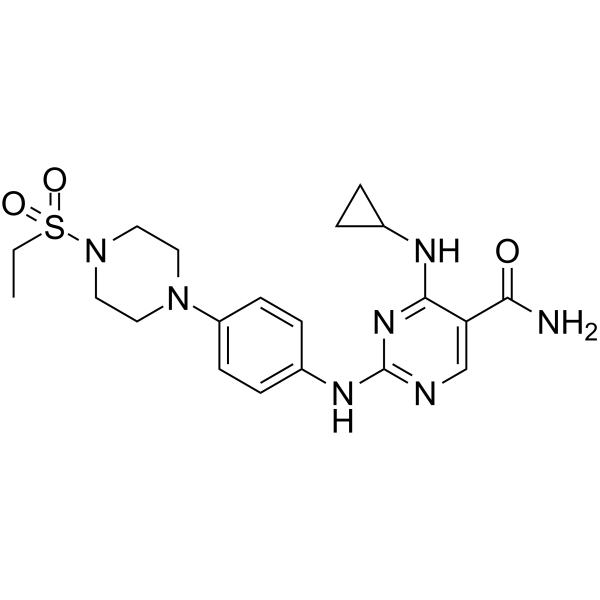
HY-15999
CerdulatinibTyk2 Inhibitor -
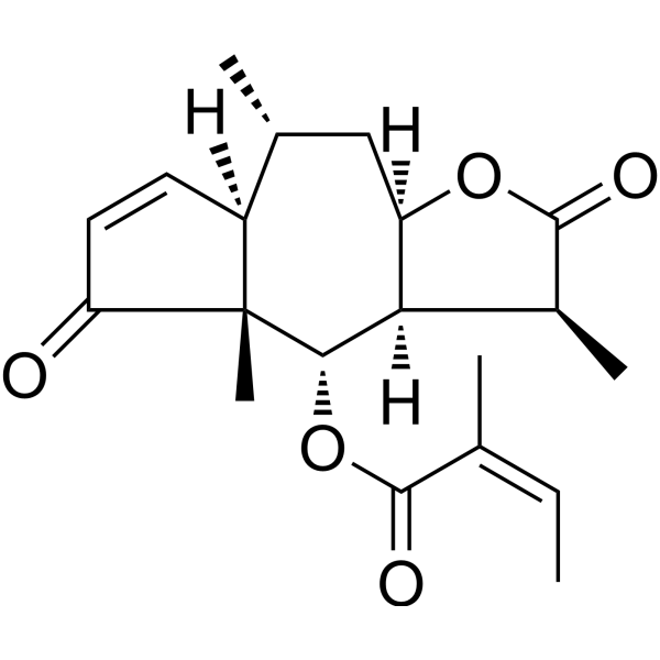
HY-N2959
Brevilin ASTAT3/JAK Inhibitor -
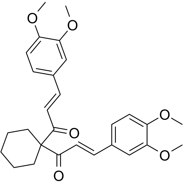
HY-100544
FLLL32JAK2/STAT3 Inhibitor -
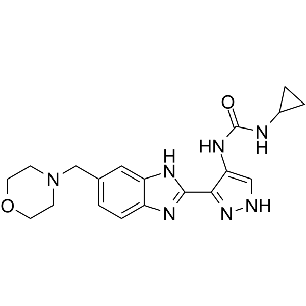
HY-50514
AT9283Aurora/JAK Inhibitor


Biological Activity
Description
IC50 & Target
[1]|
JAK2 |
In Vitro
G5-7 (0-5 μM) inhibits EGFR tyrosine phosphorylation and downstream mTOR signaling and arrests the cell cycle at G2 phase[1].
G5-7 does not directly inhibit EGFR activation[1].
G5-7 (0-10 μM) comparably increases the abundance of markers (cleved-PARP and caspase 3) of apoptosis in parental LN229 cells and U87MG/EGFRvIII cells[1].
G5-7 interacts with full-length JAK2[1].
G5-7 significantly inhibits EGFR Tyr1068 phosphorylation but had no effect on EGFR Tyr1045 phosphorylation[1].
G5-7 downregulates the downstream signaling of JAK by mTOR[1].
MedChemExpress (MCE) has not independently confirmed the accuracy of these methods. They are for reference only.
-
Cell Line:U87MG/PTEN cells.
-
Concentration:0-5 μM.
-
Incubation Time:6 hours.
-
Result:Blocked EGFR phosphorylation and cell cycle at G2 phase to inhibit cell proliferation.
In Vivo
MedChemExpress (MCE) has not independently confirmed the accuracy of these methods. They are for reference only.
-
Animal Model:Cells (4 × 10⁶) in 100 μl of serum-free DMEM were inoculated subcutaneously into 5- to 6-week-old female nude mice[1].
-
Dosage:10 and 50 mg/kg.
-
Administration:Oral gavage.
-
Result:Suppresses angiogenesis in tumors.
- Kinase activity and phosphorylation assays
- Western Blot
- Detection of Apoptosis
- TUNEL staining for apoptotic DNA fragmentation
- Flow cytometric DNA-content cell-cycle staining
- Annexin V plus membrane-impermeant dye apoptosis staining
- BrdU Incorporation Assay
- Apoptosis Solutions
- Protocol for Cell Cycle
- Protocol for Kinase activity and phosphorylation assays


Chemical Information
-
CAS No. 939681-36-4
-
Appearance Solid
-
Molecular Weight 383.39
-
Formula C22H19F2NO3
-
Color Off-white to light yellow
-
SMILES
O=C(N1C/C(C(/C(C1)=C\C2=CC=CC=C2F)=O)=C/C3=CC=CC=C3F)OCC
-
Shipping
Room temperature in continental US; may vary elsewhere.
-
Storage
Powder -20°C 3 years In solvent -80°C 6 months -20°C 1 month


Solvent & Solubility
In Vitro:
DMSO : 83.33 mg/mL (217.35 mM; Need ultrasonic; Hygroscopic DMSO has a significant impact on the solubility of product, please use newly opened DMSO)
Please refer to the solubility information to select the appropriate solvent. Once prepared, please aliquot and store the solution to prevent product inactivation from repeated freeze-thaw cycles.
Storage method and period of stock solution: -80°C, 6 months; -20°C, 1 month. When stored at -80°C, please use it within 6 months. When stored at -20°C, please use it within 1 month.
Please refer to the solubility information to select the appropriate solvent. Once prepared, please aliquot and store the solution to prevent product inactivation from repeated freeze-thaw cycles.
Storage method and period of stock solution: -80°C, 6 months; -20°C, 1 month. When stored at -80°C, please use it within 6 months. When stored at -20°C, please use it within 1 month.
Concentration (start) × Volume (start) = Concentration (final) × Volume (final)


Purity & Documentation
-
Data Sheet (275 KB)
-
SDS (394 KB)
- English - EN (394 KB)
- Français - FR (394 KB)
- Deutsch - DE (394 KB)
- Norwegian - NO (394 KB)
- Español - ES (394 KB)
- Swedish - SV (394 KB)
- Italian - IT (394 KB)
- Korean - KR (394 KB)
- Portuguese - PT (394 KB)
-
Handling Instructions (2659 KB)
References


Complete Stock Solution Preparation Table
Please refer to the solubility information to select the appropriate solvent. Once prepared, please aliquot and store the solution to prevent product inactivation from repeated freeze-thaw cycles.
Storage method and period of stock solution: -80°C, 6 months; -20°C, 1 month. When stored at -80°C, please use it within 6 months. When stored at -20°C, please use it within 1 month.
| Optional Solvent | Concentration Solvent Mass | 1 mg | 5 mg | 10 mg | 25 mg |
|---|---|---|---|---|---|
| DMSO | 1 mM | 2.6083 mL | 13.0415 mL | 26.0831 mL | 65.2077 mL |
| 5 mM | 0.5217 mL | 2.6083 mL | 5.2166 mL | 13.0415 mL | |
| 10 mM | 0.2608 mL | 1.3042 mL | 2.6083 mL | 6.5208 mL | |
| 15 mM | 0.1739 mL | 0.8694 mL | 1.7389 mL | 4.3472 mL | |
| 20 mM | 0.1304 mL | 0.6521 mL | 1.3042 mL | 3.2604 mL | |
| 25 mM | 0.1043 mL | 0.5217 mL | 1.0433 mL | 2.6083 mL | |
| 30 mM | 0.0869 mL | 0.4347 mL | 0.8694 mL | 2.1736 mL | |
| 40 mM | 0.0652 mL | 0.3260 mL | 0.6521 mL | 1.6302 mL | |
| 50 mM | 0.0522 mL | 0.2608 mL | 0.5217 mL | 1.3042 mL | |
| 60 mM | 0.0435 mL | 0.2174 mL | 0.4347 mL | 1.0868 mL | |
| 80 mM | 0.0326 mL | 0.1630 mL | 0.3260 mL | 0.8151 mL | |
| 100 mM | 0.0261 mL | 0.1304 mL | 0.2608 mL | 0.6521 mL |


G5-7 Related Classifications
HY-115452 Related Classifications
 Powered by Bioz