



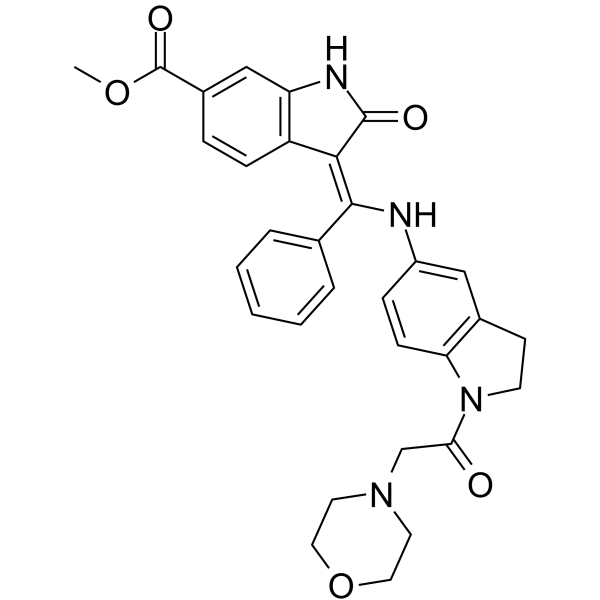
KBP-7018
KBP-7018 is an orally active, tyrosine kinase-selective multi-kinase inhibitor with an IC50 value of 10 nM against c-KIT, 7.6 nM against RET, and 25 nM against human PDGFR. KBP-7018 improves survival rates in a mouse model of pulmonary fibrosis. KBP-7018 can be used in research related to idiopathic pulmonary fibrosis.
For research use only. We do not sell to patients.
- CAS No.: 1613437-66-3
- Formula: C31H30N4O5
- Molecular Weight:538.59
-
Storage:
Please store the product under the recommended conditions in the Certificate of Analysis.

To place orders, for customer services and technical support, please contact: MedChemExpress USA
Tel: 609-228-6898 E-mail: [email protected] [email protected]


-
 Biological Activity
-
Chemical Information
- Protocols
- Purity & Documentation
- References
-
 Help & FAQs




Biological Activity
Description
IC50 & Target
[2]|
PDGFRβ 34 nM (IC50) |
PDGFRα 26 nM (IC50) |
CYP3A4 0.087 μM (IC50) |
In Vitro
KBP-7018 potently and selectively inhibits c-KIT (IC50 = 10 nM), RET (IC50 = 7.6 nM) and PDGFR (IC50 = 25 nM) in cell-free kinase assays. Meanwhile, it shows extremely weak inhibitory activity against hERG K+ and exhibits high kinome selectivity[1].
KBP-7018 inhibits h-PDGF-bb-stimulated proliferation of 3T3 fibroblasts in cell-based assays, with an IC50 of 100 nM[1].
KBP-7018 potently inhibits purified PDGFRα (IC50 = 26 nM) and PDGFRβ (IC50 = 34 nM)[2].
KBP-7018 (0.05-80 μM; 10 min) potently inhibits human CYP3A4 (testosterone probe, IC50 = 0.087 μM), weakly inhibits human CYP2C9 (IC50 = 35.2 μM), and shows no inhibitory effect on human CYP1A2, CYP2C19 or CYP2D6 at concentrations up to 80 μM[2].
MedChemExpress (MCE) has not independently confirmed the accuracy of these methods. They are for reference only.
Parmacokinetics
| Species | Dose | Route | AUC0-t | CLplasma | T1/2 | Tmax | AUC | F | Bioavailability | Cmax |
|---|---|---|---|---|---|---|---|---|---|---|
| Mice[1] | 50 mg/kg | p.o. | / | / | 4.3 h | / | 17004 ng·h/mL | 51 % | / | / |
| Rat[1] | 10 mg/kg | p.o. | / | / | 4.8 h | / | 6406 ng·h/mL | 68 % | / | / |
| Dog[1] | 20 mg/kg | p.o. | / | / | 3.3 h | / | 2964 ng·h/mL | 29 % | / | / |
| Mice[2] | 10 mg/kg | i.v. | 6636 ng·h/mL | 1.50 L/h/kg | 0.80 h | 0.25 h | / | / | / | / |
| Mice[2] | 50 mg/kg | p.o. | 17004 ng·h/mL | / | 4.29 h | / | / | / | 51 % | / |
| Rat[2] | 2 mg/kg | i.v. | 1877 ng·h/mL | 1.06 L/h/kg | 1.30 h | 1.00 h | / | / | / | / |
| Rat[2] | 10 mg/kg | p.o. | 6406 ng·h/mL | / | 4.81 h | / | / | / | 68 % | / |
| Dog[2] | 2 mg/kg | i.v. | 1007 ng·h/mL | 1.87 L/h/kg | 2.33 h | 2.00 h | / | / | / | / |
| Dog[2] | 50 mg/kg | p.o. | 5178 ng·h/mL | / | 6.71 h | / | / | / | 21 % | / |
| Monkey[2] | 2 mg/kg | i.v. | 2031 ng·h/mL | 0.49 L/h/kg | 6.82 h | 6 h | / | / | / | / |
| Monkey[2] | 5 mg/kg | p.o. | 2578 ng·h/mL | / | 4.56 h | 6.0 h | / | / | 25 % | 73 ng/mL |
In Vivo
MedChemExpress (MCE) has not independently confirmed the accuracy of these methods. They are for reference only.
-
Animal Model:C57 mice[1]
-
Dosage:10 mg/kg; 30 mg/kg; 100 mg/kg
-
Administration:p.o.; daily; 28 days
-
Result:Significantly improved the 28-day survival rate in a dose-dependent manner.
Demonstrated sufficient tolerance at all tested doses (10 to 100 mg/kg).


Chemical Information
-
CAS No. 1613437-66-3
-
Molecular Weight 538.59
-
Formula C31H30N4O5
-
SMILES
O=C(C1=CC(NC/2=O)=C(C=C1)C2=C(NC3=CC4=C(N(C(CN5CCOCC5)=O)CC4)C=C3)/C6=CC=CC=C6)OC
-
Shipping
Room temperature in continental US; may vary elsewhere.
-
Storage
Please store the product under the recommended conditions in the Certificate of Analysis.


Purity & Documentation
References
[1]. Huang Z, et al. Discovery of Indolinone-Based Multikinase Inhibitors as Potential Therapeutics for Idiopathic Pulmonary Fibrosis. ACS medicinal chemistry letters. 2017 Nov 09;8(11):1142-1147. [Content Brief]
[2]. Huang Z, et al. Characterization of preclinical in vitro and in vivo pharmacokinetics properties for KBP-7018, a new tyrosine kinase inhibitor candidate for treatment of idiopathic pulmonary fibrosis. Drug design, development and therapy. 2015;9:4319-28. [Content Brief]

Calculators
Concentration (start) × Volume (start) = Concentration (final) × Volume (final)


KBP-7018 Related Classifications
HY-117873 Related Classifications
 Powered by Bioz