



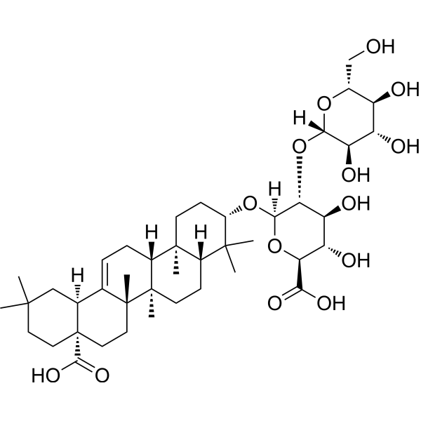
Zingibroside R1
Based on 1 publication(s) in Google Scholar
Zingibroside R1 is an orally active triterpene saponin with multiple biological activities including antioxidant, anti-inflammatory, antiviral, and metabolic regulatory properties. Zingibroside R1 reduces the expression of PIN family members, inhibits the expression of PLT1/PLT2, WOX5, SHR, and SCR, disrupts auxin transport and distribution, triggers plant ROS responses, and inhibits root growth. Zingibroside R1 extends the lifespan of Caenorhabditis elegans, enhances its heat stress resistance, and improves its motor ability. Hydrogel derivatives of Zingibroside R1 inhibit the proliferation of Candida albicans by binding to its β-1,3-glucan and exhibit antifungal activity. Zingibroside R1 inhibits GLUT1-mediated uptake and alleviates liver injury. Zingibroside R1 can be used in research related to neurodegenerative diseases, vulvovaginal candidiasis, acute liver injury, Ehrlich ascites tumor and HIV-1 infection.
For research use only. We do not sell to patients.
- Purity : 99.47%
- CAS No.: 80930-74-1
- Formula: C42H66O14
- Molecular Weight:794.97
-
Storage:
4°C, protect from light
* In solvent : -80°C, 6 months; -20°C, 1 month (protect from light)

To place orders, for customer services and technical support, please contact: MedChemExpress USA
Tel: 609-228-6898 E-mail: [email protected] [email protected]


-
 Biological Activity
-
Chemical Information
-
Solvent & Solubility
- Protocols
- Purity & Documentation
- References
-
 Help & FAQs


Publications Citing Use of MedChemExpress (MCE) Zingibroside R1
More

Biological Activity
Description
IC50 & Target
[5]|
HIV-1 |
GLUT1 |
In Vitro
Zingibroside R1 (ZR1) (20-100 μM; 15-45 days) dose-dependently inhibits the adventitious root density and root apical meristem length of ginseng adventitious roots[1].
Zingibroside R1 (5-10 μM; 2-7 days) dose-dependently inhibits primary root elongation, meristematic zone length, and elongation zone length in Arabidopsis thaliana Col-0 seedlings[1].
Zingibroside R1 (5-10 μM; 2-7 days) reduces endogenous IAA levels in Arabidopsis thaliana and disrupts its polar auxin transport by decreasing the fluorescence intensity of specific PIN transporter markers[1].
Zingibroside R1 (5-10 μM; 2-4 days) regulates the expression of core root stem cell regulators in Arabidopsis thaliana[1].
Zingibroside R1 (5-10 μM; 7-10 days) induces oxidative stress in Arabidopsis thaliana Col-0 seedlings by promoting ROS accumulation, impairing the activity of ROS-scavenging enzymes, and altering the GSH/GSSG redox balance[1].
Zingibroside R1 (2.0 wt%; 1 h heating, 30 min cooling, 6-24 h cell incubation) forms a stable hydrogel with shear recovery property, and exhibits excellent biocompatibility with human Caco-2 cells[3].
Hydrogel containing 2.0 wt% Zingibroside R1 (ZR1), upon incubation for 2-24 h, potently inhibits the proliferation of Candida albicans in vitro by disrupting cell membrane integrity[3].
Nanofibrils of Zingibroside R1 (100 ns simulation) exhibit high binding affinity for β-1,3-glucan in the cell wall of Candida albicans, and disrupt the integrity of fungal cell membranes through hydrogen bonding and structural perturbation[3].
Zingibroside R1 (2.0 wt%; 24 h incubation) hydrogel can stably encapsulate Lactobacillus rhamnosus GG and achieve sustained release, while the LGG@ZR1 formulation exerts synergistic inhibitory effects on the proliferation of Candida albicans in vitro and acidifies the culture medium[3].
Zingibroside R1 (compound 13) (10-100 μM; 10 min preincubation, 1 min uptake measurement) inhibits the uptake of 2-deoxyglucose (2-DG) by Ehrlich ascites tumor (EAT) cells, with an IC50 of 91.3 μM, and acts as a partial competitive inhibitor of glucose transport[5].
Zingibroside R1 (5 days) exhibits cytotoxicity (CC50 = 46.2 μM) and anti-HIV-1 activity against the human T-cell line (MT-4)[5].
MedChemExpress (MCE) has not independently confirmed the accuracy of these methods. They are for reference only.
In Vivo
Zingibroside R1 (2% w/v; i.vag.; daily; 9 days) gel exhibits in vivo efficacy against vulvovaginal candidiasis in BALB/c mice, reducing fungal burden and vaginal inflammation[3].
Zingibroside R1 (R1) (15-30 mg/kg; p.o.; daily; 7 days) exerts significant hepatoprotective effects against LPS/D-GalN-induced acute liver injury in male BALB/c mice by reducing oxidative stress, suppressing inflammation, modulating immune cell populations, restoring gut microbiota balance, and regulating hepatic metabolite profiles including succinic acid[4].
Zingibroside R1 (30 mg/kg; oral; once daily; for 7 days) fecal microbiota transplantation in mice significantly ameliorates LPS/D-GalN-induced acute liver injury in male BALB/c mice by attenuating oxidative stress, inhibiting inflammatory responses, and restoring the hepatic metabolite profile including succinate[4].
MedChemExpress (MCE) has not independently confirmed the accuracy of these methods. They are for reference only.
-
Animal Model:BALB/c (female, 18-20 g, vulvovaginal candidiasis model induced by subcutaneous estradiol benzoate followed by intravaginal C. albicans inoculation)[3]
-
Dosage:2% w/v
-
Administration:i.vag.; daily; 9 days
-
Result:Reduced C. albicans colony counts from vaginal washes throughout the treatment period.
Lessened epithelial disruption and neutrophil infiltration in vaginal tissues compared to untreated controls.
Showed gradual improvement in vaginal redness and inflammation by day 9.
Revealed residual fungal presence in vaginal tissues via PAS staining.
-
Animal Model:BALB/c (male, 18-22 g, LPS/D-GalN-induced acute liver injury)[4]
-
Dosage:15 mg/kg; 30 mg/kg
-
Administration:p.o.; daily; 7 days
-
Result:Showed significant reduction in liver haemorrhaging, hepatocyte necrosis, nuclear shrinkage and inflammatory infiltration.
Decreased serum ALT and AST levels.
Reduced hepatic and serum ROS and MDA levels, and increased serum and hepatic SOD and GSH-Px activities.
Reduced hepatocyte apoptosis rate.
Decreased hepatic levels of TNF-α, IL-1β, IL-6 and IFN-γ, and increased IL-2 level.
Upregulated hepatic and peripheral blood Th cells, Tc cells, DCs and NK cells, and downregulated Tregs, macrophages (F4/80) and neutrophils (LY-6G).
Restored Shannon and Simpson indices, increased relative abundance of Ligilactobacillus, and decreased relative abundances of Clostridium and Anaerotignum.
Normalized liver metabolic profile, increased taurine, allose, L-carnitine, hypoxanthine, glycerol and 4-trimethylammoniobutanoic acid, and decreased succinic acid and D-lysine.
-
Animal Model:BALB/c (male, 18-22 g, LPS/D-GalN-induced acute liver injury with faecal microbiota transplantation)[4]
-
Dosage:Faecal microbiota from mice treated with 30 mg/kg Zingibroside R1
-
Administration:p.o.; daily; 6 days
-
Result:Showed reduced hepatocyte necrosis, nuclear shrinkage and inflammatory cell infiltration compared to the M-FMT group.
Decreased serum ALT and AST levels relative to the M-FMT group.
Reduced hepatic ROS generation rate and hepatocyte apoptosis rate compared to the M-FMT group.
Normalized the liver metabolic profile, increased hypoxanthine and glycerophosphocholine, and decreased succinic acid relative to the M-FMT group.
Decreased hepatic levels of pro-inflammatory cytokines IL-1β, TNF-α, IL-6 and IFN-γ compared to the M-FMT group.


Chemical Information
-
CAS No. 80930-74-1
-
Appearance Solid
-
Molecular Weight 794.97
-
Formula C42H66O14
-
Color White to off-white
-
SMILES
C[C@@]12C([C@@]3([H])[C@](C(O)=O)(CCC(C)(C)C3)CC2)=CC[C@]4([H])[C@@](C)(CC[C@H](O[C@@]5([H])[C@@H]([C@H]([C@H](O)[C@@H](C(O)=O)O5)O)O[C@]6([H])O[C@@H]([C@@H](O)[C@H](O)[C@H]6O)CO)C7(C)C)[C@@]7([H])CC[C@@]14C
-
Structure Classification
-
Shipping
Room temperature in continental US; may vary elsewhere.
-
Storage
4°C, protect from light
* In solvent : -80°C, 6 months; -20°C, 1 month (protect from light)


Publications (1)
-
Journal Impact Factor
-
Most Recent
-
Vet Microbiol
The Chinese medicine monomer Schisandrin C inhibits PRRSV infection by regulating the OGT-PI3K/AKT/mTOR signaling pathway. [Abstract]2026 May:316:110992. PMID: 41865607


Solvent & Solubility
In Vitro:
DMSO : 100 mg/mL (125.79 mM; Need ultrasonic; Hygroscopic DMSO has a significant impact on the solubility of product, please use newly opened DMSO)
Please refer to the solubility information to select the appropriate solvent. Once prepared, please aliquot and store the solution to prevent product inactivation from repeated freeze-thaw cycles.
Storage method and period of stock solution: -80°C, 6 months; -20°C, 1 month (protect from light). When stored at -80°C, please use it within 6 months. When stored at -20°C, please use it within 1 month.
Please refer to the solubility information to select the appropriate solvent. Once prepared, please aliquot and store the solution to prevent product inactivation from repeated freeze-thaw cycles.
Storage method and period of stock solution: -80°C, 6 months; -20°C, 1 month (protect from light). When stored at -80°C, please use it within 6 months. When stored at -20°C, please use it within 1 month.
Concentration (start) × Volume (start) = Concentration (final) × Volume (final)
In Vivo:
Select the appropriate dissolution method based on your experimental animal and administration route.
- For the following dissolution methods, please ensure to first prepare a clear stock solution using an In Vitro approach and then sequentially add co-solvents:
- To ensure reliable experimental results, the clarified stock solution can be appropriately stored based on storage conditions. As for the working solution for In Vivo experiments, it is recommended to prepare freshly and use it on the same day.
- The percentages shown for the solvents indicate their volumetric ratio in the final prepared solution. If precipitation or phase separation occurs during preparation, heat and/or sonication can be used to aid dissolution.
Add each solvent one by one: 10% DMSO 40% PEG300 5% Tween-80 45% Saline
Solubility: ≥ 2.5 mg/mL (3.14 mM); Clear solution
This protocol yields a clear solution of ≥ 2.5 mg/mL (saturation unknown).
Taking 1 mL working solution as an example, add 100 μL DMSO stock solution (25.0 mg/mL) to 400 μL PEG300, and mix evenly; then add 50 μL Tween-80 and mix evenly; then add 450 μL Saline to adjust the volume to 1 mL.
Preparation of Saline: Dissolve 0.9 g sodium chloride in ddH₂O and dilute to 100 mL to obtain a clear Saline solution.
Add each solvent one by one: 10% DMSO 90% (20% SBE-β-CD in Saline)
Solubility: ≥ 2.5 mg/mL (3.14 mM); Clear solution
This protocol yields a clear solution of ≥ 2.5 mg/mL (saturation unknown).
Taking 1 mL working solution as an example, add 100 μL DMSO stock solution (25.0 mg/mL) to 900 μL 20% SBE-β-CD in Saline, and mix evenly.
Preparation of 20% SBE-β-CD in Saline (4°C, storage for one week): 2 g SBE-β-CD powder is dissolved in 10 mL Saline, completely dissolve until clear.
In Vivo Dissolution Calculator
Please enter the basic information of animal experiments:
-
-
-
-
Recommended: Prepare an additional quantity of animals to account for potential losses during experiments.
Please enter your animal formula composition:
-
%DMSO +
Recommended: Keep the proportion of DMSO in working solution below 2% if your animal is weak.
-
%+
-
+%Tween-80 + +
-
%Saline +
The co-solvents required include: DMSO, . All of co-solvents are available by MedChemExpress (MCE). , Tween 80. All of co-solvents are available by MedChemExpress (MCE).
Working solution concentration: 0.22 mg/mL
Method for preparing stock solution: mg drug dissolved in μL DMSO. Stock solution concentration: mg/mL. * In solvent : -80°C, 6 months; -20°C, 1 month (protect from light)
1. Take μL DMSO stock solution;
2. Add μL .
μL , mix evenly;
3. Then add μL Tween 80, mix evenly;
4. Then add μL
Please ensure that the stock solution in the first step is dissolved to a clear state, and add co-solvents in sequence. You can use ultrasonic heating (ultrasonic cleaner, recommended frequency 20-40 kHz), vortexing, etc. to assist dissolution.


Purity & Documentation
-
Data Sheet (302 KB)
-
SDS (252 KB)
- English - EN (252 KB)
- Français - FR (252 KB)
- Deutsch - DE (252 KB)
- Norwegian - NO (252 KB)
- Español - ES (252 KB)
- Swedish - SV (252 KB)
- Italian - IT (252 KB)
- Korean - KR (252 KB)
- Portuguese - PT (252 KB)
-
Handling Instructions (2659 KB)
References
[3]. Peng M, et al. Atomic Insights Into Self-Assembly of Zingibroside R1 and its Therapeutic Action Against Fungal Diseases. Adv Mater. 2025;37(26):e2503283. [Content Brief]
[4]. Guo P, et al. Zingibroside R1 Isolated From Achyranthes bidentata Blume Ameliorates LPS/D-GalN-Induced Liver Injury by Regulating Succinic Acid Metabolism via the Gut Microbiota. Phytother Res. 2025;39(10):4520-4534. [Content Brief]
[5]. Hasegawa H, et al. Inhibitory effect of some triterpenoid saponins on glucose transport in tumor cells and its application to in vitro cytotoxic and antiviral activities. Planta Med. 1994;60(3):240-243. [Content Brief]


Complete Stock Solution Preparation Table
Please refer to the solubility information to select the appropriate solvent. Once prepared, please aliquot and store the solution to prevent product inactivation from repeated freeze-thaw cycles.
Storage method and period of stock solution: -80°C, 6 months; -20°C, 1 month (protect from light). When stored at -80°C, please use it within 6 months. When stored at -20°C, please use it within 1 month.
| Optional Solvent | Concentration Solvent Mass | 1 mg | 5 mg | 10 mg | 25 mg |
|---|---|---|---|---|---|
| DMSO | 1 mM | 1.2579 mL | 6.2895 mL | 12.5791 mL | 31.4477 mL |
| 5 mM | 0.2516 mL | 1.2579 mL | 2.5158 mL | 6.2895 mL | |
| 10 mM | 0.1258 mL | 0.6290 mL | 1.2579 mL | 3.1448 mL | |
| 15 mM | 0.0839 mL | 0.4193 mL | 0.8386 mL | 2.0965 mL | |
| 20 mM | 0.0629 mL | 0.3145 mL | 0.6290 mL | 1.5724 mL | |
| 25 mM | 0.0503 mL | 0.2516 mL | 0.5032 mL | 1.2579 mL | |
| 30 mM | 0.0419 mL | 0.2097 mL | 0.4193 mL | 1.0483 mL | |
| 40 mM | 0.0314 mL | 0.1572 mL | 0.3145 mL | 0.7862 mL | |
| 50 mM | 0.0252 mL | 0.1258 mL | 0.2516 mL | 0.6290 mL | |
| 60 mM | 0.0210 mL | 0.1048 mL | 0.2097 mL | 0.5241 mL | |
| 80 mM | 0.0157 mL | 0.0786 mL | 0.1572 mL | 0.3931 mL | |
| 100 mM | 0.0126 mL | 0.0629 mL | 0.1258 mL | 0.3145 mL |


Zingibroside R1 Related Classifications
HY-N6924 Related Classifications
 Powered by Bioz