



GQ-16

Based on 1 Customer Validation
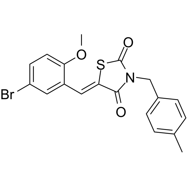
GQ-16 is an orally active PPARγ partial agonist with an IC50 of 1.84 μM and a Ki of 160 nM against human PPARγ. GQ-16 inhibits Cdk5-mediated Ser-273 phosphorylation. GQ-16 improves insulin sensitivity and glucose tolerance in obese and diabetic mice. GQ-16 also exhibits certain cytotoxicity against tumor cells. GQ-16 can be used in research related to obesity, diabetes and cancer.
For research use only. We do not sell to patients.
- Purity : 98.12%
- CAS No.: 870554-67-9
- Formula: C19H16BrNO3S
- Molecular Weight:418.30
-
Storage:Powder -20°C, 3 years ; In solvent -80°C, 6 months , -20°C, 1 month

To place orders, for customer services and technical support, please contact: MedChemExpress USA
Tel: 609-228-6898 E-mail: [email protected] [email protected]


-
 Biological Activity
-
Chemical Information
-
Solvent & Solubility
- Protocols
- Purity & Documentation
- References
-
 Help & FAQs


-
HY-100428
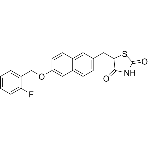
NetoglitazonePPARγ Ligand -
HY-13956
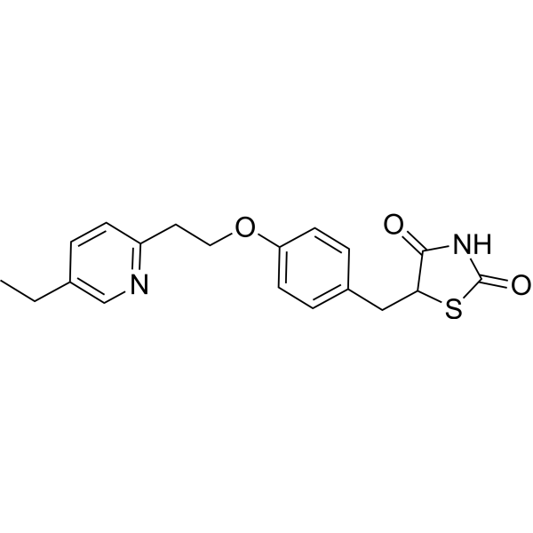
PioglitazonePPARγ Agonist -
HY-17386
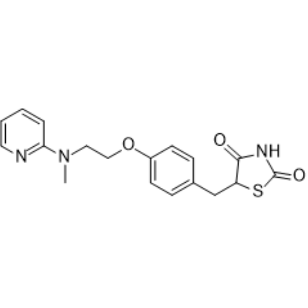
RosiglitazonePPARγ Agonist -
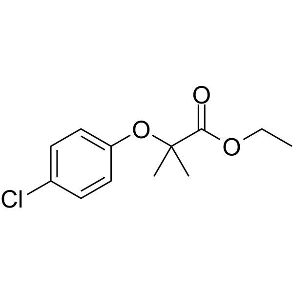
HY-B0287
ClofibratePPAR Agonist -
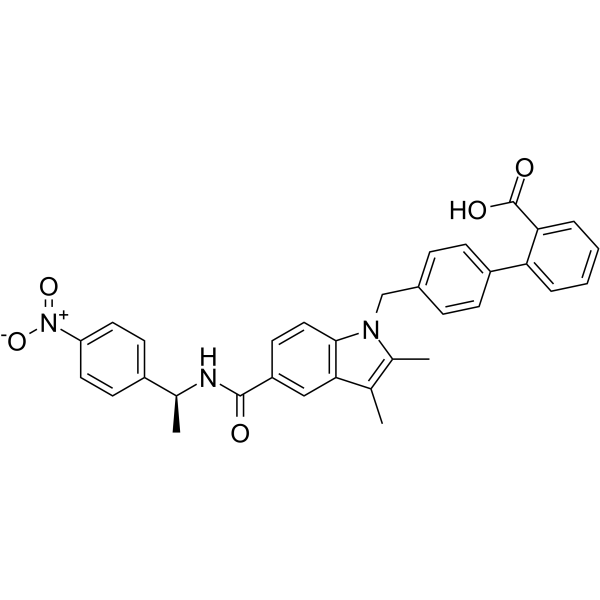
HY-12483
SR1664PPARγ Antagonist -
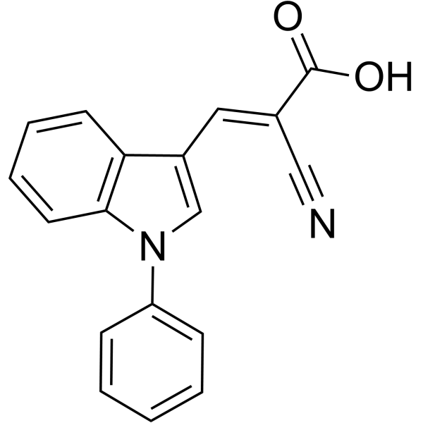
HY-15475
UK-5099Mitochondrial Pyruvate Carrier Inhibitor -
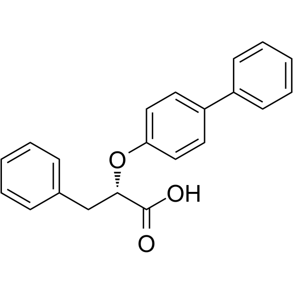
HY-121900
LT175PPARα/γ Ligand -
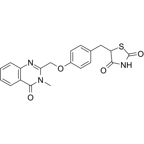
HY-16086
Balaglitazone -
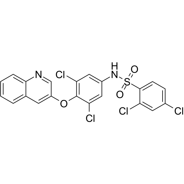
HY-117103
AMG131PPARγ Agonist -
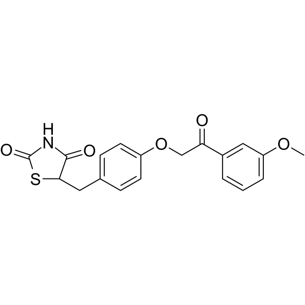
HY-108022
AzemiglitazoneMPC Inhibitor -
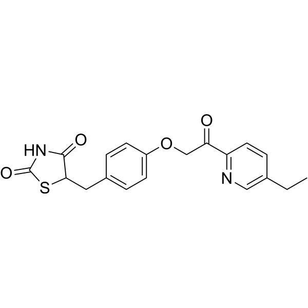
HY-100550
MSDC 0160Insulin Sensitizer -
HY-14928
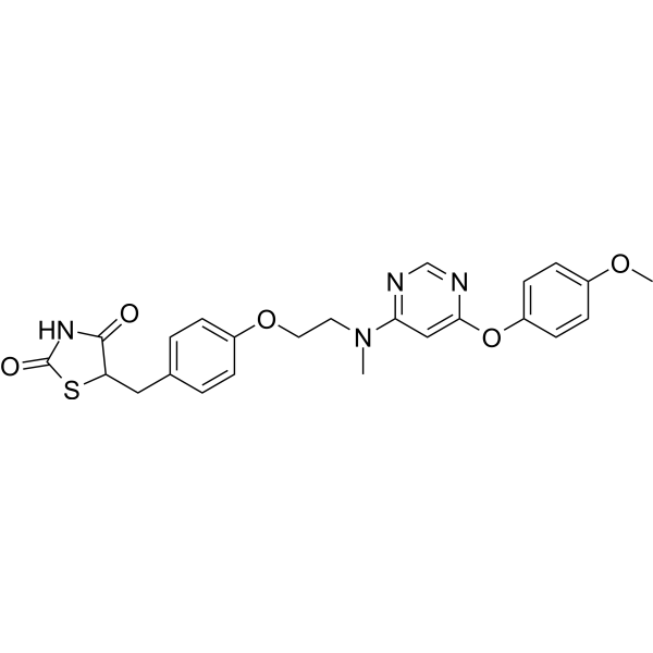
LobeglitazonePPAR Agonist


Biological Activity
Description
In Vitro
GQ-16 (7 days) promotes the differentiation of 3T3-L1 preadipocytes and upregulates the expression of adipogenic genes, but its efficacy is lower than that of Rosiglitazone (HY-17386)[1].
GQ-16 (10 nM-10 μM; 24 h) weakly activates the expression of PPARγ target genes acca1b and fgf21, and inhibits the expression of vcam1 and dcn in mature 3T3-L1 adipocytes[1].
GQ-16 (10 μM; 6-48 h) weakly and transiently upregulates the expression of OLR1 in mature 3T3-L1 adipocytes, with its activity peak occurring at 10 h post-treatment[1].
GQ-16 (10 μM; 4 days) exhibits reduced adipogenic potential in C3H10T1/2 and NIH-3T3L1 cells, with less intracellular lipid accumulation and lower aP2 expression levels compared to Rosiglitazone (HY-17386)[2].
GQ-16 (10 nM-100 μM; 24-48 h) reduces the viability of MCF-7 cells[3].
MedChemExpress (MCE) has not independently confirmed the accuracy of these methods. They are for reference only. Further protocols information, click here.
-
Cell Line:Mature 3T3-L1 adipocytes
-
Concentration:0.01, 0.1, 1, 10 μM
-
Incubation Time:24 h
-
Result:Dose-dependently increased expression of acca1b and fgf21.
Dose-dependently repressed expression of vcam1 and dcn.
In Vivo
MedChemExpress (MCE) has not independently confirmed the accuracy of these methods. They are for reference only.
-
Animal Model:Swiss mice (male, 12 weeks old, fed high fat diet from 4 weeks of age)[2]
-
Dosage:20 mg/kg/day
-
Administration:oral gavage; daily; 10, 12, or 15 days (depending on endpoint)
-
Result:Reversed high fat diet-mediated impairments in insulin signaling, including increased phosphorylation of the insulin receptor, insulin receptor substrate 1, and protein kinase B in adipose tissue, liver, and muscle.
Reversed high fat diet-dependent suppression of inhibitor of nuclear factor κB (IκBα) protein and reduced high fat diet-dependent phosphorylation of JNK in adipose tissue, liver, and muscle.
Increased the plasma glucose disappearance rate (Kitt) to improve insulin sensitivity.
Improved glucose tolerance.
Did not elicit increased weight gain relative to high fat diet-fed controls.


Chemical Information
-
CAS No. 870554-67-9
-
Appearance Solid
-
Molecular Weight 418.30
-
Formula C19H16BrNO3S
-
Color Off-white to light yellow
-
SMILES
O=C(N(CC1=CC=C(C)C=C1)C/2=O)SC2=C/C3=CC(Br)=CC=C3OC
-
Shipping
Room temperature in continental US; may vary elsewhere.
-
Storage
Powder -20°C 3 years In solvent -80°C 6 months -20°C 1 month


Solvent & Solubility
In Vitro:
DMSO : 10 mg/mL (23.91 mM; ultrasonic and warming and heat to 60°C; Hygroscopic DMSO has a significant impact on the solubility of product, please use newly opened DMSO)
Please refer to the solubility information to select the appropriate solvent. Once prepared, please aliquot and store the solution to prevent product inactivation from repeated freeze-thaw cycles.
Storage method and period of stock solution: -80°C, 6 months; -20°C, 1 month. When stored at -80°C, please use it within 6 months. When stored at -20°C, please use it within 1 month.
Please refer to the solubility information to select the appropriate solvent. Once prepared, please aliquot and store the solution to prevent product inactivation from repeated freeze-thaw cycles.
Storage method and period of stock solution: -80°C, 6 months; -20°C, 1 month. When stored at -80°C, please use it within 6 months. When stored at -20°C, please use it within 1 month.
Concentration (start) × Volume (start) = Concentration (final) × Volume (final)
In Vivo:
Select the appropriate dissolution method based on your experimental animal and administration route.
- For the following dissolution methods, please ensure to first prepare a clear stock solution using an In Vitro approach and then sequentially add co-solvents:
- To ensure reliable experimental results, the clarified stock solution can be appropriately stored based on storage conditions. As for the working solution for In Vivo experiments, it is recommended to prepare freshly and use it on the same day.
- The percentages shown for the solvents indicate their volumetric ratio in the final prepared solution. If precipitation or phase separation occurs during preparation, heat and/or sonication can be used to aid dissolution.
In Vivo Dissolution Calculator
Please enter the basic information of animal experiments:
-
-
-
-
Recommended: Prepare an additional quantity of animals to account for potential losses during experiments.
Please enter your animal formula composition:
-
%DMSO +
Recommended: Keep the proportion of DMSO in working solution below 2% if your animal is weak.
-
%+
-
+%Tween-80 + +
-
%Saline +
The co-solvents required include: DMSO, . All of co-solvents are available by MedChemExpress (MCE). , Tween 80. All of co-solvents are available by MedChemExpress (MCE).
Working solution concentration: 0.22 mg/mL
Method for preparing stock solution: mg drug dissolved in μL DMSO. Stock solution concentration: mg/mL.
1. Take μL DMSO stock solution;
2. Add μL .
μL , mix evenly;
3. Then add μL Tween 80, mix evenly;
4. Then add μL
Please ensure that the stock solution in the first step is dissolved to a clear state, and add co-solvents in sequence. You can use ultrasonic heating (ultrasonic cleaner, recommended frequency 20-40 kHz), vortexing, etc. to assist dissolution.

Protocols
-
Kinase activity and phosphorylation assays
Kinase activity assays measure the ability of kinases to transfer phosphate groups from ATP to specific substrates, while phosphorylation assays detect the presence and levels of phosphorylated proteins. Common methods include radiolabeled ATP incorporation (e. g. ,), ADP release detection via bioluminescence (e. g. ,[3]), enzyme-linked immunosorbent assays (ELISA) for phospho-specific epitopes (e. g. ,[6]), and microtiter-based formats for high-throughput screening (e. g. ,[8]). The ADP-Glo assay quantifies kinase activity by measuring ADP produced during phosphorylation using a luciferase-based system. Radiometric assays involve autoradiography or scintillation counting after incorporation of 32P-labeled ATP into substrate proteins. ELISA-based approaches rely on phospho-specific antibodies to detect activated kinases in cell lysates or purified samples.
-
Western Blot
Western blotting (WB) is a commonly used experimental method in molecular biology, biochemistry, and immunogenetics for identifying and quantifying target proteins. It combines gel electrophoresis with immunoassay, enabling researchers to analyze protein expression, post-translational modifications, and molecular weight.
-
Cell Cytotoxicity Assay
Cytotoxicity assays are usually based on the assessment of cell membrane damage, which can also be indirectly detected by measuring cell viability. Detection methods include MTT assay, CKK-8 assay, LDH assay and ATP assay, etc.
-
Mammalian live/dead viability and cytotoxicity staining
Live/dead viability and cytotoxicity staining assays are based on the simultaneous detection of intracellular esterase activity in metabolically active (viable) cells and membrane integrity loss in non-viable cells. In commonly used dual-staining approaches, membrane-permeant fluorogenic substrates are converted by intracellular esterases into fluorescent products in live cells, while impermeant DNA-binding dyes selectively enter cells with compromised plasma membranes and label nucleic acids in dead or dying cells, enabling discrimination between viable and non-viable populations by fluorescence microscopy or flow cytometry.
-
Research Protocol for Endocrine Diseases
Endocrine diseases often arise from disrupted hormone production, hormone signaling, or target-tissue responsiveness; for diabetes-focused endocrine disease models, insulin signaling regulates glucose uptake, hepatic glucose output, lipid metabolism, and β-cell compensation. Type 2 diabetes develops through interacting defects in insulin resistance, β-cell dysfunction, adipose inflammation, hepatic glucose overproduction, altered incretin signaling, and ectopic lipid metabolism. A major unresolved question is whether endocrine dysfunction is driven primarily by target-tissue insulin resistance, intrinsic β-cell failure, immune/inflammatory stress, or combined multi-organ failure that differs by disease stage.
-
Protocol for Kinase activity and phosphorylation assays
Kinase activity assays measure transfer of phosphate from ATP to a protein or peptide substrate, generating phosphorylated substrate, ADP, or incorporated radiolabeled phosphate as the readout; phosphorylation assays measure site-specific phosphorylation in cells or tissues as a proxy for kinase-pathway activation, inhibition, or substrate regulation. Phosphorylation can be detected by phospho-specific Western blot, immunoprecipitation kinase assay, phospho-immunofluorescence, phospho-flow cytometry, luminescent ADP detection, radiolabeled ATP incorporation, or reporter-based pathway assays, and these readouts can be applied to cancer cells, primary neurons, mouse tumors, organoids, inflammatory macrophages, ferroptosis studies, and mitophagy studies when the kinase target is biologically relevant.
-
Research Protocol for Metabolic Diseases
AMP-activated protein kinase, AMPK, is a conserved cellular energy sensor that responds to reduced cellular energy status and coordinates metabolism by increasing ATP-generating catabolic pathways while suppressing ATP-consuming anabolic processes. In metabolic disease research, the AMPK pathway is experimentally relevant because it regulates hepatic lipid synthesis, fatty acid oxidation, glucose production, skeletal-muscle glucose disposal, mTORC1-linked biosynthesis, autophagy, mitochondrial homeostasis, and whole-body energy balance. The central pathway logic is that energy stress, metformin, exercise-like stimulation, or direct AMPK activators increase AMPKα Thr172 phosphorylation and downstream substrate phosphorylation, including ACC and RAPTOR. Phosphorylation of ACC suppresses lipogenesis and supports fatty acid oxidation, whereas phosphorylation of RAPTOR suppresses mTORC1 signaling and links cellular energy status to growth and protein synthesis control. The pathway is linked
-
Cell Viability Determination by MTT Colorimetric Assay
The following protocol uses the MTT colorimetric assay as a classic literature-established method for assessing cell viability/metabolic activity in cultured mammalian cells. MTT[3-(4,5-dimethylthiazol-2-yl)-2,5-diphenyltetrazolium bromide] is reduced by metabolically active cells to a colored formazan product; the amount of formazan is quantified spectrophotometrically and provides an indirect measure of metabolically active viable cells. Importantly, MTT reduction reflects cellular oxidoreductase/metabolic activity rather than an absolute direct count of living cells, so changes in cellular metabolism can alter the signal independently of cell number.


Purity & Documentation
-
Data Sheet (278 KB)
-
SDS (393 KB)
- English - EN (393 KB)
- Français - FR (393 KB)
- Deutsch - DE (393 KB)
- Norwegian - NO (393 KB)
- Español - ES (393 KB)
- Swedish - SV (393 KB)
- Italian - IT (393 KB)
- Korean - KR (393 KB)
- Portuguese - PT (393 KB)
-
Handling Instructions (2659 KB)
References
[2]. Milton FA, et al. PPARγ partial agonist GQ-16 strongly represses a subset of genes in 3T3-L1 adipocytes. Biochem Biophys Res Commun. 2015 Aug 28;464(3):718-23. [Content Brief]
[3]. Amato AA, et al. GQ-16, a novel peroxisome proliferator-activated receptor γ (PPARγ) ligand, promotes insulin sensitization without weight gain. J Biol Chem. 2012;287(33):28169-28179. [Content Brief]


Complete Stock Solution Preparation Table
Please refer to the solubility information to select the appropriate solvent. Once prepared, please aliquot and store the solution to prevent product inactivation from repeated freeze-thaw cycles.
Storage method and period of stock solution: -80°C, 6 months; -20°C, 1 month. When stored at -80°C, please use it within 6 months. When stored at -20°C, please use it within 1 month.
| Optional Solvent | Concentration Solvent Mass | 1 mg | 5 mg | 10 mg | 25 mg |
|---|---|---|---|---|---|
| DMSO | 1 mM | 2.3906 mL | 11.9531 mL | 23.9063 mL | 59.7657 mL |
| 5 mM | 0.4781 mL | 2.3906 mL | 4.7813 mL | 11.9531 mL | |
| 10 mM | 0.2391 mL | 1.1953 mL | 2.3906 mL | 5.9766 mL | |
| 15 mM | 0.1594 mL | 0.7969 mL | 1.5938 mL | 3.9844 mL | |
| 20 mM | 0.1195 mL | 0.5977 mL | 1.1953 mL | 2.9883 mL |


GQ-16 Related Classifications
HY-111254 Related Classifications
 Powered by Bioz